rom j morphol embryol 2016, 57(3):1069–1073 r j m e original paper romanian journal of morphology...
TRANSCRIPT
Rom J Morphol Embryol 2016, 57(3):1069–1073
ISSN (print) 1220–0522 ISSN (online) 2066–8279
OORRIIGGIINNAALL PPAAPPEERR
Micro-CT and optical microscopy imagistic investigations of root canal morphology
EMANUELA LIDIA CRĂCIUNESCU1)#, MARIUS BOARIU2), CIPRIAN IONIŢĂ3), DANIELA MARIA POP1), COSMIN SINESCU1), MIHAI ROMÎNU1)#, MEDA LAVINIA NEGRUŢIU1)
1)Department of Prostheses Technology and Dental Materials, Faculty of Dental Medicine, “Victor Babeş” University of Medicine and Pharmacy, Timisoara, Romania
2)Department of Dental Medicine 3 – Restorative Dentistry and Endodontics, Faculty of Dental Medicine, “Victor Babeş” University of Medicine and Pharmacy, Timisoara, Romania
3)Department of Stroke and Vascular Research Center, University at Buffalo, The State University of New York, Buffalo, New York, USA
#These authors contributed equally to this paper.
Abstract Protecting the root’s internal morphology is the first key toward the success of the endodontic treatment. Due to the vast diversity of endodontic space, it is difficult to visualize and to establish the shape and limits of the root canal, especially the morphology of apical area and lateral root canals. Optical microscopy is a classical imagistic investigation method, widely used along classical methods like radiographs that also offer limited information about root morphology and extension of decay. Micro-computed tomography (micro-CT), a modern imagistic investigation method can provide detailed three-dimensional reconstructions of root canal. Micro-CT is a non-invasive method that has the possibility to offer cross-sectional and axial images of the endodontic space. The success of root canal treatment is based on cleaning and shaping. Beyond these two procedures, sealing the endodontic space by respecting its limits is another prerequisite for long-term success of endodontic therapy. Micro-CT can perform three-dimensional reconstruction of the root canal, root canal filling and can provide accurate images of the endodontic space. The assessment of root morphology can be obtained through imagistic invasive optical microscopy and already mentioned non-invasive methods (micro-CT). The aim of this study is to illustrate and analyze the endodontic space, according to its diversity by using micro-CT, a non-invasive imagistic investigation method an also optical microscopy. The two techniques can also provide the extension of carries or demineralized substance on different levels of the root.
Keywords: micro-CT, optical microscopy, root canal morphology.
Introduction
In endodontic therapy, besides cleaning and mechanical preparation of endodontic space, it is mandatory for the practitioner to identify and preview the morphology of the root canal and its limits. The classical periapical X-ray investigations give poor information about the root canal morphology and no information about the lateral root canals. Considering the group of teeth – upper, lower, incisors, molars, the angled radiographs are parallel radio-graphs providing information only about major aspects of root canal morphology. The aspects related to size, orientation and even number of the root canals are difficult to be spotted by currently used periapical X-ray investigation [1, 2]. The era of non-invasive imagistic investigation methods and three-dimensional reconstruc-tions has changed the expectations and perspectives of researchers and practitioner. The X-ray micro-computed tomography (micro-CT) is a non-destructive, non-invasive investigation method, which provides two-dimensional (2D) and three-dimensional (3D) images [3–5]. The development of these systems emphasizes the practical aspects of micro-CT, which become one of the standard techniques in three-dimensional evaluations for in vitro studies. The standard periapical X-rays still remain the one used in daily clinical practice [6]. The 3D analysis
of micro-CT images is strongly correlated with 2D cross-sections of internal root morphology. One major advantage of micro-CT is the possibility to repeat the scanning and to evaluate the image using the specific software. This investigation method shows its limits for the in vivo studies especially due to the high radiation dose. Micro-CT imagistic system was used in different investigations like micro-structural analysis of osteo-conductive bio-materials and interface evaluation between dental fillings and dental hard tissue [7–9]. Micro-CT investigation method can scan the entire tooth structures: root canal, dentin, enamel, internal and external anatomy of the root, root canal and the teeth’s tissues demineralized or affected by carries.
The aim of this in vivo study is to investigate through micro-CT and optical microscopy the endodontic mor-phology and to identify demineralized tissue, which may also be present on the root.
Materials and Methods
For this in vitro study, 27 human extracted permanent teeth (premolars and molars) were selected. The teeth were collected from dental clinics and were extracted either for orthodontic purposes or due to periodontal disease. After extraction and debridement, the teeth were
R J M ERomanian Journal of
Morphology & Embryologyhttp://www.rjme.ro/
Emanuela Lidia Crăciunescu et al.
1070
stored in 5% sodium hypochlorite solution for 24 hours, then washed under tap water and dried. The selected teeth had a fully formed apex, no restorations and no endodontic treatment. For each tooth, a silicone holder was manufactured. The holder assures a precise and stable positioning and undesired rotating possibility of the sample in the scanning system.
The samples were scanned using a custom-built cone beam micro-CT for dental investigations. This system consists of an X-ray detector, a circular/rotary positioning stage, an X-ray shutter and a micro-focus X-ray tube. The X-ray detector is made up of a charge-coupled device (CCD) chip cemented on a minifying fiber optic tape. The larger end of the taper is coupled with a 300 μm CsI (Cesium Iodide) phosphor. The effective detector pixel size is 45 μm. The focal spot of the X-ray tube is between 10–20 μm, depending on the current value.
The X-ray exposure parameters were 40 kVp, 1 mA and 300 ms per frame. Each sample was precisely positioned into the silicone holder on the circular/rotary positioning stage. The sample was exposed at a magni-fication between 2 and 1.1, depending by the sample size. The sample was rotated for 3600, in one-degree strep increments. The projections were reconstructed using a standard Feldkamp–Davis–Kress (FDK) algorithm, with a Shepp–Logan filter.
Each root was scanned and the morphology of the root canal was made after registering the cross-sectional images of the scanned roots. The cross-sections of the root canal give the possibility to evaluate its characteristics and individual morphology in high detail. The 3D reconstruc-tion of the root canal gives the possibility to evaluate its characteristics and individual morphology in great detail.
The area of interest of each sample was scanned. The frames obtained during scanning of the root canal were analyzed in detail in order to establish the particu-larities of the root canal.
In opposition to micro-CT, optical microscopy inves-tigation is an invasive imagistic investigation method,
which involves the preparation in terms of sectioning the samples. The roots of permanent teeth were cut into slices of 2 mm thickness following a transversal direction (similar with the micro-CT investigation) and the shape of the root canal was then analyzed using a Krüss optic micro-scope, at a magnification of ×10. Optical microscopy is difficult to be used on non-planar preparation analysis of sharp and irregular cuts. The image acquisition is difficult to be obtained because of the small focus depth of the optical microscope. The proper method for solving this issue is the acquisition of a sequence of microscopic images with a constant change of the focal length between images. A non-planar sample cannot be displayed sharp in a full version. The samples are scanned gradually with changing focal length.
Results
The images obtained through micro-CT showed the detailed morphology of the root canal and apical region. Elements, which might have been remained unnoticed on periapical X-ray, were detected.
The shape of the root canal on transversal section is well defined and is varying in diameter and shape. The registered images of the main root canal show an oval, triangle flatten or round shape and it is not always localized in the centre of the root. The shape of the root canal is constantly changing, at different cross-sectional levels. Curvature of the root and root canal are positioning the apical delta and the apex mesially or distally.
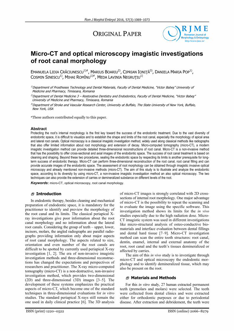
Moreover, secondary fine root canals could be identified and usually these cannot be detected on classical 2D radiographs. Micro-CT spotted that the shape of the same root canal is changing from a regular, round shape into an oval shape into the apical third (Figure 1, a–c). The walls of the root canal may display irregular margins, which generate an irregular shape of the root canal in the middle third area (Figure 1, d–f).
Figure 1 – Micro-CT aspect (a–d) of upper first premolar 1.4– transversal section. The diameter of the two root canals is increasing in size and shape is changing at different levels:apical third (a), middle third (b and c), cervical third (d and e).In the cervical area, a discontinuity of the contour (arrow –e and f) may be probably attributed to root caries.
Micro-CT and optical microscopy imagistic investigations of root canal morphology
1071
The apex and apical delta may have an unpredictable position and morphology. Very fine located root canals are almost impossible to be investigated by radiographs and almost impossible to be prepared (Figure 1, a–c). It is mandatory for the success of the endodontic treatment to always consider and to seal both these lateral and fine root canals. Their identification and sealing is possible only by associating an adequate preparation and sealing technique.
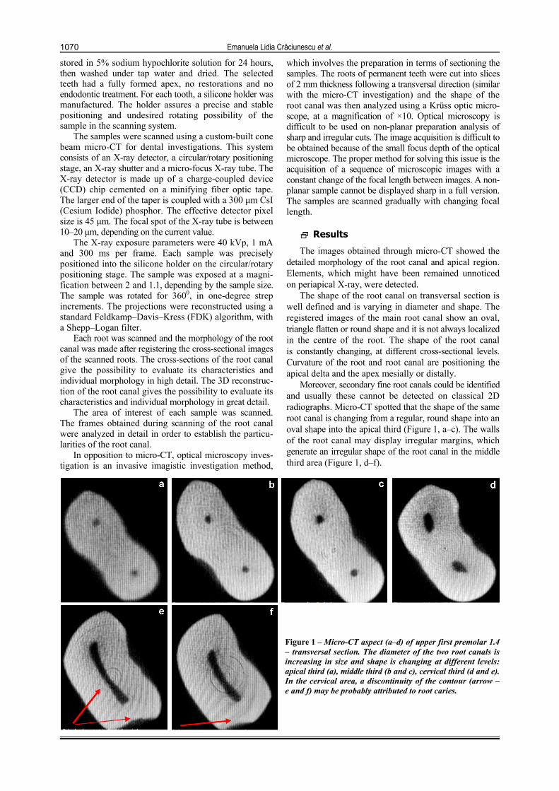
The micro-CT investigation also detected lateral root canals, which are positioned at different levels of the root (Figure 2, a–g). Lateral root canals are localized in the neighborhood of the root canal and may be missed by periodontal X-rays. Carries and the demineralized dentin around the active process are favorable for fissure-like defects (Figure 3). The demineralized dentin by carries and other associated factors may initiate fissures into the tooth structure.
Figure 2 – Micro-CT aspect of tooth 3.5-transversal section: (a) In the third apical area, the forked root canals are consisting into one root canal; (b and c) The same root canal is forked in the middle third area of the root; (d and e) Only one root canal is observed into third cervical area of the second upper premolar.
Figure 3 – Micro-CT aspect of an upper premolar. The
secondary root canal is observed in the apical area and a demineralized area
of the proximal face of the root.
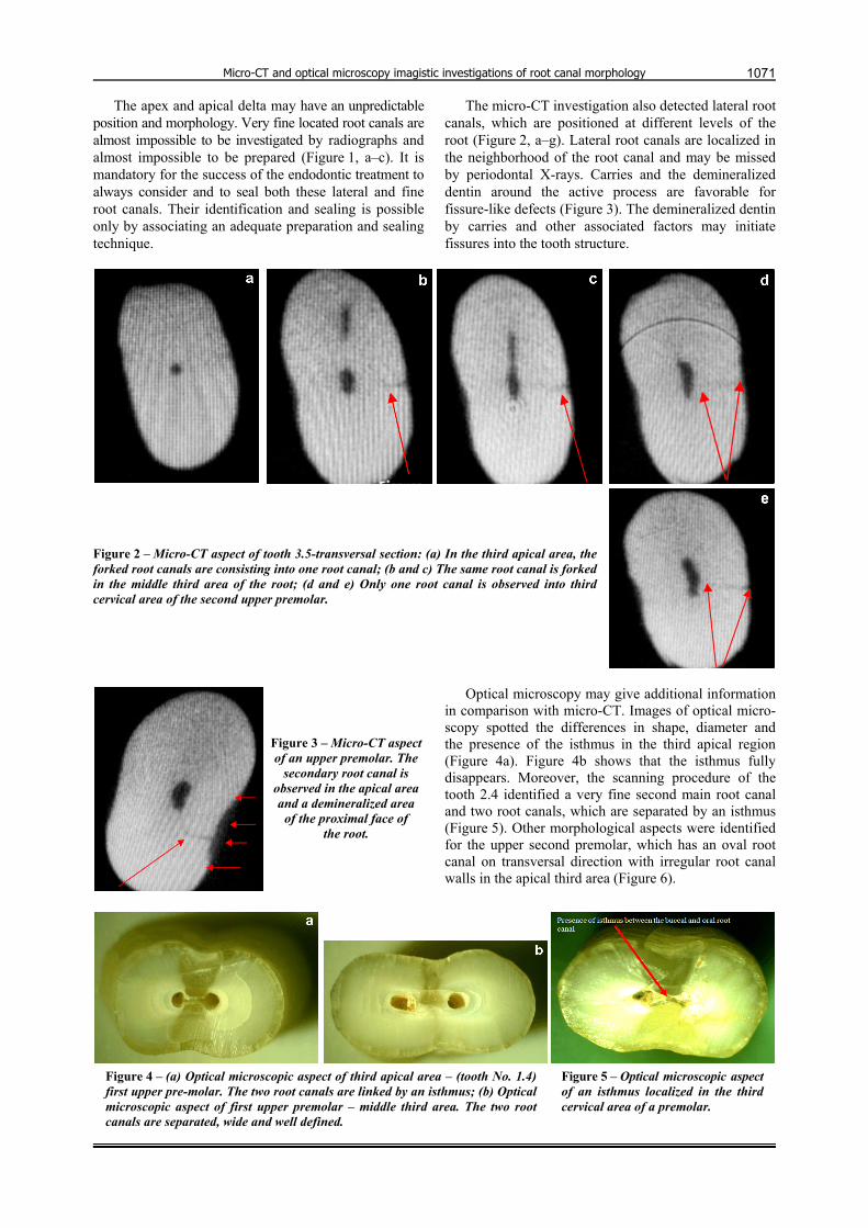
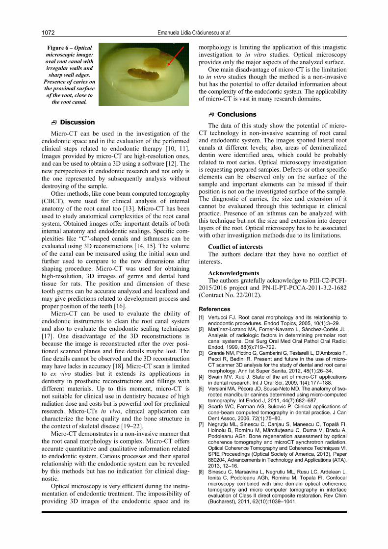
Optical microscopy may give additional information in comparison with micro-CT. Images of optical micro-scopy spotted the differences in shape, diameter and the presence of the isthmus in the third apical region (Figure 4a). Figure 4b shows that the isthmus fully disappears. Moreover, the scanning procedure of the tooth 2.4 identified a very fine second main root canal and two root canals, which are separated by an isthmus (Figure 5). Other morphological aspects were identified for the upper second premolar, which has an oval root canal on transversal direction with irregular root canal walls in the apical third area (Figure 6).
Figure 4 – (a) Optical microscopic aspect of third apical area – (tooth No. 1.4) first upper pre-molar. The two root canals are linked by an isthmus; (b) Optical microscopic aspect of first upper premolar – middle third area. The two root canals are separated, wide and well defined.
Figure 5 – Optical microscopic aspect of an isthmus localized in the third cervical area of a premolar.
Emanuela Lidia Crăciunescu et al.
1072
Figure 6 – Optical microscopic image: oval root canal with irregular walls and sharp wall edges.
Presence of caries on the proximal surface of the root, close to
the root canal.
Discussion
Micro-CT can be used in the investigation of the endodontic space and in the evaluation of the performed clinical steps related to endodontic therapy [10, 11]. Images provided by micro-CT are high-resolution ones, and can be used to obtain a 3D using a software [12]. The new perspectives in endodontic research and not only is the one represented by subsequently analysis without destroying of the sample.
Other methods, like cone beam computed tomography (CBCT), were used for clinical analysis of internal anatomy of the root canal too [13]. Micro-CT has been used to study anatomical complexities of the root canal system. Obtained images offer important details of both internal anatomy and endodontic sealings. Specific com-plexities like “C”-shaped canals and isthmuses can be evaluated using 3D reconstructions [14, 15]. The volume of the canal can be measured using the initial scan and further used to compare to the new dimensions after shaping procedure. Micro-CT was used for obtaining high-resolution, 3D images of germs and dental hard tissue for rats. The position and dimension of these tooth germs can be accurate analyzed and localized and may give predictions related to development process and proper position of the teeth [16].
Micro-CT can be used to evaluate the ability of endodontic instruments to clean the root canal system and also to evaluate the endodontic sealing techniques [17]. One disadvantage of the 3D reconstructions is because the image is reconstructed after the over posi-tioned scanned planes and fine details maybe lost. The fine details cannot be observed and the 3D reconstruction may have lacks in accuracy [18]. Micro-CT scan is limited to ex vivo studies but it extends its applications in dentistry in prosthetic reconstructions and fillings with different materials. Up to this moment, micro-CT is not suitable for clinical use in dentistry because of high radiation dose and costs but is powerful tool for preclinical research. Micro-CTs in vivo, clinical application can characterize the bone quality and the bone structure in the context of skeletal disease [19–22].
Micro-CT demonstrates in a non-invasive manner that the root canal morphology is complex. Micro-CT offers accurate quantitative and qualitative information related to endodontic system. Carious processes and their spatial relationship with the endodontic system can be revealed by this methods but has no indication for clinical diag-nostic.
Optical microscopy is very efficient during the instru-mentation of endodontic treatment. The impossibility of providing 3D images of the endodontic space and its
morphology is limiting the application of this imagistic investigation to in vitro studies. Optical microscopy provides only the major aspects of the analyzed surface.
One main disadvantage of micro-CT is the limitation to in vitro studies though the method is a non-invasive but has the potential to offer detailed information about the complexity of the endodontic system. The applicability of micro-CT is vast in many research domains.
Conclusions
The data of this study show the potential of micro-CT technology in non-invasive scanning of root canal and endodontic system. The images spotted lateral root canals at different levels; also, areas of demineralized dentin were identified area, which could be probably related to root caries. Optical microscopy investigation is requesting prepared samples. Defects or other specific elements can be observed only on the surface of the sample and important elements can be missed if their position is not on the investigated surface of the sample. The diagnostic of carries, the size and extension of it cannot be evaluated through this technique in clinical practice. Presence of an isthmus can be analyzed with this technique but not the size and extension into deeper layers of the root. Optical microscopy has to be associated with other investigation methods due to its limitations.
Conflict of interests The authors declare that they have no conflict of
interests.
Acknowledgments The authors gratefully acknowledge to PIII-C2-PCFI-
2015/2016 project and PN-II-PT-PCCA-2011-3.2-1682 (Contract No. 22/2012).
References [1] Vertucci FJ. Root canal morphology and its relationship to
endodontic procedures. Endod Topics, 2005, 10(1):3–29. [2] Martínez-Lozano MA, Forner-Navarro L, Sánchez-Cortés JL.
Analysis of radiologic factors in determining premolar root canal systems. Oral Surg Oral Med Oral Pathol Oral Radiol Endod, 1999, 88(6):719–722.
[3] Grande NM, Plotino G, Gambarini G, Testarelli L, D’Ambrosio F, Pecci R, Bedini R. Present and future in the use of micro-CT scanner 3D analysis for the study of dental and root canal morphology. Ann Ist Super Sanita, 2012, 48(1):26–34.
[4] Swain MV, Xue J. State of the art of micro-CT applications in dental research. Int J Oral Sci, 2009, 1(4):177–188.
[5] Versiani MA, Pécora JD, Sousa-Neto MD. The anatomy of two-rooted mandibular canines determined using micro-computed tomography. Int Endod J, 2011, 44(7):682–687.
[6] Scarfe WC, Farman AG, Sukovic P. Clinical applications of cone-beam computed tomography in dental practice. J Can Dent Assoc, 2006, 72(1):75–80.
[7] Negruţiu ML, Sinescu C, Canjau S, Manescu C, Topală FI, Hoinoiu B, Romînu M, Mărcăuţeanu C, Duma V, Bradu A, Podoleanu AGh. Bone regeneration assessment by optical coherence tomography and microCT synchrotron radiation. Optical Coherence Tomography and Coherence Techniques VI, SPIE Proceedings (Optical Society of America, 2013), Paper 880204, Advancements in Technology and Applications (ATA), 2013, 12–16.
[8] Sinescu C, Marsavina L, Negrutiu ML, Rusu LC, Ardelean L, Ionita C, Podoleanu AGh, Rominu M, Topala FI. Confocal microscopy combined with time domain optical coherence tomography and micro computer tomography in interface evaluation of Class II direct composite restoration. Rev Chim (Bucharest), 2011, 62(10):1039–1041.
Micro-CT and optical microscopy imagistic investigations of root canal morphology
1073
[9] Jung M, Lommel D, Klimek J. The imaging of root canal obturation using micro-CT. Int Endod J, 2005, 38(9):617–626.
[10] Villas-Bôas MH, Bernardineli N, Cavenago BC, Marciano M, Del Carpio-Perochena A, de Moraes IG, Duarte MH, Bramante CM, Ordinola-Zapata R. Micro-computed tomography study of the internal anatomy of mesial root canals of mandibular molars. J Endod, 2011, 37(12):1682–1686.
[11] Hammad M, Qualtrough A, Silikas N. Evaluation of root canal obturation: a three-dimensional in vitro study. J Endod, 2009, 35(4):541–544.
[12] Shibuya E, Matsubayashi T, Shida T. Experimental study for the accuracy micro-CT apparatus. Shika Gakuho, 2000, 100(12):1221–1226.
[13] Baratto Filho F, Zaitter S, Haragushiku GA, de Campos EA, Abuabara A, Correr GM. Analysis of the internal anatomy of maxillary first molars by using different methods. J Endod, 2009, 35(3):337–342.
[14] Fan B, Pan Y, Gao Y, Fang F, Wu Q, Gutmann JL. Three-dimensional morphologic analysis of isthmuses in the mesial roots of mandibular molars. J Endod, 2010, 36(11):1866–1869.
[15] Bergmans L, Van Cleynenbreugel J, Wevers M, Lambrechts P. A methodology for quantitative evaluation of root canal instru-mentation using microcomputed tomography. Int Endod J, 2001, 34(5):390–398.
[16] Dong G, Dong Q, Liu Y, Lou B, Feng J, Wang K, Zhou X, Wu H. High-resolution micro-CT scanning as an innovative tool for evaluating dental hard tissue development. J Appl Clin Med Phys, 2014, 15(4):335–344.
[17] Karapinar-Kazandag M, Basrani BR, Friedman S. The operating microscope enhances detection and negotiation of accessory mesial canals in mandibular molars. J Endod, 2010, 36(8):1289–1294.
[18] von Stechow D, Balto K, Stashenko P, Müller R. Three-dimensional quantitation of periradicular bone destruction by micro-computed tomography. J Endod, 2003, 29(4):252–256.
[19] Burghardt AJ, Link TM, Majumdar S. High-resolution computed tomography for clinical imaging of bone microarchitecture, Clin Orthop Relat Res, 2011, 469(8):2179–2193.
[20] Negrutiu ML, Nica L, Sinescu C, Topala F, Ionita C, Bradu A, Petrescu EL, Pop DM, Rominu M, Podoleanu AGh. SEM and microCT validation for en face OCT imagistic evaluation of endodontically treated human teeth. SPIE7961 Proceedings, Medical Imaging 2011: Physics of Medical Imaging, 79614W (March 17, 2011).
[21] Lyroudia K, Mikrogeorgis G, Nikopoulos N, Samakovitis G, Molyvdas I, Pitas I. Computerized 3-D reconstruction of two “double teeth”. Endod Dent Traumatol, 1997, 13(5):218–222.
[22] Niederoest M, Niederoest J, Šcucka J. Shape from focus: fully automated 3D reconstruction and visualization of micro-scopic objects. Technical Session, 4–11. http://e-collection. library.ethz.ch/eserv/eth:26934/eth-26934-01.pdf.
Corresponding author Cosmin Sinescu, Lecturer, MD, PhD, Department of Prostheses Technology and Dental Materials, Faculty of Dental Medicine, “Victor Babeş” University of Medicine and Pharmacy, 2 Eftimie Murgu Square, 300041 Timişoara, Timiş County, Romania; Phone +40722–280 132, e-mail: [email protected] Received: March 6, 2015
Accepted: December 14, 2016