etiopatogenia bolii de reflux gastro … sindrom sjogren, diabet zaharat, mixedem, les, polimiozite,...
TRANSCRIPT
REVISTA ROMÂNÅ DE PEDIATRIE – VOLUMUL LIX, NR. 3, AN 2010172
Boala de refl ux gastro-esofagian (BRGE) repre-zintă trecerea intermitentă sau permanentă a con-ţinutului gastric în esofag, determinând un cortegiu de simptome: digestive, respiratorii şi/sau neuro-com portamentale sau absenţa oricăror acuze.
O bună cunoaştere a datelor de embriogeneză, anatomie şi fi ziologie esofagiană duce la o înţelegere corectă a patologiei de la acest nivel şi, implicit, la o abordare terapeutică adecvată.
ELEMENTE DE EMBRIOLOGIE
Tractul gastrointestinal derivă în totalitate din intestinul primitiv, un tub extins de la gură la anus, care se formează în săptămâna 3-4 de viaţă intra-uterină prin invaginaţia părţii dorsale a sacului vitelin în embrion.
Dezvoltarea se produce astfel:din endoderm derivă epiteliul şi glandele anexe ale aparatului digestiv,din mezoderm ia naştere lamina propria a sub-mucoasei şi a tunicilor musculare şi seroase.
Topografi c, intestinul primitiv se împarte în trei părţi: anterior, mijlociu şi posterior, care se afl ă la originea segmentelor tubului digestiv.
Esofagul derivă din partea anterioară a intes-tinului primitiv din care mai derivă: cavitatea
1.
2.
bucală, faringe, stomac, duodenul proximal până la ampula lui Vater, fi catul, vezicula biliară şi pan-creasul, toate irigate de trunchiul celiac (1).
La capătul cranial un diverticul ventral care va deveni traheea formează o gaură în peretele intes-tinului primitiv la aproximativ 3 săptămâni de la fertilizare. Pe măsură ce ambele structuri cresc, apar nişte creste pe pereţii laterali ai diverticulului şi ai intestinului primitiv care se unesc la aproximativ 5 săptămâni formând canale separate pentru esofag şi trahee (2).
ANATOMIA ESOFAGULUI
Esofagul este un conduct musculo-membranos, suplu şi dilatabil, ce uneşte faringele cu stomacul, extinzându-se de la nivelul vertebrei C6 (joncţiunea faringo-esofagiană) până la nivelul vertebrei T11 (orifi ciul cardiei) (3).
Direcţia esofagului nu este rectilinie. În traiectul său, prezintă curburi în plan sagital şi frontal.
La naştere măsoară 8-10 cm lungime şi 0,5 cm diametru, după care creşte cu 0,65 cm/an, ajungând să măsoare la adult 25 cm lungime şi 2-3 cm dia-metru (4).
Joncţiunea eso-gastrică (complexul esofagian inferior) este un sfi ncter fi ziologic fără individuali-
REFERATE GENERALE3ETIOPATOGENIA BOLII DE REFLUX GASTRO-ESOFAGIANDr. V.V. Lupu, Prof. Dr. M. BurleaClinica V Pediatrie Gastroenterologie, Disciplina Pediatrie UMF „Gr. T. Popa“, Iaşi
REZUMAT Boala de refl ux gastroesofagian (BRGE), „boala mileniului 3“, este mult mai frecvent întâlnită în practica pediatrică decât s-a crezut până în prezent. Autorii fac o incursiune scurtă în embriologia, anatomia, fi ziologia esofagului şi în istoria naturală a bolii pentru a explora, identifi ca şi explica mecanismele patogenice care stau la baza dezvoltării BRGE. Sunt descrise cele 3 verigi patogenice: disfuncţia sfi ncterului esofagian inferior, disfuncţia esofagiană şi disfuncţia gastrică, la care se adaugă hernia hiatală care are o relaţie patogenică specială cu BRGE.
Cuvinte cheie: joncţiunea esogastrică, refl ux gastroesofagian, imunohistochimie
Adresa de corespondenţă:Dr. V.V. Lupu, Universitatea de Medicină şi Farmacie „Gr. T. Popa“, Str. Universităţii, Nr. 16, Iaşi
REVISTA ROMÂNÅ DE PEDIATRIE – VOLUMUL LIX, NR. 3, AN 2010 173
tate anatomică. Elementele componente ale acestei zone sunt:
ampula epifrenică – concept radiologic – o zonă de expansiune luminală care apare dea-supra sfi ncterului vestibular în inspir;sfi ncterul vestibular – corespunde sfi ncterului esofagian inferior (SEI);vestibulul esofagian (ampula esofagiană);linia Z – linia de tranziţie epitelială – linie de demarcaţie anatomo-histologică între mu-coasa esofagiană pavimentoasă şi mucoasa gastrică, cilindrică – reper foarte util endos-copic (4, 5).
În condiţii normale, joncţiunea esogastrică repre-zintă un dispozitiv anatomic antirefl ux care îm piedică refl uarea conţinutului acid gastric în esofag.
Perturbări ale mecanismului contenţiei gastro-esofagiene determină:
apariţia RGE cu esofagite peptice secundare pâna la stenoză;hernii hiatale diafragmatice – când hiatusul esofagian este mai larg.
Inervaţia esofagului:a) inervaţia extrinsecă – asigurată de fi brele
simpatice, nervii vagi şi nervul laringial recurent (cerebrospinal);
b) inervaţia intrinsecă – constituită din plexul intramuscular al lui Auerbach şi din plexul submucos al lui Meissner. (2)
Nocicepţia şi proiecţia centrală comună esofag-trahee sunt incriminate în manifestările „asthma-like“ ale BRGE. (6)
Prin studii imunohistochimice este posibilă pre-cizarea siturilor de elaborare a substanţelor ne uro-hormonale (7):
VIP – prezent din viaţa intrauterină în plexuri şi SEI;Substanţa P – în fi brele musculare şi în plexuri, determină contracţia SEI;Enkefalina – în fi brele striate;Motilina – controlează SEI.
DATE DE FIZIOLOGIE A ESOFAGULUI
Esofagul îndeplineşte rol de:transport al bolului alimentar din cavitatea bucală în stomac;a preveni refl uxul gastroesofagian şi esofago-faringian, cu concursul sfi ncterului esofagian superior (SES) şi SEI.
SES menţine extremitatea proximală a esofagului şi previne refl uarea eso-faringiană. Presiunea SES este 40-100 mmHg, iar în timpul deglutiţiei, SES se relaxează tranzitoriu (8).
1.
2.
3.4.
1.
2.
1.
2.
3.4.
1.
2.
Tranzitul esofagian al bolului alimentar se face prin intervenţia a doi factori:
undele peristaltice;forţa gravitaţională.
Participarea celor doi factori depinde de poziţia corpului (3):
în clinostatism pasajul bolusului alimentar se face numai prin activitate peristaltică;în ortostatism, contribuie variabil şi forţa gra-vi ta ţională.
Undele peristaltice de la nivelul esofagului sunt:unde primare – iau naştere în faringe, sunt în relaţie cu deglutiţia, favorizează propulsia bolului alimentar;unde secundare – induse de distensia esofa-gului, iritanţi chimici sau mecanici şi nu se corelează cu deglutiţia.
Prevenirea leziunilor esofagiene determinate de re-fl uxul acid gastric se face prin contracţiile peri staltice se cundare ce realizează clearance-ul eso fagian.
Presiunea exercitată de undele peristaltice înving tonusul SEI, îl relaxează. Tonusul acestui sfi ncter creşte cu vârsta:
la copilul mic şi sugar, tonusul muscular scăzut face ca alimentele să treacă repede „ca prin tub“ şi să cadă în stomac;la copilul mare presiunea exercitată de bolul alimentar învinge tonusul muscular şi re-laxează cardia.
Funcţia principală a sfi ncterului esofagian in-ferior este de a preveni RGE. SEI nu se comportă în totdeauna ca o barieră antirefl ux, chiar la persoane sănătoase, astfel, postalimentar, putând apare o re-laxare tranzitorie a SEI neprecedată de unda peri-staltică.
ISTORIE NATURALĂ
Este binecunoscut faptul că refl uxul este fi zio-logic în primele 3 luni de viaţă, manifestat foarte precoce, de obicei în primele 6 săptămâni de viaţă (9).
De asemeni, în mare parte, regurgitaţiile dispar pâna la vârsta de 12-24 luni, persistenţa lor după această vârstă ridică problema apariţiei consecin-ţelor refl uxului – esofagita de refl ux – şi încadrarea ca zurilor în boala de refl ux gastro-esofagian (10).
Copiii care au prezentat în perioada de sugar episoade frecvente de regurgitaţii şi/sau vărsături sunt mai predispuşi de a dezvolta boala de refl ux în perioada următoare, mai ales dacă se asociază fu-matul şi prezenţa simptomelor de refl ux la mamă în timpul sarcinii respective (10).
1.2.
1.
2.
1.
2.
1.
2.
REVISTA ROMÂNÅ DE PEDIATRIE – VOLUMUL LIX, NR. 3, AN 2010174
Astfel, există diferenţe între refl uxul prezent în primele 12-24 luni, manifestat prin regurgitaţii şi vărsături şi refl uxul apărut după această vârstă (de obicei peste 18 luni), când predomină manifestările de tip respirator (mai ales wheezing recurent), iar descoperirea refl uxului este, de multe ori, întâm-plătoare sau prin excludere.
De asemeni, o categorie aparte o constituie copiii taraţi neurologic, encefalopaţii, care prezintă în mod aproape constant refl ux gastro-esofagian (11).
Dacă ar trebui să concluzionăm, populaţia cu risc pentru apariţia BRGE include:
prematuri şi dismaturi;nou-născuţi care au suferit de hipoxie sau asfi xie perinatală;sugari care au benefi ciat de sondă gastrică ,,a demeure“;sugari cu retard al ,,vidanjării gastrice“, (pri-mitiv sau secundar);nou-născuţi operaţi pentru atrezie esofagiană cu sau fără fi stulă;fi broză chistică;encefalopaţi, etc. (12)
ETIOPATOGENIE
Bariera anti-refl uxDupă cum am precizat la secţiunea privind ana-
tomia esofagului, în condiţii normale, joncţiunea esogastrică funcţionează ca un veritabil dispozitiv anatomic antirefl ux care împiedică refl uarea conţi-nutului gastric în esofag.
Elemente componente ale dispozitivului anti-refl ux (3):
calibrul normal al hiatusului esofagian;deschiderea oblică a esofagului în stomac prin orifi ciul cardiei – unghiul lui His;valvula lui Gubarow – vizibilă endoscopic – ce închide ca o clapă cardia când presiunea intra gastrică creşte;prezenţa şi dispoziţia „călare“ sau „în cravată“ a fi brelor oblice ale stomacului;inervaţia comună a cardiei şi esofagului ab-dominal.
Dar rolul major în crearea barierei împotriva refl uării conţinutului gastric în esofag îl joacă sfi nc-terul esofagian inferior (SEI), un sfi ncter virtual, el existând numai datorită jocurilor pre sionale de la nivelul joncţiunii eso-gastrice şi care prezintă parti-cularităţi morfologice, fi ziologice, far macologice şi metabolice (13). Acest sfi ncter se maturează înainte şi după naştere, începând să aibă rol antirefl ux efi cace abia din a 5-7 săptămână de viaţă.
1.2.
3.
4.
5.
6.7.
1.2.
3.
4.
5.
La apariţia refl uxului gastro-esofagian patologic participă în proporţii variabile trei verigi patogenice (11):
disfuncţia sfi ncterului esofagian inferior;disfuncţia esofagiană;disfuncţia gastrică.
a. Disfuncţia sfi ncterului esofagian inferiorÎn mod normal, SEI menţine o presiune restantă
mai mare de 12mmHg faţă de presiunea intragastrică, ceea ce determină crearea unei bariere presionale ce se opune întoarcerii conţinutului gastric în esofag (5).
Presiunea intragastrică este de 6-10 mm Hg, iar presiunea intratoracică variază între –6 şi +10 mm Hg, în funcţie de timpii respiratori, la această di-ferenţă de presiune refl uxul gastro-esofagian nu este posibil (14).
Deci, nu presiunea în sine a SEI are rol în pre-venirea RGE, ci diferenţa dintre presiunea gastrică şi cea esofagiană, aşa-zisa „presiune de barieră“ (15).
Când unda peristaltică plecată de la nivelul joncţiunii faringiene ajunge la nivelul sfi ncterului esofagian inferior se produce relaxarea acestuia, iar apoi tonusul sfi ncterului revine la nivelul său bazal. Relaxarea SEI care determină trecerea bolului ali-mentar are o durată de 5-10 secunde (16).
Relaxarea tranzitorie reprezintă mecanismul major de producere a RGE, în timp ce relaxarea ba-zală (de fond) a SEI constituie mecanismul minor de producere a refl uxului gastro-esofagian (11,17). Relaxarea tranzitorie a SEI poate fi spontană şi apare de obicei după mişcările peristaltice normale.
Studiile din literatură au încercat individualizarea unor factori responsabili de relaxările tranzitorii ale SEI, dar datele obţinute sunt insufi ciente. Mai multe studii afi rmă implicarea unui mecanism neural, de tip refl ex vagal cu punct de plecare de la nivelul me-canoreceptorilor din peretele gastric stimulaţi de distensia gastrică (13).
Producerea unei presiuni anormale la nivelul SEI sau modifi cări ale lungimii sau poziţiei acestuia, crează premizele apariţiei refl uxului gastro-eso-fagian:
disfuncţii mecanice ale SEI – scurtarea seg-mentului intraabdominal al esofagului (18) – determină modifi carea presiunii nor male a SEI, astfel încât, acesta nu-şi mai poate men-ţine tonusul în anumite condiţii: modi fi cări de poziţie ale corpului, situaţii în care creşte presiunea intraabdominală, favorizând apa-riţia refl uxului;distonii ale SEI – apar în caz de:– modifi cări ale raporturilor anatomice ale
structurilor ce asigură fi xarea eso-cardio-
1.2.3.
1.
2.
REVISTA ROMÂNÅ DE PEDIATRIE – VOLUMUL LIX, NR. 3, AN 2010 175
tuberozitară (ligamentul freno-esofagian şi ligamentul gastro-frenic) (5);
– afecţiuni ale musculaturii sfi ncteriene şi esofagiene în boli sistemice ce modifi că peristaltismul şi presiunea SEI: sclero-dermie, sindrom Sjogren, diabet zaharat, mixedem, LES, polimiozite, etc.
– diverse incoordonări, vagotomie, sclero-terapie (15).
hormoni, peptide, agenţi farmacologici (14) – care acţionează prin:– creşterea presiunii sfi ncterului esofagian
inferior – acetilcolina, histamina, ence-falina, bombezina, motilina, agonişti α-adrenergici, etc.
– scăderea presiunii SEI – agoniştii β-adre-nergici, serotonina, progesteron, glucagon, dopamina, VIP, AMPc, etc.
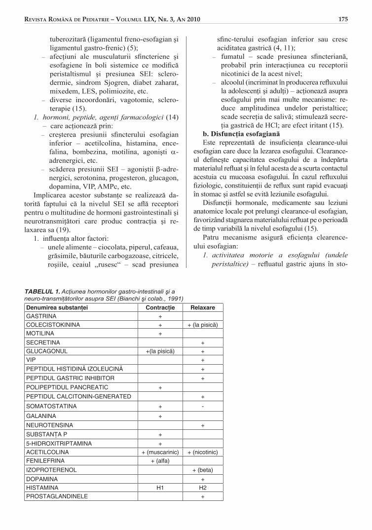
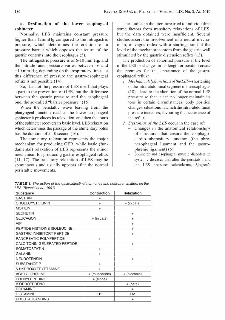
Implicarea acestor substanţe se realizează da-torită faptului că la nivelul SEI se afl ă receptori pentru o multitudine de hormoni gastrointestinali şi neurotransmiţători care produc contracţia şi re-laxarea sa (19).
infl uenţa altor factori:– unele alimente – ciocolata, piperul, cafeaua,
grăsimile, băuturile carbogazoase, citricele, roşiile, ceaiul ,,rusesc“ – scad presiunea
1.
1.
sfi nc -terului esofagian inferior sau cresc aciditatea gastrică (4, 11);
– fumatul – scade presiunea sfi ncteriană, pro babil prin interacţiunea cu receptorii nicotinici de la acest nivel;
– alcoolul (incriminat în producerea refl ux ului la adolescenţi şi adulţi) – acţionează asupra esofagului prin mai multe meca nisme: re-duce amplitudinea undelor peri staltice; scade secreţia de salivă; stimulează secre-ţia gastrică de HCl; are efect iritant (15).
b. Disfuncţia esofagianăEste reprezentată de insufi cienţa clearance-ului
esofagian care duce la lezarea esofagului. Clearance-ul defi neşte capacitatea esofagului de a îndepărta materialul refl uat şi în felul acesta de a scurta contactul acestuia cu mucoasa esofagului. În cazul refl uxului fi ziologic, constituienţii de refl ux sunt rapid evacuaţi în stomac şi astfel se evită leziunile esofagului.
Disfuncţii hormonale, medicamente sau leziuni anatomice locale pot prelungi clearance-ul esofagian, favorizând stagnarea materialului refl uat pe o perioadă de timp variabilă la nivelul esofagului (15).
Patru mecanisme asigură efi cienţa clearence-ului esofagian:
activitatea motorie a esofagului (undele peristaltice) – refl uatul gastric ajuns în sto-
1.
TABELUL 1. Acţiunea hormonilor gastro-intestinali şi a neuro-transmiţătorilor asupra SEI (Bianchi şi colab., 1991)Denumirea substanţei Contracţie RelaxareGASTRINA +COLECISTOKININA + + (la pisică)MOTILINA +
SECRETINA +GLUCAGONUL +(la pisică) +VIP +
PEPTIDUL HISTIDINĂ IZOLEUCINĂ +
PEPTIDUL GASTRIC INHIBITOR +
POLIPEPTIDUL PANCREATIC +
PEPTIDUL CALCITONIN-GENERATED +
SOMATOSTATINA + -
GALANINA +
NEUROTENSINA +
SUBSTANŢA P +
5-HIDROXITRIPTAMINA +ACETILCOLINA + (muscarinic) + (nicotinic)FENILEFRINA + (alfa)
IZOPROTERENOL + (beta)
DOPAMINA +HISTAMINA H1 H2PROSTAGLANDINELE +
REVISTA ROMÂNÅ DE PEDIATRIE – VOLUMUL LIX, NR. 3, AN 2010176
mac în timpul relaxării tranzitorii a SEI este retrimis în stomac cu ajutorul undelor peri-staltice (20). Perturbări ale motilităţii eso-fagiene determină apariţia RGE; odată insta-late, aceste tulburări motorii tind să devină ireversibile şi să favorizeze apariţia compli-caţiilor refl uxului, respectiv a leziunilor de esofagită (13);forţa gravitaţională – în ortostatism, contri-buie la retrimiterea refl uatului gastric înapoi în stomac; în clinostatism sau în timpul som-nului, în absenţa gravitaţiei, clearance-ul eso-fagian este întârziat, favorizând stagnarea re-fl u atului în esofag, cu iritarea mucoasei acestuia.secreţia salivară – prin conţinutul în bicar-bonat salivar, tamponează acidul refl uat din stomac, eliminând efectul iritant al acestuia asupra mucoasei esofagului. În condiţii de scă dere a secreţiei salivare: tutun, medicaţie anticolinergică, radioterapie, xerostomie, sin-drom Sjogren,etc., lipsa tamponării acidităţii gastrice refl uate duce la iritarea chimică a mucoasei esofagului (21).secreţia glandelor esofagiene (mucoase sau nemucoase) – rol protector asupra mucoasei esofagiene.
c. Disfuncţia gastricăReprezintă o altă verigă patogenică (15) care
intervine în producerea refl uxului gastro-easofagian prin:
creşterea volumului gastric – în condiţii de supraalimentaţie sau slaba funcţionare a vi da-njei gastrice (golire întârziată) determină re-laxări tranzitorii repetate ale SEI, favorizând refl uxul;distensia gastrică – prin aerofagie sau supra-alimentaţie prelungită, ducând la scur tarea lungimii SEI în porţiunea intra abdo minală;creşterea presiunii intraabdominale – la valori ce depăşesc presiunea bazală a sfi nc-terului esofagian inferior, determinând relax-area acestuia; există anumite situaţii în care se produce creşterea presiunii intraabdominale, favorizând astfel producerea refl uxului: obe-zitate, purtarea de îmbrăcăminte prea strâmtă, corsete, efortul de tuse, strănut, defecaţie.evacuare gastrică întârziată – la peste 40% din bolnavii cu RGE evacuarea gastrică este prelungită; cei cu stază gastrică au RGE mai abundent şi leziuni esofagiene mai severe.
Studiile cu izotopi explică întârzierea evacuării stomacului prin mecanismele:
2.
3.
4.
1.
2.
3.
4.
– motilitate antrală anormală;– refl uxul duodeno-gastric;– combinarea acestor două mecanisme (15).
refl uxul duodeno-gastric – ar sta la baza le-ziunilor mai severe în caz de refl ux mixt gastro-esofagian acid şi alcalin, prin acţiunea com-binată a HCl, pepsinei, enzimelor pan creatice şi acizilor biliari (Fiorucci, 1992);hipersecreţia gastrică – creşterea concentra-ţiei de acid agresionează mucoasa esofagiană în timpul relaxărilor tranzitorii ale SEI; de asemeni, aciditatea determină perturbări ale peristalticii normale esofagiene (studiul per-fuzării endo-esofagiene cu acid clorhidric diluat 0,1N efectuat de Triadafi lopoulos, in 1991) (22).
În ultimul timp se acordă o importanţă deosebită infecţiei gastrice cu Helicobacter pylori, aşa cum reiese din studii efectuate pe loturi de adulţi (23) şi care concluzionează că infecţia ar scădea rata BRGE prin scăderea acidităţii gastrice. Astfel, s-a avansat ideea conform căreia, pe de o parte, H. pylori ar favoriza apariţia de boli gastrice severe (inclusiv esofag Barret şi adenocarcinom esofa-gian), iar pe de altă parte, ar exercita rol protector faţă de boala de refl ux gastro-esofagian (13).
RELAŢIA BRGE – HERNIA HIATALĂ
Se insistă foarte mult pe relaţia patogenică dintre hernia hiatală şi BRGE, deoarece malpoziţia seg-mentului abdominal al esofagului la bolnavii cu hernie hiatală are ca rezultat – în mod obişnuit – incompetenţa joncţiunii eso-gastrice cu consecinţa sa directă – refl uxul gastro-esofagian (24).
Hernia hiatală reprezintă pătrunderea constantă sau intermitentă în torace a unei părţi din stomac prin hiatusul esofagian. Deşi presiunea intra abdo-minală este superioară presiunii intratoracice, există o serie de factori care se opun angajării stomacului în cutia toracică, printre care şi mijloa cele anti-refl ux. Relaxarea/pierderea elasti cităţii structurilor de fi xare eliberează puţin câte puţin stomacul, per-miţând angajarea sa în torace.
Hernia prin alunecare esta cea mai frecventă formă de hernie hiatală şi evoluează cel mai adesea cu refl ux.
Studii recente endoscopice, de ph-metrie şi ma-nometrie esofagaină au arătat rolul herniei hiatale în patogeneza bolii de refl ux, demonstrând relaţia existentă între dimensiunile herniei hiatale şi dis-funcţionalitatea SEI, pe de o parte, şi reducerea clearance-ului esofagian acid, pe de altă parte.
1.
2.
REVISTA ROMÂNÅ DE PEDIATRIE – VOLUMUL LIX, NR. 3, AN 2010 177
Prezenţa unei hernii hiatale de mici dimensiuni la copiii cu boală de refl ux, nu se soldează cu mo difi cări importante de tonus a SEI sau modifi cări de clearance esofagian, similar cu copiii fără hernie hiatală. În cazul asocierii unei hernii hiatale de mari dimensiuni se înregistrează o diminuare a tonusului SEI şi o încetinire a clearance-ului esofagian, deter minând şi un grad mai mare a leziunilor esofagitice postrefl ux.
Deci, doar herniile de dimensiuni medii şi mari ar interveni în patogeneza RGE.
La concluzii similare au ajuns şi cei care au efectuat studii ph-metrice sau manometrice, su-gerând că herniile hiatale de mici dimensiuni sunt un slab factor de predicţie în ceea ce priveşte pre-zenţa refl uxului gastro-esofagian (24).
Oricum, boala de refl ux gastro-esofagian nu se însoţeşte întotdeauna de hernie hiatală, iar hernia hiatală nu determină intotdeauna refl ux (25). La pa-cienţii cu BRGE descoperirea unei hernii hiatale este un factor de gravitate, mai ales dacă hernia este de mari dimensiuni. Unele studii au demonstrat că asocierea herniei hiatale şi a esofagitei de refl ux se însoţeşte de o întârziere a evacuării gastrice.
Rezumând, hernia hiatală joacă un rol important în patogeneza BRGE prin următoarele elemente:
diminuarea tonusului sfi ncterului esofagian inferior;încetinirea clearance-ului esofagian;reducerea peristaltismului esofagian;
1.
2.3.
întârzierea golirii esofagiene;hernia hiatală per se, de dimensiuni medii şi mari.
CONCLUZII
Boala de refl ux gastroesofagian este o entitate frecventă în practica pediatrică, dar încă sub-diagnosticată, iar tratatele româneşti şi străine îi dedică prea puţin spaţiu. Ea îşi are rădăcini încă din viaţa intrauterină, important este ca familia să ştie că refl uxul gastroesofagian este un fenomen mai mult sau mai puţin fi ziologic la sugarul din primul semestru de viaţă.
BRGE se manifestă sub incidenţa a numeroase comorbidităţi. Anemia, falimentul creşterii şi infec-ţiile repetate respiratorii fac diferenţierea între re-fl uxul fi ziologic şi cel patologic.
Boala de refl ux gastroesofagian are un tablou clinic complex, adesea fi ind o capcană de diagnostic în bolile respiratorii.
Metodele moderne de investigaţie au pus în evidenţă boala de refl ux asociată unor tablouri cli-nice nespecifi ce, dezvăluind incidenţa reală a bolii.
Etiopatogenia complexă este reprezentată prin cele trei verigi patogenice: disfuncţia sfi ncterului eso fagian inferior, disfuncţia esofagiană şi disfuncţia gastrică, fi ecare cu multiple mecanisme care duc în fi nal la apariţia bolii.
4.5.
Gastro-esophageal reflux disease ethiopathogeny
V.V. Lupu, M. Burlea5th Pediatrics Clinic-Gastroenerology, University of Medecine and Pharmacy “Gr. T. Popa” Iasi
ABSTRACT The gastro-esophageal refl ux disease (GERD), “the disease of the 3rd millennium”, is more frequently met in the pediatric practice than it has been thought. The authors make a short incursion into the embryology, anatomy, physiology of the esophagus and into the natural history of the disease to explore, identify and explain the pathogenic mechanisms that are the base of the GERD development. They describe the 3 pathogenic links: the dysfunction of the lower esophageal sphincter, the esophageal dysfunction and the gastric dysfunction, and also the hiatal hernia which has a special pathogenic relation to GERD.
Key words: esogastric junction, gastresophageal refl ux, lower esophageal sphincter
The gastro-esophageal refl ux disease (GERD), represents the intermittent or permanent passage of the gastric content into the esophagus, determining a series of symptoms: digestive, respiratory and/or neuro-behavioural or absence of any symptoms.
The good knowledge of the embryogenesis, anatomy and esophageal physiology data leads to the correct understanding of the pathology at this level and, implicitly, to a proper therapeutic approach.
REVISTA ROMÂNÅ DE PEDIATRIE – VOLUMUL LIX, NR. 3, AN 2010178
ELEMENTS OF EMBRYOLOGY
The gastrointestinal tract derives entirely from the primitive bowel, a tube extended from the mouth to the anus, which is formed in the 3rd-4th week of intrauterine life through the invagination of the dorsal side of the vitelline sac into the embryo.
The development takes place as follows: from the endoderm derive the epithelium and the annex glands of the digestive apparatus,the mesoderm creates the lamina propria of the submucosae and of the muscular and serous tunics.
Topographically, the primitive bowel is divided into three parts: anterior, middle and posterior, which is at the origin of the segments of the digestive tube.
The esophagus derives from the anterior part of the primitive bowel from which also derive: the oral cavity, the pharynx, the stomach, the proximal duodenum up to the ampulla of Vater, the liver, the gall-bladder and the pancreas, all of them irrigated by the celiac trunk (1).
At the cranial end, a ventral diverticulum which will become the trachea forms a groove in the foregut cavity fl oor about 3 weeks from the fertilization. As the structures elongates, ridges appear on the lateral walls of the foregut cavity and the diverticulum, which will unite about 5 weeks of age, forming separate tracheal and esophageal channels (2)
ANATOMY OF ESOPHAGUS
The esophagus is a musculo-membraneous conduct, supple and dilatable, which links the pharynx to the stomach, extending from the C6 vertebra (pharynx-esophageal junction) to the T11 vertebra (the cardia) (3).
The esophagus is not straight. It has curves in the sagittal and frontal plane.
At birth it is 8-10 cm long and 0.5 cm in diameter, and then it grows 0.65 cm/year, reaching at an adult person 25 cm long and 2-3 cm in diameter (4).
The esogastric junction (lower esophageal com-plex) is a physiological sphincter without anatomic individuality. The constituent elements of this area are:
epiphrenic ampulla – radiological concept – an area of luminal expansion which appears above the vestibular sphincter in inspirationvestibular sphincter – corresponds to the lower esophageal sphincter (LES)esophageal vestibulum (esophageal ampulla)Z line – the line of epithelial transition – an ana-tomo-histological demarcation line between
1.
2.
1.
2.
3.4.
the stratifi ed squamous epithelium of the esophagus and the columnar epithelium of the stomach – a very useful endoscopic mark (4, 5).
In normal conditions, the esogastric junction represents an anti-refl ux anatomic device which prevents the refl ux of the gastric acid content into the esophagus.
The dysfunctions of the mechanism of gastro-esophageal retention determine:
the appearance of GERD with secondary peptic esophagitis up to stenosis,diaphragmatic hiatal hernias– when the esophageal hiatus is larger.
The esophagial innervation:a) extrinsic innervation – sympathetic fi bers,
vagus nerves and recurrent laryngeal nerve (cerebro-spinal)
b) intrinsec innervation – compose of myenteric Auerbach’s plexus and submucosal Meissner’s plexus. (2)
The common nociception and central projection of the esophagus and trachea sre responsible for the asthma-like of the GERD. (6)
Using imunohistochemistry studies it was possible to identify the sites of neurohormonal substances secretion (7):
VIP – present since intrauterin period in plexuses and LESP substance – in the muscular fi bers and plexuses, determines the contraction of LESEnkefalina – in the striated muscular fi bersMotilin – controls the LES.
DATA ABOUT THE PHYSIOLOGY OF THE ESOPHAGUS
The esophagus plays the role of:transport of the alimentary bolus from the oral cavity to the stomach to prevent the gastro-esophageal and eso-pharyngeal refl ux, by means of the upper esophageal sphincter (UES) and LES.
UES maintains the proximal extremity of the esophagus and prevents the eso-pharyngeal refl ux. The UES pressure is of 40-100 mmHg, and during the deglutition, UES transitorily relaxes itself (8).
The esophageal transit of the alimentary bolus is carried out by means of two factors:
peristaltic wavesgravity force.
The participation of the two factors depends on the position of the body (3):
in clinostatism the passage of the alimentary bolus is made only through peristaltic activity
1.
2.
1.
2.
3.4.
A.
B.
1.2.
1.
REVISTA ROMÂNÅ DE PEDIATRIE – VOLUMUL LIX, NR. 3, AN 2010 179
in orthostatism, the gravity force also contri-butes to a variable extent.
The peristaltic waves at the esophagus level are:
primary waves – are originated in the pharynx, are in relation with the deglutition, they favour the propulsion of the alimentary bolus;secondary waves – induced by the distension of the esophagus, chemical or mechanical irritants and don’t correlate with the deglutition.
The prevention of the esophageal lesions de-termined by the gastric acid refl ux is done by the se condary peristaltic contractions which perform the esophageal clearance.
The pressure exercised by the peristaltic waves get over the tonus of LES, they relax it. The tonus of that sphincter increases with age:
at the young child and baby, the low muscular tonus makes the food pass fast “just like through a tube” and to fall into the stomach at the older child the pressure exercised by the alimentary bolus overcomes the muscular tonus and relax the cardia.
The main function of the lower esophageal sphincter is to prevent GER. LES doesn’t always act as an anti-refl ux barrier, even at healthy people, so after meal, there may appear a transitory relaxa-tion of LES not preceded by a peristaltic one.
NATURAL HISTORY
It is well known that the refl ux is physiological in the fi rst 3 months of life, manifested very pre-cociously, usually in the fi rst 6 weeks of life (9).
Also, most regurgitations disappear until the age of 12-24 months, their persistence after this age raising the problem of the appearance of refl ux consequences – refl ux esophagitis – and the classifi cation of the cases in the gastro-esophageal refl ux disease (10).
Children who had as babies frequent episodes of regurgitations and/or vomiting are more likely to develop the refl ux disease in the following period, especially if it is associated with smoking and the presence of refl ux symptoms at the mother during the respective pregnancy (10).
Thus, there are differences between the refl ux existing in the fi rst 12-24 months, manifested by regurgitations and vomiting and the refl ux appeared after that age (usually over 18 months), when the respiratory manifestations are predominant (espe-cially recurrent wheezing), and the discovery of the refl ux is, many times, random or by exclusion.
Also, another special category is represented by the children with neurological defects, the
2.
1.
2.
1.
2.
encephalopates, who have almost constantly the gastro-esophageal refl ux (11).
If we had to draw a conclusion, the population with risk to develop GERD includes:
premature and dismature babies,new-born who suffered from hypoxia or perinatal axphyxia,babies who benefi ted from ,,a demeure,, gastric probebabies with (primitive or secondary) delay of “gastric emptying”,new-born operated for an esophageal atresia with or without fi stula,cystic fi brosis,encepahlopates, etc. (12)
ETIOPATHOGENY
The anti-refl ux barrier As we have mentioned in the section on the
esophagus anatomy, in normal conditions, the eso-gastric junction functions as a true anatomic anti-refl ux device which prevents the refl ux of the gastric contents into the esophagus.
The constituent elements of the anti-refl ux device (3):
the normal calibre of the esophageal hiatus;the oblique opening of the esophagus to the stomach through the cardiac orifi ce – angle of His;valvule of Gubarow – visible at endoscope – which closes as a fl ap valve the cardia when the intragastric pressure increases;presence and disposition ″on horse back″ or ″in tie″ of the oblique fi bres of the stomach;commune innervation of cardia and abdominal esophagus.
But the major role in creating the barrier against the refl ux of the gastric contents into the esophagus is played by the lower esophageal sphincter (LES), a virtual sphincter, existing only due to the pressure movements at the level of the eso-gastric junctions and which has morphological, physiological, pharmacological and metabolic particularities (13). This sphincter matures before and after birth, starting to have an effi cacious anti-refl ux role only from the 5-7th week of life.
Three pathogenic links contribute to the appearance of the gastro-esophageal refl ux (11):
dysfunction of lower esophageal sphincter,esophageal dysfunction,gastric dysfunction.
1.2.
3.
4.
5.
6.7.
1.2.
3.
4.
5.
1.2.3.
REVISTA ROMÂNÅ DE PEDIATRIE – VOLUMUL LIX, NR. 3, AN 2010180
a. Dysfunction of the lower esophageal sphincter
Normally, LES maintains constant pressure higher than 12mmHg compared to the intragastric pressure, which determines the creation of a pressure barrier which opposes the return of the gastric contents into the esophagus (5).
The intragastric pressure is of 6-10 mm Hg, and the intrathoracic pressure varies between –6 and +10 mm Hg, depending on the respiratory times, at this difference of pressure the gastro-esophageal refl ux is not possible (14).
So, it is not the pressure of LES itself that plays a part in the prevention of GER, but the difference between the gastric pressure and the esophageal one, the so-called “barrier pressure” (15).
When the peristaltic wave leaving from the pharyngeal junction reaches the lower esophageal sphincter it produces its relaxation, and then the tonus of the sphincter recovers its basic level. LES relaxation which determines the passage of the alimentary bolus has the duration of 5-10 second (16).
The transitory relaxation represents the major mec hanism for producing GER, while basic (fun-damental) relaxation of LES represents the minor mechanism for producing gastro-esophageal refl ux (11, 17). The transitory relaxation of LES may be spontaneous and usually appears after the normal peristaltic movements.
The studies in the literature tried to individualize some factors from transitory relaxations of LES, but the data obtained were insuffi cient. Several studies assert the involvement of a neural mecha-nism, of vagus refl ex with a starting point at the level of the mechanoreceptors from the gastric wall stimulated by the gastric distension refl ex (13).
The production of abnormal pressure at the level of the LES or changes in its length or po sition create the premises for the appearance of the gastro-esophageal refl ux:
Mechanical dysfunctions of the LES – shortening of the intra-abdominal segment of the esophagus (18) – lead to the alteration of the normal LES pressure so that it can no longer maintain its tone in certain cir cumstances: body position changes, situa tions in which the intra-abdominal pressure increases, favouring the occurrence of the refl ux.Dystonias of the LES occur in the case of:– Changes in the anatomical relationships
of structures that ensure the esophago-cardio-tuberositary junction (the phre-nesophageal ligament and the gastro-phrenic ligament) (5),
– Sphincter and esophageal muscle disor ders in systemic diseases that alter the peristalsis and the LES pressure: sclero derma, Sjogren’s
1.
2.
TABLE 1. The action of the gastrointestinal hormones and neurotransmitters on the LES (Bianchi et al., 1991)Substance Contraction RelaxationGASTRIN +CHOLECYSTOKININ + + (in cats)MOTILIN +SECRETIN +GLUCAGON + (in cats) +VIP +PEPTIDE HISTIDINE ISOLEUCINE +GASTRIC INHIBITORY PEPTIDE +PANCREATIC POLYPEPTIDE +CALCITONIN-GENERATED PEPTIDE +SOMATOSTATIN + -GALANIN +NEUROTENSIN +SUBSTANCE P +5-HYDROXYTRYPTAMINE +ACETYLCHOLINE + (muscarinic) + (nicotinic)PHENYLEPHRINE + (alpha)ISOPROTERENOL + (beta)DOPAMINE +HISTAMINE H1 H2PROSTAGLANDINS +
REVISTA ROMÂNÅ DE PEDIATRIE – VOLUMUL LIX, NR. 3, AN 2010 181
syndrome, diabetes mellitus, myxedema, SLE, polymyositis, etc.
– various miscoordinations, vagotomy, scle ro- therapy (15).
Hormones, peptides, pharmacological agents (14) - which act by:– Increasing lower esophageal sphincter
pressure – acetylcholine, histamine, enke-phalin, bombesin, motilin, α-adre nergic agonists, etc.
– Decreasing the LES pressure – β-adre nergic agonists, serotonin, proges terone, glucagon, dopamine, VIP, cAMP, etc.
The involvement of these substances is achieved due to the fact that at the level of the LES there are receptors for a variety of gastro intestinal hormones and neurotransmitters that cause its contraction and relaxation (19).
Infl uence of other factors:– Certain foods – chocolate, pepper, coffee,
fats, carbonated beverages, citrus fruits, tomatoes, “Russian” tea, – de crease the lower esophageal sphincter pre ssure or increase gastric acidity (4, 11);
– Smoking – reduces sphincter pressure, pro-bably by means of the interaction with the nicotinic receptors at this level;
– Alcohol (incriminated for producing the refl ux in adolescents and adults) – acts on the esophagus through several mecha nisms:
– It decreases the amplitude of peristaltic waves,
– It reduces the secretion of saliva,– It stimulates the gastric secretion of HCl– It has an irritating effect (15).
b. The esophageal dysfunction It is represented by the insuffi ciency of eso phageal
clearance resulting in damage to the eso phagus. Esophageal clearance defi nes the capacity of the esophagus to remove the refl uxed material and thus to shorten its contact with the esophageal mucosa. In physiological refl ux, refl ux constituents are quickly evacuated to the stomach and thereby esophageal lesions are avoided.
Hormonal dysfunctions, drugs or local anatomic lesions may prolong esophageal clearance, favouring the stagnation of the refl uxed material in the esophagus over a variable period of time (15).
There are four mechanisms which ensure the effi ciency of esophageal clearance:
Motor activity of the esophagus (peristaltic waves) – the gastric refl uxed material that reached the stomach during the transient LES
1.
1.
1.
relaxation is returned to the stomach by peristaltic waves (20). Esophageal motility dis-tur bances cause GER; once installed, these motor disorders tend to become irreversible and to favour the development of refl ux com-plications, namely esophagitis lesions (13);Gravity – in orthostatism, it contributes to resending of the gastric refl uxed material back into the stomach, in clinostatism or during sleep, in the absence of gravity, the esophageal clearance is delayed, favouring the stagnation of the refl uxed material in the esophagus, with its subsequent mucosal irritation.Saliva secretion – by means of its salivary bicarbonate content, it counteracts the refl uxed acid from the stomach, eliminating its irritating effect on the esophageal mucosa. In the case of a decrease of salivary secretion: tobacco, anti-cholinergic medication, radiotherapy, xero sto-mia, Sjogren’s syndrome, etc., lack of counter-action of the gastric acidity leads to chemical irritation of the esophageal mucosa (21). Secretion of esophageal glands (mucous or non-mucous) – protective role of the esopha-geal mucosa.
c. Gastric dysfunctionIt is another pathogenic link (15) that takes part in
the production of gastro-esophageal refl ux by:Increasing the gastric volume – in case of overnutrition or poor gastric emptying function (delayed emptying) causes repeated transient LES relaxations, favouring the refl ux;Gastric distension – by aerophagia or prolonged overnutrition, leading to shortening the length of the LES in its intra-abdominal segment;Increase of the intra-abdominal pressure – to values exceeding the basal lower esophageal sphincter pressure, causing it to relax; there are situations where a rise in abdominal pressure occurs, thus favouring the refl ux: obesity, wearing excessively tight clothes, corsets, cough, sneezing, defecation effort.Delayed gastric emptying – in over 40% of patients suffering from GER the gastric emptying is prolonged; those with gastric stasis have a more abundant GER and more severe esophageal lesions.
Studies using isotopes explain the delay in stomach emptying by means of the following mechanisms:
Abnormal antral motility,Duodeno-gastric refl ux,Combination of these two mechanisms (15).duodeno-gastric refl ux – could be at the core of more severe injuries in case of mixed acid -
2.
3.
4.
1.
2.
3.
4.
1.2.3.4.
REVISTA ROMÂNÅ DE PEDIATRIE – VOLUMUL LIX, NR. 3, AN 2010182
alkaline gastro-esophageal refl ux through the combined action of HCl, pepsin, pancreatic enzymes and bile acids (Fiorucci, 1992);gastric hypersecretion – the increase of acid concentration aggresses the esophageal mucosa during transient LES relaxations; moreover, acidity causes alterations of the normal eso-phageal peristalsis (the study of endo-eso-phageal infusion with 0.1N hydrochloric acid performed by Triadafi lopouLES in 1991) (22).
Lately special attention is given to the gastric infection with Helicobacter pylori, as shown in studies conducted on groups of adults (23), concluding that the infection could decrease the GERD rate by decreasing gastric acidity. Thus, an idea has emerged, according to which, on the one hand, H. pylori could favour the occurrence of severe gastric diseases (including Barrett’s esophagus and esophageal adenocarcinoma), and on the other hand, it could exert a protective role against gastro-esophageal refl ux disease (13).
GERD – HIATAL HERNIA RELATIONSHIP
The pathogenic relationship between hiatal hernia and GERD is highly emphasized, as the malposition of the abdominal segment of the esophagus in patients with hiatal hernia usually has as a result the incom-petence of esophago-gastric junction, with its direct consequence – gastro-esophageal refl ux (24).
Hiatal hernia is the constant or intermittent pene-tration of a part of the stomach into the thorax through the esophageal hiatus. Although intra-abdominal pres-sure is higher than the intra-thoracic pressure, there are several factors which prevent the stomach from penetrating into the thoracic cavity, including anti-refl ux means. Relaxation/loss of elasticity of fi xation structures gradually releases the stomach, allowing its protrusion into the thorax.
The sliding hernia is the most common form of hiatal hernia and it develops with GER most often.
Recent endoscopic, pH-metric and esophageal manometric studies have shown the role of the hiatal hernia in the pathogenesis of refl ux disease, demons-trating, on the one hand, the relationship between the size of hiatal hernia and LES malfunctioning and, on the other hand, the reduction of the esophageal acid clearance. The presence of small hiatal hernias in children with refl ux disease does not lead to signifi cant altering of the LES tone or of the esophageal clearance, similar to children without hiatal hernia. In the case of a large hiatal hernia there is a reduction of the LES tone
5.
and a slowdown in esophageal clearance, resul ting in a higher degree of post-refl ux esophagitis lesions.
Therefore, only medium sized and large hernias could intervene in the pathogenesis of the GER.
Similar conclusions were also reached by those who have conducted pH-metric or manometric studies, suggesting that small hiatal hernias are a weak pre-dicting factor of the existence of the gastro-esophageal refl ux (24).
However, gastro-esophageal refl ux disease does not always accompany hiatal hernia and hiatal hernia does not always determine GER (25). In patients with GER, the discovery of a hiatal hernia is an aggravating factor, especially if the hernia is large. Some studies have shown that the combination of hiatal hernia and refl ux esophagitis is accompanied by a delayed gastric emptying.
Summarizing, hiatal hernia plays an important role in the pathogenesis of GER by means of the following elements:
Decrease of the lower esophageal sphincter tone,Slowdown of the esophageal clearance,Reduction of the esophageal peristalsis,Delay in esophageal emptying,Medium sized and large hiatal hernia per se.
CONCLUSIONS
GERD is a frequent disease in the pediatric prac tice, but still underdiagnosed, and the Romanian and foreign literature has too little space for it. GERD has roots from the intrauterine period, important is that the family knows that the gastroesophagian refl ux is more or less physiolo gical in the fi rst semester of life.
GERD manifests as a lot of comorbidities. The anemia, failure to thrive and recurrent respiratory infections makes the difference between the physiological and pathological refl ux.
The gastroesophageal refl ux disease has a complex clinical picture, being often a diagnostic trap especially in respiratory diseases.
The modern investigation methods reveals the refl ux disease associated with unspecifi c clinical symptoms, showing the real incidence of the disease.
The complex etiopathogeny is represented by the three pathogenic links: dysfunction of lower eso phageal sphincter, esophageal dysfunction, gastric dysfunction, each of them with multiple mechanisms which leads fi nally to the development of the GERD.
1.
2.3.4.5.
REVISTA ROMÂNÅ DE PEDIATRIE – VOLUMUL LIX, NR. 3, AN 2010 183
Anghelescu V – Elemente de embriologie. Ed. Medicală, 1963; 298-299.DiMarino AJ, Benjamin SB – Gastrointestinal Disease: An Endoscopic Approach, second edition, Ed. Slack Incorporated, New Jersey, 2002; 159-172Angelescu N – Tratat de patologie chirurgicală. Ed. Medicală, Bucureşti, 2003.Behrman RE, Kliegman RM, Jenson HB – Nelson Textbook of Pediatrics – 18-th edition. Ed. Saunders, Philadelphia, 2007; 1541-1542, 1547-1549.Burlacu M – Actualităţi în patologia esofagiană la copil, Teză doctorat, U.M.F. Iaşi, 2005.Cannin BJ, Mazzone SB – Refl ex Mechanisms in Gastroesophageal Refl ux Disease and Asthma, The American Journal of Medicine, vol. 115, nr. 3, supl. 1, 2003, 45-48Aprodu G, Munteanu V, Botez C, Goţia DG – Patologia chirurgicală a esofagului la copil. Ed. Gr. T. Popa, Iaşi, 2003; 9.Hăulică I – Fiziologie umană, ediţia 2. Ed. Medicală, Bucureşti, 2000; 537-540.Ciofu EP, Ciofu C – Esenţialul în pediatrie, ediţia a II-a. Ed. Medicală Amaltea, Bucureşti, 2002; 337-339.Walker WA et al – Pediatric Gastrointestinal Disease, ediţia a IV-a Ed. BC Decker Inc, Hamilton, 2004; 384-399.Ciofu EP, Ciofu C – Pediatria - ediţia I. Ed. Medicală, Bucureşti, 2001; 453-454, 469-474.Geormăneanu M – Pediatrie, Ed. Didactică şi Pedagogică, Bucureşti, 1997; 155-168.Navarro J, Schmitz J – Gastroenterologie Pédiatrique. Ed. Médicine-Sciences, Fammarion, Paris, 2000; 131-167.
1.2.
3.4.
5.
6.
7.
8.
9.
10.
11.
12.
13.
Burlea M – Pediatrie pentru studenţii Facultăţii de Medicină Stomatologică. Ed. Apollonia, Iaşi, 1999; 159-167.Pascu O, Grigorescu M – Tratat de gastroenterologie clinică, vol. I. Ed. Tehnică, Bucureşti, 1996; 216-221.Roy CC, Silverman A, Allagile A – Pediatric Clinical Gastroenterology, 4-th edition. Mosby-Year Book Inc. 1995; 163-168.Georgescu A – Compendiu de pediatrie, ediţia a II-a. Ed. Bic All, Bucureşti, 2005; 389-391.Bufo AJ, Chen MK, Lobe TE, Shah RS, Gross E, Hixon SD, Hollabaugh RS, Schropp KP – Laparoscopic Funduloplication in Children: A Superior Technique. Pediatric Endosurgery & Innovative Techniques, 1997; 1(2): 71-76.Gherasim L – Medicina internă, vol. III. Ed. Medicală, Bucureşti, 1999.Robinson M – Gastroesophageal refl ux disease: selecting optimal therapy. Postgrad Med, 1994; 95:88-102.Peters JH, DeMeester TR – Gastroesophageal refl ux. Surg Clin North Am, 1993; 73: 1119-1144.Triadafi lopoulos G, Castilio T – No propulsive esophageal contractions and gastroesophageal refl ux. Am J Gastroenterol, 1991; 86: 153-159.Koike T, Masamune A – Helicobacter pylori Infection in Refl ux Esophagitis and Atrophic Gastritis: Clinical Implications. Gastroenterology, 2001; 3(2)[Medscape].Miu N – Tratat de medicină a adolescentului, Ed. Casa Cărţii de Ştiinţă, Cluj, 1999; 302-303.Jocu IC, Popescu EA – Patologia joncţiunii esogastrice, Fiziopatologie, diagnostic şi tratament, Ed.Medicală, Bucureşti, 1982.
14.
15.
16.
17.
18.
19.20.
21.
22.
23.
24.
25.
REFERENCES