rolul calciului În fiziologia ªi patologia …301 dermatovenerol.(buc.),55:299-312...
TRANSCRIPT
299
* Spitalul Clinic de Dermato-Venerologie „Prof. Dr. Scarlat Longhin“, Bucureºti.
ROLUL CALCIULUIÎN FIZIOLOGIA ªI PATOLOGIA MELANOCITULUI
CALCIUM ROLEIN MELANOCYTE PHYSIOLOGY AND PATHOLOGY
AMALIA ELENAANGHEL*, ILINCANICOLAE*, SIMONA ROXANAGEORGESCU*, J.D. DIACONU*
Bucureºti
Introducere
Prezenþa calciului în melanocit influenþeazãnumeroase cãi metabolice, inclusiv menþinereaechilibrului redox în celulã ºi reglarea melano-genezei, în principal prin modularea disponibi-litatãþii de L-tirozinã via fenilalanin hidroxilaza.Pigmentul conþinut în melanocite, melanina, esteimplicat în homeostazia calciului în celulã,
Introduction
The presence of calcium in melanocytesaffects numerous metabolic pathways, includingthe maintenance of cell redox balance andregulating melanogenesis, primarily throughmodulation the supply of substrate, L-tyrosine,via phenylalanine hydroxylase. The pigmentcontained in the melanocytes, melanin, has been
REFERATE GENERALEGENERAL ISSUES
Summary
Calcium is an important ion in the body throughmany and essential processes involving, regulated bybinding protein, in intracellular and extracellularenvironments. It has been shown that melanosomes serveas intracellular mediators of calcium homeostasisthrough melanin, here, in this context, a protein withbinding activity. The ion homeostasis in the melanocyt isparticularly important for maintaining redox balanceand regulation of cell biological response to oxidativestress. Calcium also participates actively in the centralprocess of melanocyt, melanogenesis, by regulating thesustrate, L-tyrosine. Melanocyte-keratinocyte interactionprocess is regulated by the level of this ion. Calciumvariations in context of cutaneous melanoma, inparticular hypercalcemia is considered a worseprognostic factor.
Keywords: calcium, melanocytes, melanogenesis,redox status.
Rezumat
Calciul reprezintã un ion important la nivelulorganismului prin procesele numeroase ºi esenþiale la careparticipã, sub reglementarea unor proteine de legare, înmediile intracelulare cât ºi extracelulare. S-a dovedit cãmelanozomii servesc drept mediatori intracelulari aihomeostaziei calciului, prin intermediul melaninei, în acestcontext funcþionând ca proteinã cu activitate de legare.Homeostazia acestui ion la nivelul melanocitului estedeosebit de importantã pentru menþinerea echilibrului redoxºi reglementarea rãspunsului biologic al celulei la stresuloxidativ. De asemenea calciul participã activ la procesulcentral desfãºurat la nivelul melanocitului, ºi anumemelanogeneza, în primul rând prin reglarea substratuluiacesteia, L-tirozina. Procesul de interacþiune melanocit-keratinocit este reglat de nivelul acestui ion. Variaþiilecalciului în contextul melanomului cutanat, în specialhipercalcemia, este consideratã factor de prognostic nefast.
Cuvinte cheie: calciu, melanocit, melanogeneza,status redox.
DermatoVenerol. (Buc.), 55: 299-312
300
DermatoVenerol. (Buc.), 55: 299-312
susþinându-se ipoteza conform cãreia melano-zomii servesc drept mediatori intracelulari aihomeostaziei calciului. Implicarea acestuia înmenþinerea echilibrului redox în melanocitepoate juca un rol în reglementarea rãspunsuluibiologic al celulei la stresul oxidativ. Calciulinhibã tioredoxin reductaza, o enzimã importantãcu funcþie antioxidantã în celulã (SchallreuterandWood, 1999; Wood et al., 1999). Menþinereahomeostaziei calciului în melanocitele epider-mice este de asemenea esenþialã, deoarece acesteasunt expuse în mod constant razelor UV, ce potinduce stres oxidativ prin fotogenerarea de speciireactive de oxigen. De exemplu, tioredoxinreductaza este exprimatã în piele la întuneric înconcentraþie de cinci ori mai mare decât în modnormal în piele(Schallreuter ºi Wood, 2001).Deoarece nivelul calciului moduleazã activitateaacestei enzime, controlul acestuia este crucialpentru reglementarea echilibrului redox ºiprevenirea deteriorarii ADN-ului în celule.
Am urmãrit în acest articol sã expunemaspecte legate de relaþia calciu-melanocit, cuefecte asupra unor procese extrem de importanteîn metabolismul celular.
Melanozomii - rol în reglarea homeostazieicalciului
Calciul are un rol important la nivelulorganismului prin procesele la care participã, subreglementarea unor proteine de legare(CBP-calciu binding protein), responsabile pentrutrafic, semnalizare, precum ºi tamponare, atât înmediile intracelulare cât ºi extracelulare. Totuºi,multe dintre aceste roluri pentru ionii de calciu,cât ºi pentru proteinele de legare rãmânincomplet înþelese ca urmare a complexitãþiiinteracþiunilor pe care le stabilesc ºi reþelelor desemnalizare ce le guverneazã. În acest context,melanina reprezintã un chelator cunoscut alionilor de calciu(Liu ºi Simon, 2005; Liu et al.,2004), implicatã în reglementarea homeostazieiacestuia la nivelul melanocitelor (Hoogduijn etal., 2003).
S-a demonstrat cã abilitatea melanocitelor dea funcþiona ca tampon împotriva expuneriitranzitorii la niveluri ridicate de calciu depindede cantitatea de melaninã prezentã în celulã(Hoogduijn et al., 2003). Într-un studiu separat
implicated in maintaining calcium homeostasisin cell, supporting the hypothesis thatmelanosomes serve as mediators of intracellularcalcium homeostasis. Its involvement inmaintaining redox balance in melanocytes mayplay a role in regulating the cell’s biologicalresponse to oxidative stress. Calcium inhibitsthioredoxin reductase, an importan enzyme withantioxidant function in cell(Schallreuter andWood, 1999; Wood et al., 1999). Maintainingcalcium homeostasis ?n epidermal melanocytes isalso essential because they are constantlyexposed to UV rays that can cause oxidativestress by generating reactive oxygen species. Forexample, thioredoxin reductase is expressed inthe dark skin in concentrations five times graterthen in normall ?n skin (Schallreuter and Wood,2001). Since calciummodulates the activity of thisenzyme, its control is crucial for the regulation ofredox balance and preventing DNA damage incells.
I’m proposed in this article to expose aspectsabout calcium- melanocytes relationship, witheffects on crucial processes in cell metabolism.
Melanosomes - role in regulating calciumhomeostasis
Calcium plays an important role in the bodythrough the processes involved, under regulationof binding protein (CBP-calcium bindingprotein), responsible for traffic, signaling, andbuffering, both in intracellular and extracellularenvironments. However, many of these roles forcalcium ions and the binding proteins remainincompletely understood due to complexity oftheir interactions and signaling networks thatgovern them. In this context, melanin is a knownas a chelator of calcium ions (Liu and Simon,2005, Liu et al., 2004), involved in the regulationof its homeostasis within melanocytes(Hoogduijn et al., 2003).
It has been shown that the ability ofmelanocytes to act as a buffer against transientexposure to high levels of calcium depends onthe amount of melanin present in the cell(Hoogduijn et al., 2003). In a separate studyinvolving the inner ear melanocytes, melanin hasbeen shown to act as a biological reservoir forcalcium, interfering in regulation of manycalcium dependent cellular processes (Gill and
301
DermatoVenerol. (Buc.), 55: 299-312
care implicã melanocitele din urechea internã, s-ademonstrat cã melanina poatea acþiona ca unrezervor biologic pentru calciu, intervenind înreglementarea mai multor procese celulare calciudependente (Gill ºi Salt, 1997; Meyer zumGottesberge, 1988). În plus, la nivelul pielii,calciul extracelular este cunoscut cã poate juca unrol critic în reglarea proliferarii ºi diferenþieriikeratinocitelor (Boyce ºi Ham, 1983). S-a postulatcã melanina joacã un rol în aceastã cale, princapacitatea sa de a lega ºi tampona calciu ( Buffeyet al., 1993; Drager 1985; Hoogduijn et al., 2004;Panessa ºi Zaduainsky, 1981; Salceda si Riesgo-Escovar, 1991; Salceda ºi Sanchez-Chavez, 2000).În schimb, calciu a fost dovedit a reglementaproducþia de melaninã în melanocitele epi-dermice acþionând ca un cofactor pentrufenilalanin hidroxilazã, enzima responsabilã desinteza L-tirozinei, un substrat critic pentrumelanogenezei, din L-fenilalaninã (Schallreuter ºiWood, 1999). În plus, calciu este esenþial pentruprotejarea echilibrul redox în melanociteleepidermice ºi pentru a preveni deteriorareaADN-ului în interiorul acestor celule(Schallreuter ºi Wood, 2001; Wood et al., 1999).
Importanþa primordialã biologicã a calciuluica mesager secund necesitã o reglementare strictãa homeostaziei acestuia pentru ca o celulã sãfuncþioneze în mod normal. Concentraþia decalciu liber citosolic în cele mai multe celule „înrepaus” este de ordinul a 10-7M, aproximativ 100nM (Carafoli, 1987). Aceastã concentraþie estemenþinutã în primul rând prin existenþa unorcanale de ioni ºi pompe, sistemele de tampon acalciului citosolic, ºi depozitarele de calciulintracelular (Tsien ºi Tsien, 1990). Concentraþiileintracelulare de calciu = 10-5 M vor duce laactivarea de proteaze calciu-dependente,rezultând o cascadã de evenimente ce conduc lamoarte celularã (Carafoli et al., 1984; Waring,2005). Absorbþia, depozitarea, ºi eliberareareglementatã a calciului sunt, prin urmare,riguros controlate de proteine specializate, cumar fi calmodulina, calbindina, anexine, parval-bumina, ºi S-100. Cele mai multe dintre acesteproteine de legare a calciului au prezentatconstante de legare de 105-107 / M (Carafoli etal., 1984; Frausto da Silva ºi Williams, 1991), darexistã un numãr de proteine de stocare a calciului
Salt, 1997, Meyer zum Gottesberge, 1988). Inaddition, in skin, extracellular calcium is knownto play a critical role regulating the keratinocyteproliferation and differentiation (Boyce andHam, 1983). It was postulated that melanin playsa role ?n this way, by its ability to bind calciumand buffer (Buffey et al., 1993, Drager 1985;Hoogduijn et al., 2004; Panessa and Zaduainsky,1981; Salceda and Riesgo-Escovar, 1991; Salcedaand Sanchez-Chavez, 2000). However, calciumhas been shown to regulate production ofmelanin in epidermal melanocytes by acting as acofactor for phenylalanine hydroxylase, theenzyme responsible for synthesis of L-tyrosine, acritical substrate for melanogenesis, from L-phenylalanine (Schallreuter and Wood, 1999). Inaddition, calcium is essential for protecting theredox balance in epidermal melanocytes and toprevent damage to DNA within these cells(Schallreuter and Wood, 2001, Wood et al., 1999).
Primary biological importance of calcium as asecond messenger requires strict regulation of itshomeostasis for a cell to function normally.Citosolic free calcium concentration in mostresting cells is in the order of 10-7M,approximately 100 nM (Carafoli, 1987). Thisconcentration is maintained primarily by theexistence of calcium ion channels and pumps,citosolic calcium buffer systems, and intracellularcalcium stores (Tsien and Tsien, 1990).Intracellular calcium concentrations=10-5 M willlead to activation of calcium-dependentproteases, resulting in a cascade of events leadingto cell death (Carafoli et al., 1984, Waring, 2005).The uptake, storage, and regulated release ofcalcium are therefore strictly controlled byspecialized proteins such as calmodulin,calbindin, anexins, parvalbumin and S-100. Mostof these calcium-binding protein have bindingconstants 105-107 / M (Carafoli et al., 1984,Frausto da Silva and Williams, 1991), but thereare a number of proteins with high capacity andlow affinity of calcium storage, involved insequestration and buffering calciumwith bindingconstant of about 103 / M (Pozzan et al., 1994).Two good examples are calsequestrin andcalreticulin, each with a binding constant (KB) ofapproximately 1 * 103 / M. These are known to becalcium storage protein in the lumen ofsarcoplasmic reticulum of skeletal and cardiac

302
DermatoVenerol. (Buc.), 55: 299-312
cu capacitate crescutã ºi afinitate scãzutã,implicate în sechestrarea ºi tamponarea calciului,cu constante de legare de aproximativ 103 / M(Pozzan et al., 1994). Douã exemple concludentesunt calsechestrina ºi calreticulina, fiecare cu oconstantã de legare (KB) de aproximativ 1 * 103 /M. Acestea sunt cunoscute a fi proteine destocare a calciului în lumenul reticululuisarcoplasmic din celulele musculare scheletice ºicardiace, precum ºi în lumenul reticululuiendoplasmatic în þesuturile non-musculare.(Opasºi Michalak, 1992). Pe baza proprietãþilor delegare a calciului a tuturor proteinelor menþio-nate mai sus, existã o puternicã corelaþie întrefuncþia specificã a fiecãrei proteine ºi constantade legare a calciului(KB). Proteinele cu afinitatemare de legare (> 106 / M) au roluri specializateîn transportul calciului ºi de semnalizare, în timpce proteinele cu afinitate micã de legare (< 104 /M) au roluri în sechestrarea, tamponarea ºistocarea de calciu. Prin urmare, melanina sedovedeste ºi ea importantã prin rolul sãufuncþional în cadrul reþelei de semnalizare acalciului.
În acest scop relatãm un studiu realizat recentfolosind melanina Sepia care prezintã proprietãþisimilare cu eumelanina de mamifere (Hong et al.,2004; Liu ºi Simon, 2005; Liu et al ., 2004; Potts ºiUA, 1976; Szekeres, 1975). Calciul se leagã degrupãri carboxil din granule de pigment (Hong ºiSimon, 2006), fenomen observat în toate cazurilede ataºare calciu proteine de legare(Carafoli et al.,1984; Carafoli, 1987). S-a emis ipoteza cã acestegrupãri carboxil ar apãrea în urma oligomerizãriiacidului 5,6-dihidroxiindol-2-carboxilic (DHICA)(Liu et al., 2004). Mai mult decat atât, melaninaSepia are o capacitate mare de legare a calciuluicu timpi de echilibrare rapizi.(Hong et. Al. 2004),cuantificând legarea calciului prin titrareacalorimetricã izotermic(ITC) ºi spectroscopiefluorescentã. Melanina Sepia, sãrãcitã de metale,la o concentraþie de 1 mg / ml, a fost tratatã cuCaCl2 în eºantioane celulare în microcalori-meteru, înregistrandu-se cãldura emanatã, cucare se traseazã o curbã. Aceasta depinde denumarul situsurilor de legare, nivelul ocupanþeiºi constantele de legare asociate. Constanta delegare determinatã prin acest studiu a fost de3.3(+-0.2)*103/M, susþinând rolul postulat
muscle cells and the lumen of endoplasmaticreticulum in non-muscle tissues. (Opas andMichalak, 1992). Based on the calcium bindingproperties of all proteins mentioned above, thereis a strong correlation between the specificfunction of each protein and calcium bindingconstant (KB). High-affinity binding protein (>106 / M) have specialized roles in calciumtransport and signaling, whereas low-affinitybinding proteins (<104 / M) have roles insequestration, buffering and storing calcium.Therefore, melanin also proves importantfunctional role in calcium signaling network.In this way, we mention about a recent studyusing Sepia melanin with similar properties tomammalian eumelanina (Hong et al., 2004, Liuand Simon, 2005, Liu et al., 2004, Potts and AU,1976, Szekeres, 1975) . Calcium binds to carboxylgroups of pigment granules (Hong and Simon,2006), a phenomenon observed in all cases ofattachment of calcium binding proteins (Carafoliet al., 1984; Carafoli, 1987). It was hypothesizedthat the carboxyl groups would arise from theoligomerization of 5,6-dihydroxyindole-2-carboxylic acid (DHICA) (Liu et al., 2004).Moreover, Sepia melanin has a high capacity ofcalcium binding with fast equilibration times.(Hong et. Al. 2004), quantifying the binding ofcalcium by isothermal titration calorimetric (ITC)and fluorescence spectroscopy. Sepia melanin,poverty in metal at a concentration of 1 mg / ml,was treated with CaCl2 in microcalorimeter cellsamples, the heat emanating recording and tracea curve. It depends on the number of bindingsites, the occupants and associated bindingconstants. Binding constant determined by thisstudy was 3.3 (+/-0.2) * 103 / M, supporting thepreviously postulated role of melanin to act as areservoir of intracellular calcium and contributeto its homeostasis regulation in the melanocytes.At this level of binding, affinity for calcium is lessthan at least 100 times more ,compared tointracellular calcium transport proteins, such ascalmodulin (KB 106-107 / M), calbindin (107 KB /M) , parvalbumin (108 KB / M), anexin (106 KB /M), and calpain (105 KB / M) (Frausto da Silvaand Williams, 1991; Carafoli et al., 1984). Abinding constant of 103 /M is desirable to dab theflow of calcium. Almost all proteins that bindintracellular calcium directly involved in

303
DermatoVenerol. (Buc.), 55: 299-312
anterior al melaninei de a acþiona ca un rezervorintracelular de calciu ºi de a contribui la reglareahomeostaziei acestuia la nivelul melanocitelor. Laacest grad de legare, afinitatea pentru calciu estemai micã de cel puþin 100 de ori în comparaþie cumai multe proteine de transport intracelular alcalciului, cum ar fi calmodulina (KB 106-107 / M),calbindina (KB 107 / M), parvalbumina (KB 108 /M), anexina (KB 106 / M), ºi calpaina (KB 105 / M) (Frausto da Silva ºi Williams, 1991; Carafoli etal., 1984). O constantã de legare de 103 / M estede dorit pentru a tampona fluxul de calciu.Aproape toate proteinele intracelulare ce leagãcalciu, implicate direct în semnalizare, nu suntactivate, pânã când concentraþiile de calciu ajungla 10-6-10-8 M, iar concentraþia extracelularã acalciului este de obicei 10-3 M. Mai mult, cascadaproteoliticã asociatã cu moarte celularã estedeclanºatã în cazul în care concentraþiaintracelularã a calciului depãºeºte 10-5 M în celemai multe celule. Se postuleazã cã eumelaninareglementeazã homeostazia calciului înmelanocite prin sechestrarea ºi tamponareaconcentraþiilor de calciu intracelular, deoareceproprietãþile de legare a calciului(afinitatescãzutã, capacitate crescuã) sunt bine adaptate înacest scop.
Calciul - reglator al melanogenezei
Pigmentarea cutanatã este reglementatã deun complex genetic compus din mai mult de 150alele rãspandite în aproximativ 90 locusuri cecodificã sinteza a numeroase enzime, proteinestructurale, reglatori de transcripþie, transportori,receptori, factori de creºtere etc. Sunt descrisedouã tipuri de melaninã, eumelanina ºifeomelanina, ce diferã structural, prin compoziþiachimicã, caracteristicele structurale ºi proprie-tãþile fizice.
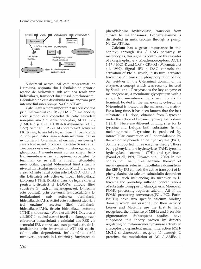
Prima etapã obligatorie, limitativã, estesinteza din L-tirozinã a L-DOPA, printr-o reactiede hidroxilare, catalizatã de tirozinazã, enzimacheie a întregului proces. Ulterior apar multiplereacþii de oxidoreducere ºi transformãriintracelulare(vezi imagine). (Hideya Ando,Hirofumi Kondoh, Masamitsu Ichihashi andVincent J Hearing. Approaches to IdentifyInhibitors of Melanin Biosynthesis via theQuality Control of Tyrosinase, 2007)
signaling are not activated until calciumconcentrations reach 10-6-10-8 M and extracellularcalcium concentration is usually 10-3 M. Inaddition, the proteolytic cascade associated withcell death is triggered when the intracellularconcentration of calcium exceeds 10-5 M in mostcells. It postulates that eumelanin regulate calciuhomeostasis in melanocytes by sequestration andbuffering intracellular calcium concentrationsbecause of calcium binding properties(lowaffinity, high capacity) well being suited for thispurpose.
Calcium - regulator of melanogenesis
Skin pigmentation is genetically governed bya complex composed of more than 150 allelesspread in approximately 90 binding sites thatencode the synthesis of many enzymes, structuralproteins, transcription regulators, transporters,receptors, growth factors etc.. Are described twotypes of melanin, eumelanin and pheomelanin,which differ structurally, by chemical composition,structural characteristics and physical properties.
The first phase is mandatory, exhaustive, isthe synthesis from L-tyrosine of L-dopa, ahydroxylation reaction, catalyzed by tyrosinase, akey enzyme of all process. Subsequently multiplereactions occur oxidoreductions and intracellularprocessing(see picture). (Hideya Ando, HirofumiKondoh, Masamitsu Ichihashi and Vincent JHearing. Approaches to Identify inhibitor ofmelanin Biosynthesis via the Quality Control oftyrosine, 2007)
Substrate of this way is represented by L-tyrosine, obtained from L-phenylalanine byhydroxylation reaction under the action of
304
DermatoVenerol. (Buc.), 55: 299-312
Substratul acestei cãi este reprezentat deL-tirozinã, obþinutã din L-fenilalaninã printr-oreactie de hidroxilare sub acþiunea fenilalaninhidroxilazei, transport din citosol în melanozomi.L-fenilalanina este distribuitã în melanozom prinintermediul unei pompe Na-Ca-ATPaza.
Calciul are o mare importanþã în acest contextprin intermediul cãii IP3 / DAG. În melanocite,acest semnal este controlat de cãtre cascadelenorepinefrinã / α1-adrenoreceptori, ACTH 1-17/ MC1-R ºi CRF / CRF-R1(Wakamatsu et all,1997). Semnalul IP3 /DAG controleazã activareaPKCβ care, la rândul sãu, activeaza tirozinaza de2,5 ori, prin fosforilarea a douã reziduuri de Serîn domeniul C-terminal al enzimei, un conceptcare a fost recent promovat de cãtre Sasaki et al.Tirozinaza este enzima cheie a melanogenezei, oglicoproteinã membranarã cu un singur helixtransmembranar în apropierea capatului C-terminal, ce se aflã la nivelul citosoluluimelanocitar, capatul N-terminal fiind situat lanivelul matrixului melanosomal.Multã vreme s-acrezut cã substratul optim este L-DOPA, obþinutãdin L-tirozinã sub acþiunea tirozin hidroxilazeiizoforma 1(THI). Existã situsuri de legare diferitepentru L-tirozinã ºi L-DOPA, ambele fiindsubstrate în cadrul melanogenezei. L-tirozinaeste obþinutã prin conversia intracelularã a L-fenilalaninei sub acþiunea fenilalaninhidroxilazei(PAH). Astfel este susþinutã „teoria atrei enzime”, acestea fiind fenilalaninhidroxilaza(PAH), tirozin hidroxilaza izoforma1(THI) ºi tirozinaza.(Wood et all, 1991, Olivares etall. 2002) În cadrul acestei teorii a melanogenezei,eliberarea intracelularã a calciului din RER viasemnalul IP3, controleazã transportul activ de L-fenilalaninã prin intermediul ATP-azã calciu-calmodulin dependentã, influienþând astfelturnoverul acesteia în L-tirozinã ºi furnizarea de
phenylalanine hydroxylase, transport fromcitosol to melanosomes. L-phenylalanine isdistributed in melanosomes through a pump,Na-Ca-ATPaza.
Calcium has a great importance in thiscontext, through IP3 / DAG pathway. Inmelanocytes, this signal is controlled by cascadesof norepinephrine / α1-adrenoreceptors, ACTH1-17 / MC1-R and CRF / CRF-R1 (Wakamatsu etall, 1997). Signal IP3 / DAG controls theactivation of PKCâ, which, in its turn, activatestyrosinase 2.5 times by phosphorylation of twoSer residues in the C-terminal domain of theenzyme, a concept which was recently fosteredby Sasaki et al. Tirosynase is the key enzyme ofmelanogenesis, a membrane glycoprotein with asingle transmembrane helix near to its C-terminal, located in the melanocytic cytosol, theN-terminal is located in the malanosome matrix.For a long time, it has been known that the bestsubstrate is L -dopa, obtained from L-tyrosineunder the action of tyrosine hydroxylase isoform1 (THI). There are different binding sites for L-tyrosine and L-dopa, both substrates ?n themelanogenesis. L-tyrosine is produced byintracellular conversion of L-phenylalanine bythe action of phenylalanine hydroxylase (PAH).So it is supported „three enzymes theory”, thosebeing phenylalanine hydroxylase (PAH), tyrosinehydroxylase isoform 1 (THI) and tyrosinase(Wood et all, 1991, Olivares et all. 2002). In thiscontext of the „three enzyme theory” ofmelanogenesis, release intracellular calcium fromthe RER by IP3 controls the active transport of L-phenylalanine via calcium-calmodulin-dependentATP-ase, such influencing its turnover to L-tyrosine and providing sufficient concentrationsof substrate to support melanogenesis. Moreover,POMC processing requires calcium. All of thePOMC processing convertases(PC1, PC2, Furin,PACE4) have two specific calcium bindingdomain which are essential for their activity.Lerner and McGuire are the first to haverecognized the influence of MSHα and β on skinpigmentation. Subsequent studies havesupported this theory proven by directlyregulating on melanosomes tyrosinase activity ina receptor indepandent maner. Interaction MSH-MC1R (melanocortin receptor 1) through Gproteins, the modulation of AC / AMPc, is
305
DermatoVenerol. (Buc.), 55: 299-312
concentraþii suficiente de substrat pentru asusþine melanogeneza. Mai mult decât atât,prelucrarea POMC necesitã calciu. Toateconvertazele de prelucrare POMC (PC1, PC2,Furin, PACE4) au douã situsuri specifice decalciu, cu caracter obligatoriu, care sunt esenþialepentru activitatea lor. Lerner ºi McGuire suntprimii care au recunoscut influenþa MSH α ºi βasupra pigmentãrii cutanate. Studiile ulterioareau susþinut acestã teorie dovedind capacitatea dereglare directã în melanozomi a activitãþiitirozinazei, într-un mod receptor independent.Interacþiunea MSH-MC1R(melanocortin receptor1) prin intermediul proteinelor G, cu modulareaAC/AMPc, este reglatã in vitro de schimbãrileconcentraþiei de calciu. MSH este internalizat îninteriorul melanozomilor,prin intermediul unorsitusuri de legare. De asemenea, α/β MSH poatelega cu afinitate crescutã 6/7 BH4, recunoscuþiinhibitori ai tirozinazei, ceea ce conduce la oenzimã funcþionalã.
Rolul calciului în interacþiunea melanocit-keratinocit
Melanocitele produc melanina în interiorulmelanozomilor, ce sunt transferaþi keratinocitelorvecine din epiderm ºi rezultã pigmentarea vizbilãa pielii. Contactul fizic între melanocite ºikeratinocite este o condiþie prealabilã pentrutransferul de melanozomi, dar semnalele celulareinduse în timpul sau dupã contact nu sunt pedeplin înþelese. Se presupune cã interacþiuniledintre membranele plasmatice ale acestor douãtipuri de celule au indus un semnal intracelularde calciu la nivelul keratinocitelor care va finecesar pentru transferul de pigment. Acestsemnal intracelular de calciu are loc ca urmare aeliberãrii de calciu din depozitarele intracelulare.Transferul de pigment observat în coculturilemelnocite-keratinocite a fost inhibat, în condiþiileîn care calciul intracelular în keratinocitelor a fostchelatat. S-a lansat ideea unei interacþiuni de tip„ligand-receptor” între melanocite ºi keratinocitecare declanºeazã mobilizarea intracelularã decalciu la nivelul keratinocitelor ºi mediazãtransferul de melaninã. Procesul de transfer demelanozomi este un proces unic biologic, careeste încã neînþeles. Ipoteze diferite, cum ar fieliberarea de melanozomi de cãtre melanocite,urmatã de endocitoza lor în keratinocite,inocularea directã (injectare) de melanozomi în
regulated in vitro by calcium concentrationchanges. MSH is internalized insidemelanosomes through binding sites. Also, á / âMSH can bind with high affinity 6 / 7 BH4,tyrosinase recognized inhibitors, leading to afunctional enzyme.
Role calcium in melanocyte-keratinocyteinteraction
The melanocytes produce melanin withinmelanosomes, which is transferred to theirneighbors, keratinocytes in the epidermis,resulting visible skin pigmentation. Physicalcontact between melanocytes and keratinocytesis a prerequisite for the transfer of melanosomes,but cellular signals induced during or aftercontact are not fully understood. It is assumedthat the interactions between plasma membranesof these two cell types were induced byintracellular calcium signal in the keratinocytesthis being necessary for the transfer of pigment.This intracellular calcium signal is due to releaseof calcium from intracellular stores. Pigmenttransfer observed in melnocite-keratinocytes co-cultures was inhibited when intracellular calciumin keratinocytes was chelated. It is proposed theidea of a „ligand-receptor“ type of interactionbetween melanocytes and keratinocytes thattriggers intracellular calcium mobilization in thekeratinocytes and mediates melanin transfer. Theprocess of melanosomes transfer is a uniquebiological process, which is still uncom-prehensible. Different assumptions, such asrelease of melanosomes by melanocytes,followed by their endocytosis into keratinocytes,direct inoculation (injection) of melanosomes intokeratinocytes and keratinocyte-melanocytemembrane fusion have been proposed as possiblemechanism (Boissy, 2003, Nguyen andWei, 2004).There are methods in the literature to studypigment transfer in vitro (Aspengren et al., 2006,Berens et al., 2005). Some surface molecules havebeen identified on keratinocytes governing theprocess of melanosomes phagocytosis. Some ofthese include keratinocyte growth factor receptorand protease-activated receptor (2) (PAR-2)(Boissy, 2003; Cardinali et al., 2005). It was shownthat modulation of PAR-2 can influencemelanosomes phagocytosis, a process in whichthe melanocyte-keratinocyte contact is essential

306
DermatoVenerol. (Buc.), 55: 299-312
keratinocite ºi fuziunea membranarã melanocit-keratinocit au fost propuse ca mecanism posibil(Boissy, 2003; Nguyen ºi Wei, 2004). Existãmetode în literatura de specialitate pentru astudia transferul de pigment in vitro(Aspengrenet al., 2006; Berens et al., 2005). Anumite moleculede suprafaþã au fost identificate pe keratinocitecare reglementeazã procesul de fagocitozã amelanozomilor. Unele dintre acestea includreceptorul factorului de creºtere a keratinocitelorºi receptorul activatorului de proteazã(2)(RAP-2)(Boissy, 2003; Cardinali et al., 2005). A mai fostarãtat cã modularea RAP-2 poate influenþafagocitoza melanozomilor, un proces în carecontactul melanocit-keratinocit este esenþial(Seiberg, 2001; Seiberg et al., 2000). În interac-þiunea melanocit-keratinocit sunt implicateprobabil evenimente precum formarea de procesedendritice ºi filopode care se extind de lamelanocite spre keratinocite ºi formareahomodimerului E-cadherinã între melanocite ºikeratinocite (Scott, 2002; Scott et al., 2002; Tang etal., 1994). Mecanismele implicate în recunoaº-terea melanocit - keratinocit în prezent nu suntcunoscute. Interacþiunea celulã-celulã esteresponsabilã pentru reglementarea formeicelulare, a motilitãþii ºi structurii þesuturilor.Complexele proteice care se gãsesc la punctele decontact dintre celule sunt integrate în cãi desemnalizare intracelularã care conduc laadeziune celularã, proliferare, diferenþiere ºiapoptozã (Braga ºi Harwood, 2001). Acesteprocese celulare sunt de obicei mediate prinsemnal intracelular de calciu care este consideratevenimentul iniþial de semnalizare. Este cunoscutfaptul cã semnalizarea intracelularã generatã decalciu controaleazã procesele de creºtere,diferentiere si apoptozã a keratinocitelorepidermice (Gniadecki ºi Gajkowska, 2003).
În acest scop citatãm un studiu în care a fostcercetat rolul concentraþiei intracelulare de calciuliber în recunoaºterea keratinocit-melanocit ºitransferul de melaninã. Pentru a studia procesulde transfer a melaninei, precum ºi rolul calciuluiintracelular în acest fenomen, a fost dezvoltat untest de transfer in vitro a melaninei. S-au utilizatculturi de celule de melanom cu culturi celularede keratinocite epidermoide, observându-se oataºare a celulelor de melanom la celekeratinocitare în decurs de 2 ore, transferul depigment monitorizandu-se cu ajutorul coloraþieiFontana-Masson. Dupã 24 de ore de co-cultivare,
(Seiberg, 2001, Seiberg et al., 2000). Inmelanocyte-keratinocyte interactions areprobably involved events such are formation ofdendritic processes and filopodia that extendfrom melanocytes towards keratinocytes and E-cadherin homodimer formation betweenmelanocytes and keratinocytes (Scott 2002, Scottet al., 2002, Tang et al. , 1994). The mechanismsinvolved in recognizing melanocyte –keratinocyte is currently not known. Cell-cellinteraction is responsible for regulating cellshape, motility and tissue structure. Proteincomplexes found at points of cell contact areintegrated into intracellular signaling pathwaysleading to cell adhesion, proliferation,differentiation and apoptosis (Braga andHarwood, 2001). These cellular processes areusually mediated by intracellular calcium signalthat is considered as the initial signaling event. Itis known that intracellular calcium signalingcontrols growth, differentiation and apoptosis ofepidermal keratinocytes (Gniadecki andGajkowski, 2003).
In this way we mentioned a study in wichwas investigated the role of intracellularconcentration of free calcium to keratinocyte-melanocyte in recognition and transfer ofmelanin. To study the process of melanin transferand the role of intracellular calcium in thisphenomenon, a test was developed in vitrotransfer of melanin. Were used cell culture ofmelanoma cell, cultures of epidermoidkeratinocytes, observing the attachment ofmelanoma cells to keratinocytes within 2 hours,transfer of pigment monitoring with the Fontana-Masson coloration. After 24 hours of co-cultivation, there was a presence of pigment inthe 60 ± 5% of keratinocytes. The experiment wasrepeated with human melanocytes, observing aloading rate with pigment within 55 ± 5% ofkeratinocytes. Precondition for the transfer ofmelanin is the contact between melanocytes andkeratinocytes (Seiberg, 2001, Seiberg et al., 2000).In melanocyte-keratinocyte co-cultures,melanocytes extend filopodia to keratinocyte, buttechnically is not feasible to monitor intracellularsignaling during the actual event of physicalcontact. Therefore, it was investigated the abilityof membrane fractions isolated from melanomacells to induce signaling in the keratinocytes. By

307
DermatoVenerol. (Buc.), 55: 299-312
s-a observat o prezenþã de pigment în 60 ± 5%dintre keratinocite. Experimentul s-a repetat ºi cuajutorul melanocitelor umane, observandu-se orata de încarcare cu pigment la 55 ± 5% dintrekeratinocite. Condiþia prealabilã pentrutransferul de melaninã este reprezentatã decontactul dintre melanocite ºi keratinocite(Seiberg, 2001; Seiberg et al., 2000).In aceste co-culturi melanocit-keratinocit, s-au observatfilopode ale melanocitelor în jurul keratinocitelor,dar punct de vedere tehnic nu este fezabil a semonitoriza semnalizarea intracelularã în timpulevenimentului real de contact fizic. Prin urmare,a fost investigatã capacitatea unor fracþiuni demembranã din celulele izolate de melanom dacãpot induce semnalizare la nivelul keratinocitelor.Prin adãugarea acestor fracþiuni membranare s-aobservat o creºtere tranzitorie a calciuluiintracelular la nivelul keratinocitelor, specificacestei interacþiuni. Aceastã modificare nu s-aobservat cand se foloseºte un alt tip de fracþiunimembranare tumorale sau dacã proteinelemembranare au fost distruse prin inhibitori deproteine sau îngheþare. De aici s-a desprinsconcluzia conform cãreia semnalizarea intra-celularã a calciului la nivelul keratinocitelornecesitã prezenþa proteinelor membraneimelanocitelor într-o conformaþie activã. Creºtereacalciului la nivelul keratinocitelor se datoreazãeliberãrii intracelulare din depozite. Aceste dateconfirmã faptul cã recunoaºterea keratinocitelorde cãtre melanocite este specificã. Se sugereazã cão astfel de interacþiune fizicã duce la generareaunui semnal de calciu intrakeratinocitar, esenþialîn procesul de transfer al melanozomilor.Interacþiunea melanocit-keratinocit este practicbaza coloraþiei pielii, realizatã prin transferul demelaninã în direcþia menþionatã, în pluskeratinocitele pot afecta alte funcþii alemelanocitelor, inclusiv procesul de sintezã amelaninei, proliferarea melanocitelor, etc (Duvalet al., 2002; Nakazawa et al., 1995a, b; Valyi-Nagyet al., 1993). Acest studiu demonstreazã practic,utilizând co-culturi din sisteme diferite, ointeracþiune de tipul „ligand-receptor like”, ceconduce la o semnalizare intracelularã a calciuluila nivelul keratinocitelor, generatã de interacþiunide tipul proteinã-proteinã în urma unui contactfizic al acestor douã tipuri de celule. Acestmecanism a fost descris ºi în alte sistemecelulare(de exemplu, activarea limfocitelor B decãtre celulele T (Kim et al., 2001) ºi controlul
adding these membrane fractions was observed atransient increase in intracellular calcium in thekeratinocyte, specifically this interaction. Thischange was not observed when using anothertype of tumor membrane fractions or membraneproteins were destroyed by inhibitors of proteinor freezing. Hence the conclusion that split offintracellular calcium signaling in the keratinocytemembrane proteins requires the presence ofmelanocytes in an active conformation. Increasedcalcium in the keratinocytes is due to releasefrom intracellular stores. These data confirm thatrecognition of the melanocyte keratinocyte isspecific. It is suggested that such a physicalinteraction results in generating a calcium signalinside keratinocyte essential in the transfer ofmelanosomes. Melanocyte-keratinocyte inter-action is practically the basis of skin colour,achieved by transfer of melanin in mentioneddirection, and keratinocytes affect other functionsof melanocytes, including the process of melaninsynthesis, the proliferation of melanocytes, etc.(Duval et al., 2002, Nakazawa et al., 1995a, b;Valy-Nagy et al., 1993). This study demonstrates,using co-cultures of different systems, aninteraction oft „ligand-receptor like” type,leading to an intracellular calcium signaling inthe keratinocytes, caused by protein-proteininteractions generated by physical contactbetween these two cell types. This mechanismwas described in other cellular systems too (eg., Bcell activation by T cells (Kim et al., 2001) andcontrol of axonal outgrowth in neurons frommyelin membrane (Hunt et al., 2002). Nogo orMAG proteins present on the myelin membranebind to NOGO receptor on the neuronal growthcone membrane, and induce release of calciumand activae other signalling pathways, which inturn control axonal growth. Bush andSimon(2007) have shown that melanosomes cansequester calcium and help maintain calciumhomeostasis in melanocytes. In vivo, thepossibility exists for calcium signals to begenerated by keratinocyte through release ofcalcium from melanosomes transferred tokeratinocytes. Thus there is clear evidence thatplasma membrane interaction of melanocyteswith keratinocytes induced calcium signaling inthe keratinocytes.

308
DermatoVenerol. (Buc.), 55: 299-312
creºterii axonale în neuroni prin membrana demielinã (Hunt et al., 2002). Proteinele NOGO sauMAG prezente pe membrana de mielinã se leagãde receptorul NOGO de pe membrana neuronalãºi induc eliberarea calciului cu activarea altor cãide semnalizare, ce controleazã creºterea axonalã.Bush ºi Simon (2007) au arãtat cã melanozomi potsechestra calciu si ajutã la menþinerea homeo-staziei acestuia în melanocite. In vivo, existãposibilitatea generãrii de semnale intra-keratinocitare calciu-inductibile, prin eliberareade calciu din melanozomii transferaþi keratino-citelor. Astfel existã dovezi clare cã interacþiuneamembranei plasmatice a melano-citelor cukeratinocitele induce semnalizare de calciu lanivelul keratinocitelor.
Rolul calciului în reglarea echilibruluiRedox
Calciul este un ion cu rol important înpotenþialul antioxidant al pielii controlat prinnivelurile intracelulare ale acestuia. Concen-traþiile intracelulare de calciu ≥ 10-5 M duc laactivarea de proteaze calciu-dependente,rezultând o cascadã de evenimente corelate cumoarte celularã (Carafoli et al., 1984; Waring,2005).
Sunt descrise trei mecanisme prin care calciulinfluenþeazã echilibrul redox. Primul estereprezentat de inhibarea tioredoxin reductazei ºicreºterea oxidãrii tioredoxinei, care este ºi cel maiimportant. Schllreuter et. al. au raportat acestefect de inhibare a tioredoxin reductazei în celulemelanomului uman. Tioredoxina în mod normalconverteºte continuu punþile S-S proteice îndithioli reduºi. Pentru aceste reacþii este necesarãparticiparea unei enzime în formã redusã ce serealizeazã sub acþiunea tioredoxin reductazei.Consecinþa inhibãrii acestei enzime estereprezentatã de acumularea 2-peroxiredoxin-S2,ce are douã efecte principale, conduce la oxidarearapidã a tioredoxinei, ce participa la rândul sãu laoxidarea altor proteine cu efect intrinsec deoxidare a gruparilor thiol proteice. Pe lângãconversia sus menþionatã, tioredoxina contribuiela legarea regulatorie selectivã al thioluluitioredoxinei la alte proteine funcþionale. Prinurmare tioredoxina are douã funcþii ºi anumecatalizator redox ºi regulator redox sensibil prininteractiuni proteinã-proteinã. De asemenea,tioredoxina este asociatã porþiunii N-terminalã a
Role of calcium in Redox balancereglation
Calcium is an ion with important antioxidantpotential of skin, controlled by its intracellularlevels. Intracellular calcium concentrations ≥ 10-5
M lead to activation of calcium-dependentproteases, resulting in a cascade of events relatedto cell death (Carafoli et al., 1984, Waring, 2005).
Are described three mechanisms by whichcalcium affects the redox balance. The first is theinhibition of thioredoxin reductase and increasedoxidation of thioredoxin, which is the mostimportant. Schllreuter et al. were reported thiseffect of thioredoxin reductase inhibition inhuman melanoma cells. Thioredoxin normallyconverts continuously protein SS bridges inreduced dithiols. These reactions requires theinvolvement of an enzyme in reduced form,realized under the action thioredoxin reductase.The consequence of this enzyme inhibition is theaccumulation of 2-peroxiredoxin-S2, which hastwo main effects, lead to rapid oxidation ofthioredoxin, which in his turn participate atoxidation of other proteins with intrinsic effect ofprotein thiol groups oxidation. In addition to theaforementioned conversion, tioredoxin contributeto selective regulatory binding of tioredoxin thiolto other functional proteins. Therefore tioredoxinhas two functions, redox catalyst and redox-sensitive regulator by protein-proteininteractions. Thioredoxin also by its N-terminaldomain bind to ASK-1 in vivo and in vitro, beinga physiological inhibitor of this in its reducedform. The ASK-1 effect is conditiont by redoxstatus of thioredoxin. In this context withthioredoxin reductase inhibition dependent byincreased calcium levels and accumulation ofthioredoxin in oxidized form, there is anactivation of ASK-1 with subsequent increased ofJNK/p38 MAP kinases and cell death (DanonAvihai and Carlos Gitler, 2003).
The second mechanism is the stimulation ofNADPH oxidase, leading to an explosivesynthesis of superoxide ions with cytoplasmicaccumulation of hydrogen peroxide, whichinhibits phosphorylation of receptor kinasesthrough fosfotyrosine.
The third mechanism is the ability ofcytoplasmic calcium redox regulation to oxidateSS protein bridges.
309
DermatoVenerol. (Buc.), 55: 299-312
ASK-1 in vivo ºi in vitro, fiind un inhibitorfiziologic al acesteia în forma sa redusã. EfectulASK-1 este condiþionat de statusul redox altioredoxinei. În acest context al inhibãriitioredoxin reductazei dependente de concentraþiicrescute ale calciului ºi acumulãrii tioredoxineiîn formã oxidatã, se constatã o activare a ASK-1cu amplificarea subsecventã a cãii kinazelorJNK/p38 MAP ºi moartea celularã (Gitler Carlosand Danon Avihai, 2003)
Cel de-al doilea mecanism este reprezentat destimularea NADPH oxidazei, ce conduce la osintezã explozivã de ioni superoxid, cu acumu-larea de peroxid de hidrogen citoplasmatic, careinhibã fosforilarea receptorului kinazic prinintermediul fosfotirozinei.
Al treilea mecanism este reprezentat decapacitatea de reglare redox a calciului cito-plasmatic prin oxidarea punþilor S-S proteice.
Toate aceste mecanisme conduc la cumularede kinaze, asociate cu moarte celularã, arãtândimportanþa reglãrii homeostaziei calciului.
Perturbarea homeostaziei calciului înmelanomul uman
Tumorile cutanate maligne se caracterizeazãprin acumularea excesivã a metalelor. Prinmetode fizice(INAA, PIXE) ºi chimice (spectro-metrie) autorii au constatat existenþa unui raportsupranumerar între concentraþia calciului dinþesutul tumoral ºi þesutul sãnãtos adiacent(rezultate nepublicate).
Variaþiile nivelurilor plasmatice ale calciuluiîn malignitãþi, în cazul de faþã în melanomulcutanat, prezintã importanþã prin apreciereaasupra prognosticului. Cel mai frecvent sevorbeºte de hipercalcemie, rar manifestare iniþialãa malignitãþii, asociatã cu un prognostic sever(supravieþiure sub 6 luni, decesul survenind la50% dintre pacienþi în 30 zile). Hipercalcemiaapare la 5-10% dintre aceºti pacienþi, dupã unelestudii pânã la 20-30%(Fisken et all, 1980; Rolstonet all. 1990). Hipercalcemia inregistratã la 1.37%dintre pacienþii cu melanom cutanat se asociazãcu prognostic sumbru(Nicolae I, Anghel A.,2009). Aprecierea hipercalcemiei PTH inde-pendentã, semnalatã într-un studiu amplu,efectuat pe 950 pacienþi, în perioada 2000-2010, s-a fãcut pe baza cCaT(calciu total corectat), înfuncþie de nivelul albuminei circulante(Nicolae I.,Anghel A., 2009). Au fost descrise trei mecanisme
All these mechanisms lead to theaccumulation of kinase associated with celldeath, indicating the importance of calciumhomeostasis regulation.
Calcium homeostasis distruption inhuman melanoma
Malignant skin tumors are characterized byexcessive accumulation of metals. By physicalmethods (INAA, PIXE) and physico-chemical(spectrophotometry) the authors have foundexistence of a supernumerary betwen calciumconcentration in tumor tissue and adjacenthealthy tissue (unpublished results).
Variations in serum calcium levels inmalignancies, in our case in cutaneous melanoma,is important in assessing the prognosis. The mostfrequently speaks of hypercalcemia, a rare initialmanifestation of malignancy, associated with asevere prognosis (survival under 6 months, deathoccurring in 50% of patients in 30 days).Hypercalcemia occurs in 5-10% of these patients,as some studies up to 20-30% (Fisken et all, 1980,Rolston et all. 1990). Hypercalcemia registered at1.37% of patients with cutaneous melanoma isassociated with bad prognosis (Nicholas I,Anghel A., 2009). PTH-independent hypercal-cemia assessment, in a reported large studyconducted on 950 patients, in period 2000-2010,was based on cCaT (corrected total calcium), thelevel of circulating albumin (Nicholas I., AnghelA., 2009). Have been described three mechanismsof its occurrence, like the synthesis of PTH-likesubstances (PTHrP) identified in the tumor, boneosteolitic metastases and tumor production ofcalcitriol. The first mechanism that is mostimportant, being responsible for approx. 80% oftumor hypercalcemia. PTHrP is noted as a newhormone, without function, but with importantphysiological role, different structures of PTH,but with amino-terminal domain common, whichmakes the protein PTHrP active to the PTHreceptor, equipotente for them and the sameacute effects (Kemp et all., 1987). This featuretogether with similarities in gene structuresuggested origin in a common ancestral gene(Yasudo, 1989, Stream, 1989, Kemp et all., 1987).From the different roles of PTH and PTHrP waslaunched idea PTHrP receptor, which alsoidentifies and PTH. Note the secretion of
310
DermatoVenerol. (Buc.), 55: 299-312
de apariþie a acesteia ºi anume sinteza uneisubstanþe PTH-like(PTHrP)identificatã la niveltumoral, metastazele osoase osteolitice ºiproducþia tumoralã de calcitriol. Primul mecanismmenþionat este cel mai important, fiindresponsabil de aprox. 80% dintre hipercalcemiiletumorale. PTHrP este notat a fi un nou hormon,fãrã funcþie, dar cu rol fiziologic important,diferitã structural de PTH, dar cu capãtul amino-terminal comun, ceea ce face ca proteina PTHrPsã fie activã pe receptorii PTH fiind equipotentepentru aceºtia, având aceleaºi efecte acute(Kempet all., 1987). Aceastã caracteristicã împreunã cuasemãnãrile în structura genelor au sugeratoriginea într-o genã ancestralã comunã(Yasudo,1989; Suvo, 1989; Kemp et all., 1987). Pornind dela rolurile diferite ale PTH ºi PTHrP a fost lansatãideea receptorilor de PTHrP, ce identificã deasemenea ºi PTH. De notat ºi secreþia decitokine(TNFα, IL1β, IL6, RANKL) de cãtretumorã asociatã celei de PTHrP ce acþioneazãsinergic pentru apariþia hipercalcemiei (Suvo etall, 1987) .
Hipocalcemia este rar asociatã malignitãþii ºicunoaºte în primul rând douã mecanisme ºianume instalarea leziunilor osteoblasticemetastazice condiþie în care calciul extracelularutilizat pentru mineralizarea osului nou, ºisindromul de lizã tumoralã în care tumoraelibereaza ioni intracelulari, precum fosfat,potasiu, acid uric(Mundy et all., 1990). Creºtereafosfatului implicã scãderea rapidã a calciuluiseric. Evaluãrile experimentale ale autorilor, pe operioadã de 10 ani, au evidenþiat hipocalcemie la27,6% dintre pacienþii cu melanom (datenepublicate).
Concluzii
Calciul reprezintã un ion cu implicaþii majoreîn procesele fiziologice normale, esenþiale, ce sedesfãºoarã la nivelul melanocitului precummelanogeneza ºi echilibrul redox, participand lainteractiunea intercelularã melanocit-keratinocit.Este demonstrat cã melanocitul participã activ lahomeostazia calciului prin melaninã, în acestcontext proteinã cu funcþie de legare. Variaþiilenivelului de calciu în mod special hipercalcemiaasociatã procesului malign aduce aprecieriasupra severitãþii prognosticului.
Intrat în redacþie: 10.02.2010
cytokines (TNFÜ, IL1â, IL6, RANKL) by tumor,associated with that of PTHrP which actsynergistically for the development ofhypercalcemia (Stream et all, 1987).Hypocalcemia is rarly associated withmalignancy and known primarly twomechanisms, the installation of osteoblasticmetastatic lesions in which extracellular calciumis used for new bone mineralization, and tumorlysis syndrome in which releases intracellularions such as phosphate, potassium, uric acid(Mundy et all., 1990). Increasing phosphateinvolves rapid decrease in serum calcium.Experimental evaluations of authors, over aperiod of 10 years showed hypocalcemia in 27.6%of patients with melanoma (unpublished data).
Conclusions
Calcium is an ion with major implications innormal essential physiological processes, thattakes place in melanocytes like melanogenesisand redox balance, participating in melanocyte-keratinocyte intercellular interaction. Melanocyteis shown to actively participate in calciumhomeostasis by melanin, in this context a proteinwith binding function. Changes in the calciumlevel in specifically hypercalcemia associatedwith malignant process bring assessment aboutprognostic severity.
Received: 10.02.2010
311
DermatoVenerol. (Buc.), 55: 299-312
Bibliografie/Bibliography1. Aspengren, S., Hedberg, D., and Wallin, M. (2006). – Studies of pigment transfer between Xenopus laevis
melanophores and fibroblasts in vitro and in vivo. Pigment Cell Res. 19, 136–145.2. Berens, W., Van Den Bossche, K., Yoon, T.J., Westbroek, W.,Valencia, J.C., Out, C.J., Naeyaert, J.M., Hearing, V.J.,
and Lambert, J. (2005). – Different approaches for assaying melanosome transfer. Pigment Cell Res. 18, 370–381.3. Boissy, R.E. (2003). – Melanosome transfer to and translocation in the keratinocyte. Exp. Dermatol. 12 (Suppl. 2), 5–12.4. Boyce, S.T., and Ham, R.G. (1983). – Calcium-regulated differentiationof normal human epidermal keratinocytes
in chemically defined clonal culture and serum-free serial culture. J. Invest. Dermatol. 81, 33s–40s.5. Braga, V., and Harwood, A.J. (2001). – ‘Super glue’. Nat. Cell Biol. 3, E168–E170.6. Bush, W.D., and Simon, J.D. (2007). – Quantification of Ca2+ binding to melanin supports the hypothesis that
melanosomes serve a functional role in regulating calcium homeostasis. Pigment Cell Res. 20, 134–139.7. Buffey, J.A., Edgecombe, M., and MacNeil, S. (1993). – Calcium plays a complex role in the regulation of
melanogenesis in murine B16 melanoma cells. Pigment Cell Res. 6, 385–393.8. Carafoli, E. (1987). – Intracellular calcium homeostasis. Annu. Rev.Biochem. 56, 395–433.9. Carafoli, E., Guiseppe, I., and Rosen, B.P. (1984). – Calcium transport across biological membranes. In Metal Ions
in Biological Systems, Vol. 17, H. Sigel, ed. (New York, NY: Marcel Dekker), pp.129–186.10. Cardinali, G., Ceccarelli, S., Kovacs, D., Aspite, N., Lotti, L.V.,Torrisi, M.R., and Picardo, M. (2005). – Keratinocyte
growth factor promotes melanosome transfer to keratinocytes. J. Invest. Dermatol. 125, 1190–1199.11. Drager, U.C. (1985). – Calcium binding in pigmented and albino eyes. Proc. Natl Acad. Sci. USA 82, 6716–6720.12. Duval, C., Smit, N.P.M., Kolb, A.M., Régnier, M., Pavel, S., and Schmidt, R. (2002). – Keratinocytes control the pheo
/ eumelanin ratio in cultured normal human melanocytes. Pigment Cell Res. 15, 440–446.13. Fisken RA, Heath DA, Bold AM – Hypercalcaemia - A hospital survey. Q J Med 1980; 196: 405-415.14. Frausto da Silva, J.J.R., and Williams, R.J.P. (1991). – The Biological Chemistry of the Elements (New York, NY:
Oxford University Press), pp. 268–296.15. Gill, S.S., and Salt, A.N. (1997). – Quantitative differences in endolymphatic calcium and endocochlear potential
between pigmented and albino guinea pigs. Hear. Res. 113, 191–197.16. Gitler Carlos and Danon Avihai (2003) – Cellular implications of radox signaling, 276-284.17. Gniadecki, R., and Gajkowska, B. (2003). – Intracellular calcium pool emptying induces DNA synthesis in HaCaT
keratinocytes. Exp.Dermatol. 12, 453–459.18. Hunt, D., Coffin, R.S., and Anderson, P.N. (2002). – The Nogo receptor, its ligands and axonal regeneration in the
spinal cord; A review. J. Neurocytol. 31, 93–120.19. Hong, L., and Simon, J.D. (2006). – Insight into the binding of divalent cations to Sepia eumelanin from IR
absorption spectroscopy. Photochem. Photobiol. 82, 1265–1269.20. Hong, L., Liu, Y., and Simon, J.D. (2004). – Binding of metal ions to melanin and their effects on the aerobic
reactivity. Photochem. Photobiol. 80, 477–481.21. Hoogduijn, M.J., Smit, N.P., Van Der Laarse, A., Van Nieuwpoort,A.F., Wood, J.M., and Thody, A.J. (2003). –
Melanin has a role in Ca2+ homeostasis in human melanocytes. Pigment Cell Res. 16, 127–132.22. Hoogduijn, M.J., Cemeli, E., Ross, K., Anderson, D., Thody, A.J.,and Wood, J.M. (2004). – Melanin protects
melanocytes and keratinocytes against H2O2-induced DNA strand breaks through its ability to bind Ca2+. Exp.Cell Res. 294, 60–67.
23. Kim, S., Braunstein, N.S., Leonard, E.F., and Thomas, J.L. (2001). – Controlling duration of contact between T cellsand antigenpresenting cells. J. Immunol. Meth. 249, 73–84.
24. Kemp BE, Moseley JM, Rodda CP, et al – Parathyroid hormone-related protein of malignancy: Active syntheticfragments. Science 1987; 238:1568-1570.
25. Liu, Y., and Simon, J.D. (2003). – The effect of preparation procedures on the morphology of melanin from the inksac of Sepia officinalis. Pigment Cell Res. 16, 72–80.
26. Liu, Y., and Simon, J.D. (2005). – Metal-ion interactions and the structural organization of Sepia eumelanin. PigmentCell Res. 18,42–48.
27. Liu, Y., Hong, L., Kempf, V.R., Wakamatsu, K., Ito, S., and Simon, J.D. (2004). – Ion-exchange and adsorption ofFe(III) by Sepia melanin. Pigment Cell Res. 17, 1–8.
28. Mangin M, Ikeda K, Dreyer BE, Broadus AE – Isolation and characterization of the human parathyroid hormone-like peptide gene. Proc Natl Acad Sci USA 1989; 86:2408-2412.
29. Meyer zum Gottesberge, A.M. (1988). – Physiology and pathophysiology of inner ear melanin. Pigment Cell Res. 1,238–249.
312
DermatoVenerol. (Buc.), 55: 299-312
30. Mundy GR (1990). – Calcium hoemostasis: Hypercalcemia and hypocalcemia. London UK. Martin DunitzPublisher.
31. Nakazawa, K., Nakazawa, H., Bonnard, M., Damour, O., and Collombel, C. (1995a). – Ca2+ and UVB radiationhave no effect on E-Cadherin-mediated melanocyte–keratinocyte adhesion. Pigment Cell Res. 8, 255–262.
32. Nakazawa, K., Nakazawa, H., Collombel, C., and Damour, O. (1995b). – Keratinocyte extracellular matrix-mediated regulation of normal human melanocyte function. Pigment Cell Res. 8, 10–18.
33. Nguyen, T., and Wei, M.L. (2004). – Characterization of melanosomesin murine Hermansky–Pudlak Syndrome:mechanisms of hypopigmentation. J. Invest. Dermatol. 122, 452–460.
34. Nicolae I., Anghel A., etc. – Aspecte legate de homeostazia calciului în melanomul malign. Revista Societãþii Romanede Dermatologie, Nr. 2, 2009, 115-119.
35. Nicolae I., Anghel A., etc. – Managementul pacienþilor cu hipercalcemie tumoralã. Revista Societãþii Romane deDermatologie, Nr. 3, 2009,161-164.
36. Olivares C, Garcia-Borron J C, Solano F. – Identification of active site residues involved in metal cofactor bindingand stereospecific substrate recognition in Mammalian tyrosinase. Implications to the catalytic cycle. Biochemistry2002: 41: 679–686.
37. Panessa, B.J., and Zadunaisky, J.A. (1981). – Pigment granules: a calcium reservoir in the vertebrate eye. Exp. EyeRes. 32, 593–604.
38. Pozzan, T., Rizzuto, R., Volpe, P., and Meldolesi, J. (1994). – Molecularand cellular physiology of intracellularcalcium stores. Physiol.Rev. 74, 595–636.
39. Ralston SH, Gallacher SJ, Patel U, Campbell J, Boyle IT – Cancer-associated hypercalcemia: Morbidity andmortality-Clinical experience in 126 treated patients. Ann Intern Med 1990; 112:499-504.
40. Salceda, R., and Riesgo-Escovar, J.R. (1991). – Characterization of calcium uptake in chick retinal pigmentepithelium. Pigment Cell Res. 3, 141–145.
41. Salceda, R., and Sanchez-Chavez, G. (2000). – Calcium uptake,release and ryanodine binding in melanosomesfrom retinal pigment epithelium. Cell Calcium 27, 223–229.
42. Schallreuter, K.U., and Wood, J.M. (1999). – The importance of L-phenylalanine transport and its autocrineturnover to L-tyrosine for melanogenesis in human epidermal melanocytes. Biochem.
43. Schallreuter K U, Wood J M. – The importance of L-phenylalaninetransport and its autocrine turnover to L-tyrosine for melanogenesis in human epidermal melanocytes. Biochem Biophys Res Commun 1999: 262: 423–428.
44. Schallreuter K U, Wazir U, Kothari S, Gibbons N C, Moore J,Wood J M. – Human phenylalanine hydroxylase isactivated by H2O2: a novel mechanism for increasing the L-tyrosine supply for melanogenesis in melanocytes.Biochem Biophys Res Commun 2004: 322: 88–92.
45. Scott, G. (2002). – Rac and Rho: The story behind melanocyte dendrite formation. Pigment Cell Res. 15, 322–330.46. Scott, G., Leopardi, S., Printup, S., and Madden, B.C. (2002). Filopodia are conduits for melanosome transfer to
keratinocytes. J. Cell Sci. 115, 1441–1451.47. Seiberg, M., Paine, C., Sharlow, E., Andrade-Gordon, P., Costanzo,M., Eisinger, M., and Shapiro, S.S. (2000). The
protease-activated receptor 2 regulates pigmentation via keratinocyte–melanocyte interactions. Exp. Cell Res. 254,25–32.
48. Suva LJ, Winslow GA, Wettenhall REH, et al – A parathyroid hormonerelated protein implicated in malignanthypercalcemia: Cloning and expression. Science 1987; 237: 893-896.
49. Suva LJ, Mather KA, Gillespie MT, et al – Structure of the 5’ flanking region of the gene encoding humanparathyroid-hormone-related protein (PTHrP). Gene 1989; 77: 95-105.
50. Tsien, R.W., and Tsien, R.Y. (1990). – Calcium channels, stores, and oscillations. Annu. Rev. Cell Biol. 6, 715–760.51. Valyi-Nagy, I.T., Hirka, G., Jensen, P.J., Shih, I.M., Juhasz, I., and Herlyn, M. (1993). Undifferentiated keratinocytes
control growth, morphology, and antigen expression of normal melanocytes through cell-cell contact. Lab. Invest.69, 152–159.
52. Waring, P. (2005). – Redox active calcium ion channels and cell death. Arch. Biochem. Biophys. 434, 33–42.53. Wakamatsu K, Graham A, Cook D, Thody A J. Characterisation of ACTH peptides in human skin and their
activation of the melanocortin-1 receptor. Pigment Cell Res 1997: 10: 288–297.54. Wood J M, Schallreuter K U. – Studies on the reactions between human tyrosinase, superoxide anion, hydrogen
peroxide and thiols. Biochim Biophys Acta 1991: 1074: 378–385.55. Wood J M, Schallreuter-Wood K U, Lindsey N J, Callaghan S, Gardner L G. – A specific tetrahydrobiopterin
binding domain on tyrosinase controls melanogenesis. Biochem Biophys Res Commun 1995: 206: 480–485.56. Yasuda T, Banville D, Hendy GN, Goltzman D – Characterization of the human parathyroid hormone-like peptide
gene-Functional and evolutionary aspects. J Biol Chem 1989; 264:7720-7725