rom j morphol embryol 2014, 55(3 suppl):1215–1219 r j m ... · rom j morphol embryol 2014, 55(3...
TRANSCRIPT
Rom J Morphol Embryol 2014, 55(3 Suppl):1215–1219
ISSN (print) 1220–0522 ISSN (on-line) 2066–8279
CCAASSEE RREEPPOORRTTSS
The impact of melanocytic cell destruction in pediatric facial burns and plastic surgery therapeutic management
ANCA ROXANA UNGUREANU1), DAN IONIŢĂ2), LIVIU DRĂGHICI1,3), DANIEL ANDREI1), DAN MIRCEA ENESCU2,3), ISABELA-MAGDALENA DRĂGHICI3,4)
1)Department of General Surgery, “Sf. Ioan” Emergency Clinical Hospital, Bucharest, Romania 2)Clinic of Plastic Surgery and Reconstructive Microsurgery, “Grigore Alexandrescu” Emergency Hospital for Children, Bucharest, Romania
3)“Carol Davila” University of Medicine and Pharmacy, Bucharest, Romania 4)Department of Pediatric Surgery, “Maria Skłodowska Curie” Emergency Clinical Hospital for Children, Bucharest, Romania
Abstract The main role of the melanin production belongs to the keratinocyte–Langerhans–melanocyte complex that within a burn injury might be destroyed. A particular aspect has the pediatric patient with burns due to a deficit in pigment synthesis and particularities involving the growth process of facial structures. This article presents four eloquent cases of pediatric patients with sequelae, consequence of facial burns that varied in depth and etiology. The age of patients at time of admission was between four and 18 years, and the interval between production of the primary lesion and admission was variable (0–17 years). Conservative treatments prove to be insufficient, requiring the destroyed structures to be replaced with compatible tissue by surgical intervention. The therapeutic success of skin grafts and dermal substituents acknowledges the main role of plastic surgery. New technologies as well as new cell focused studies support the clinical proven results as well as they enlarge the spectrum of potential available therapeutic methods in order to obtain the best therapeutic results.
Keywords: growth, leucoderma, dermal substituents, skin grafts, expander, keratinocytes culture.
Introduction
Melanocytes, the specialized cells that produce melanin with two varieties pheomelanin and eumelanin [1], are situated on the basal layer of epidermis (approximate 800–1000/mm2) and through their products, they produce the skin pigmentation. Nevertheless, these cells are destroyed in facial burns that involve the basal epidermal layer.
It is well known that the children’s skin is fairer, especially less than one year of age as pigmentation modifies with age. A major element is the melanic pigment quantity and quality, both genetically linked. There are further particularities of children’s facial skin: children’s skin is very thin and highly vascularized (pink colored).
Melanocytes, there are dendritic cells that expand their extensions in the spinosum layer, in areas exposed to the sun, becoming larger and more numerous. Pituitary stimulation, influenced by solar exposure, generates MSH alpha- or beta- (stimulating hormone). This molecule binds to a specific receptor on melanocytes MC1-R, and stimulates the production of melanocytes. Alpha-MSH is initiating the immune response and generates inflammatory mediators that promote the formation of melanin. So, what once was called epidermal–melanin unit is now called keratinocyte–Langerhans–melanocyte complex, acting in a coordinated manner to ensure the functions of the skin [2].
Researchers observed that the aggression (e.g., burning) on the skin will produce discolored areas, hypo-pigmented which in most cases are surrounded by scar and are caused by the inflammatory process that destroys the melanin-producing cells [3]. They found that usually, the pigment loss is only partial and the patient’s skin
regains color within a few months by restoring the melanocyte population and their properties, less in the scarring tissue, that lack such cells and which needs special treatment, needs to be removed by physical or chemical methods.
Plastic surgery has the purpose to prevent as possible the formation of scar areas and/or bleached more so as the functional and aesthetic impact is greater for facial burns. In pediatric patients, the formation of such areas is interfering in the growth process but also has an impact on psychosocial integration. Also, the dynamic changes of cephalic extremity with age are important considerations that take into account by the surgeon in the treatment of facial burns. From birth, the facial structures begin to develop much more than the cranial ones [4]. Lund and Browder table is a widely used assessment tool for burns area in children as it compensates for variation with age [5]. It shows the percentage of the corporeal surface that the head represents: 19% for an infant and only 7% in adult. Nevertheless, those precise tools like the 3D computer display software have been developed to assist clinicians map out the burn area, to estimate the depth and as such to choose the proper treatment [6].
Patients and Methods
In this regard, the Clinic of Plastic Surgery and Reconstructive Microsurgery of “Grigore Alexandrescu” Emergency Hospital for Children, Bucharest, Romania has a great interest both in the prevention of such com-plications and the treatment of those already established. Continuous research and choosing the appropriate therapy
R J M ERomanian Journal of
Morphology & Embryologyhttp://www.rjme.ro/
Anca Roxana Ungureanu et al.
1216
for each patient had successful results. Some cases were chosen randomly for eloquent discussion on the impact of facial burn melanocytic cell destruction and surgeon’s therapeutic decision. We choose four eloquent cases for discussion about the visual impact of facial burn on cells destruction and surgeon’s therapeutic decision.
The first patient presented an extensive IIB–III degree burn, having multiple locations and involving almost totally the face. A conservative treatment was ineffective on right hemifacial aspect due to burn’s depth.
The second patient had a persistent depigmented scar because of an IIB–III degree burn. The patient had a 17-year-old scar, situated in left temporoparietal region. The excision of scar area was performed then it was covered with a synthetic skin substitute.
The aim for the third patient was to reduce the aspect of an invalidating postburn sequel on right eye angle by using cultured keratinocytes.
For the last patient, who presented terrible lesions and alopecia, an expander has been used for obtaining a flap with the same characteristics of the surrounding tissues.
Results
Case No. 1
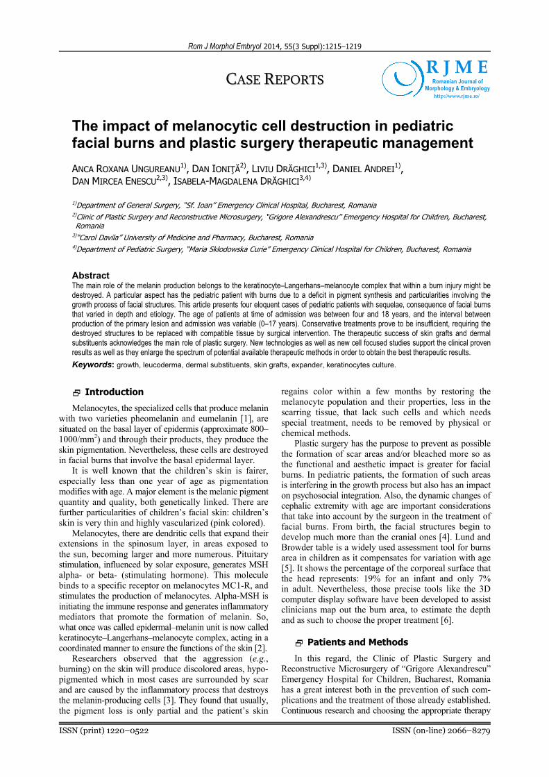
For the first patient, because the conservative treat-ment was ineffective, he required thin compatible skin grafting on the fifth day after presentation. The post-operative evolution is favorable and the excellent con-solidation of results can be seen in about four months after surgery (Figure 1). For the first case, the final result shows that the graft has same color as the patient’s normal skin but the area that was treated conservatively has a discolored aspect that implies the melanocytic cell destruction.
Figure 1 – Compatible skin grafting: (A) Post-burn aspect; (B) Conservative treatment; (C) Granulating tissue; (D) Compatible skin grafting; (E) Final result-normal pigmented grafted skin vs. depigmented area.
Case No. 2
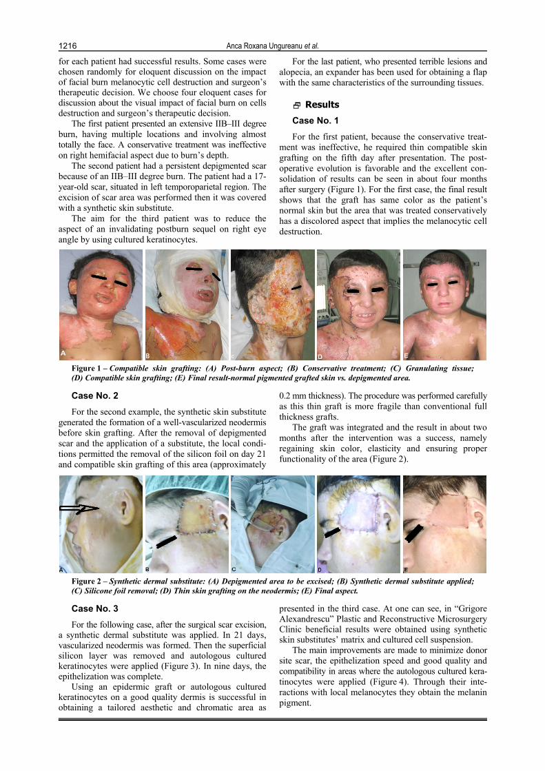
For the second example, the synthetic skin substitute generated the formation of a well-vascularized neodermis before skin grafting. After the removal of depigmented scar and the application of a substitute, the local condi-tions permitted the removal of the silicon foil on day 21 and compatible skin grafting of this area (approximately
0.2 mm thickness). The procedure was performed carefully as this thin graft is more fragile than conventional full thickness grafts.
The graft was integrated and the result in about two months after the intervention was a success, namely regaining skin color, elasticity and ensuring proper functionality of the area (Figure 2).
Figure 2 – Synthetic dermal substitute: (A) Depigmented area to be excised; (B) Synthetic dermal substitute applied; (C) Silicone foil removal; (D) Thin skin grafting on the neodermis; (E) Final aspect.
Case No. 3
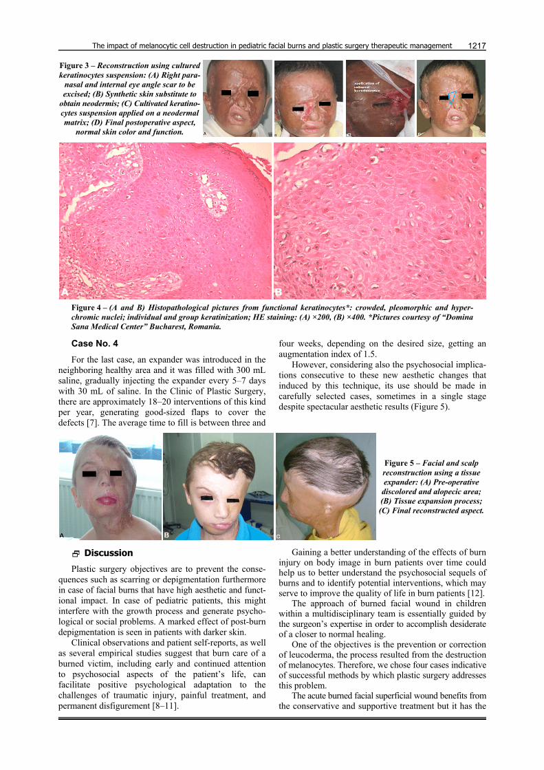
For the following case, after the surgical scar excision, a synthetic dermal substitute was applied. In 21 days, vascularized neodermis was formed. Then the superficial silicon layer was removed and autologous cultured keratinocytes were applied (Figure 3). In nine days, the epithelization was complete.
Using an epidermic graft or autologous cultured keratinocytes on a good quality dermis is successful in obtaining a tailored aesthetic and chromatic area as
presented in the third case. At one can see, in “Grigore Alexandrescu” Plastic and Reconstructive Microsurgery Clinic beneficial results were obtained using synthetic skin substitutes’ matrix and cultured cell suspension.
The main improvements are made to minimize donor site scar, the epithelization speed and good quality and compatibility in areas where the autologous cultured kera-tinocytes were applied (Figure 4). Through their inte-ractions with local melanocytes they obtain the melanin pigment.
The impact of melanocytic cell destruction in pediatric facial burns and plastic surgery therapeutic management
1217
Figure 3 – Reconstruction using cultured keratinocytes suspension: (A) Right para-
nasal and internal eye angle scar to be excised; (B) Synthetic skin substitute to
obtain neodermis; (C) Cultivated keratino-cytes suspension applied on a neodermal matrix; (D) Final postoperative aspect,
normal skin color and function.
Figure 4 – (A and B) Histopathological pictures from functional keratinocytes*: crowded, pleomorphic and hyper-chromic nuclei; individual and group keratinization; HE staining: (A) ×200, (B) ×400. *Pictures courtesy of “Domina Sana Medical Center” Bucharest, Romania.
Case No. 4
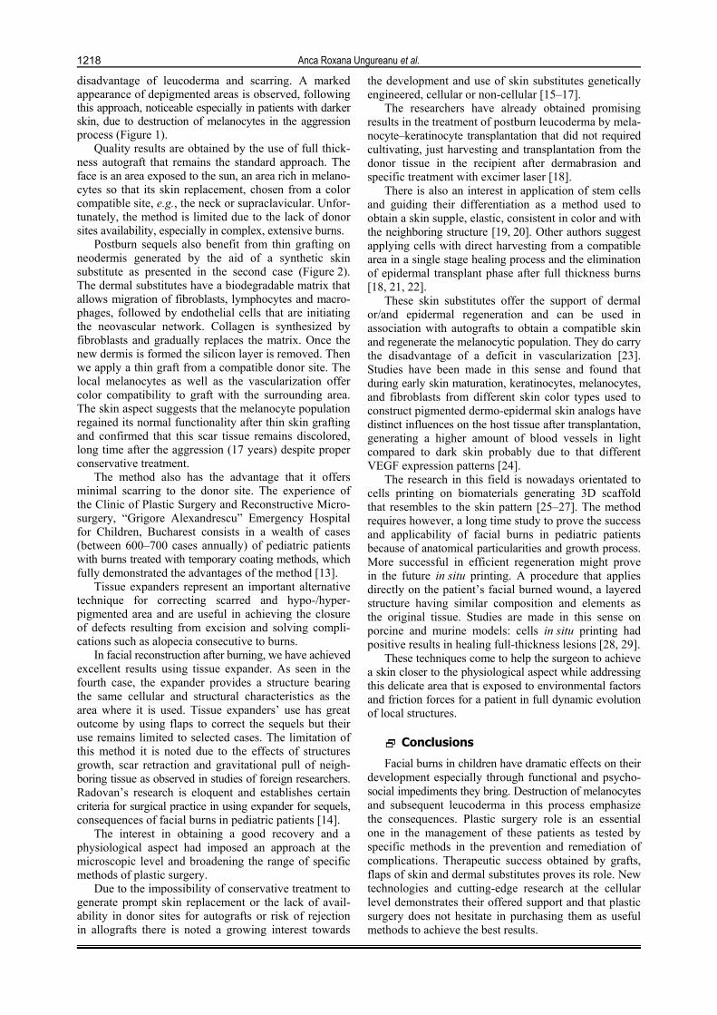
For the last case, an expander was introduced in the neighboring healthy area and it was filled with 300 mL saline, gradually injecting the expander every 5–7 days with 30 mL of saline. In the Clinic of Plastic Surgery, there are approximately 18–20 interventions of this kind per year, generating good-sized flaps to cover the defects [7]. The average time to fill is between three and
four weeks, depending on the desired size, getting an augmentation index of 1.5.
However, considering also the psychosocial implica-tions consecutive to these new aesthetic changes that induced by this technique, its use should be made in carefully selected cases, sometimes in a single stage despite spectacular aesthetic results (Figure 5).
Figure 5 – Facial and scalp reconstruction using a tissue expander: (A) Pre-operative
discolored and alopecic area; (B) Tissue expansion process; (C) Final reconstructed aspect.
Discussion
Plastic surgery objectives are to prevent the conse-quences such as scarring or depigmentation furthermore in case of facial burns that have high aesthetic and funct-ional impact. In case of pediatric patients, this might interfere with the growth process and generate psycho-logical or social problems. A marked effect of post-burn depigmentation is seen in patients with darker skin.
Clinical observations and patient self-reports, as well as several empirical studies suggest that burn care of a burned victim, including early and continued attention to psychosocial aspects of the patient’s life, can facilitate positive psychological adaptation to the challenges of traumatic injury, painful treatment, and permanent disfigurement [8–11].
Gaining a better understanding of the effects of burn injury on body image in burn patients over time could help us to better understand the psychosocial sequels of burns and to identify potential interventions, which may serve to improve the quality of life in burn patients [12].
The approach of burned facial wound in children within a multidisciplinary team is essentially guided by the surgeon’s expertise in order to accomplish desiderate of a closer to normal healing.
One of the objectives is the prevention or correction of leucoderma, the process resulted from the destruction of melanocytes. Therefore, we chose four cases indicative of successful methods by which plastic surgery addresses this problem.
The acute burned facial superficial wound benefits from the conservative and supportive treatment but it has the
Anca Roxana Ungureanu et al.
1218
disadvantage of leucoderma and scarring. A marked appearance of depigmented areas is observed, following this approach, noticeable especially in patients with darker skin, due to destruction of melanocytes in the aggression process (Figure 1).
Quality results are obtained by the use of full thick-ness autograft that remains the standard approach. The face is an area exposed to the sun, an area rich in melano-cytes so that its skin replacement, chosen from a color compatible site, e.g., the neck or supraclavicular. Unfor-tunately, the method is limited due to the lack of donor sites availability, especially in complex, extensive burns.
Postburn sequels also benefit from thin grafting on neodermis generated by the aid of a synthetic skin substitute as presented in the second case (Figure 2). The dermal substitutes have a biodegradable matrix that allows migration of fibroblasts, lymphocytes and macro-phages, followed by endothelial cells that are initiating the neovascular network. Collagen is synthesized by fibroblasts and gradually replaces the matrix. Once the new dermis is formed the silicon layer is removed. Then we apply a thin graft from a compatible donor site. The local melanocytes as well as the vascularization offer color compatibility to graft with the surrounding area. The skin aspect suggests that the melanocyte population regained its normal functionality after thin skin grafting and confirmed that this scar tissue remains discolored, long time after the aggression (17 years) despite proper conservative treatment.
The method also has the advantage that it offers minimal scarring to the donor site. The experience of the Clinic of Plastic Surgery and Reconstructive Micro-surgery, “Grigore Alexandrescu” Emergency Hospital for Children, Bucharest consists in a wealth of cases (between 600–700 cases annually) of pediatric patients with burns treated with temporary coating methods, which fully demonstrated the advantages of the method [13].
Tissue expanders represent an important alternative technique for correcting scarred and hypo-/hyper-pigmented area and are useful in achieving the closure of defects resulting from excision and solving compli-cations such as alopecia consecutive to burns.
In facial reconstruction after burning, we have achieved excellent results using tissue expander. As seen in the fourth case, the expander provides a structure bearing the same cellular and structural characteristics as the area where it is used. Tissue expanders’ use has great outcome by using flaps to correct the sequels but their use remains limited to selected cases. The limitation of this method it is noted due to the effects of structures growth, scar retraction and gravitational pull of neigh-boring tissue as observed in studies of foreign researchers. Radovan’s research is eloquent and establishes certain criteria for surgical practice in using expander for sequels, consequences of facial burns in pediatric patients [14].
The interest in obtaining a good recovery and a physiological aspect had imposed an approach at the microscopic level and broadening the range of specific methods of plastic surgery.
Due to the impossibility of conservative treatment to generate prompt skin replacement or the lack of avail-ability in donor sites for autografts or risk of rejection in allografts there is noted a growing interest towards
the development and use of skin substitutes genetically engineered, cellular or non-cellular [15–17].
The researchers have already obtained promising results in the treatment of postburn leucoderma by mela-nocyte–keratinocyte transplantation that did not required cultivating, just harvesting and transplantation from the donor tissue in the recipient after dermabrasion and specific treatment with excimer laser [18].
There is also an interest in application of stem cells and guiding their differentiation as a method used to obtain a skin supple, elastic, consistent in color and with the neighboring structure [19, 20]. Other authors suggest applying cells with direct harvesting from a compatible area in a single stage healing process and the elimination of epidermal transplant phase after full thickness burns [18, 21, 22].
These skin substitutes offer the support of dermal or/and epidermal regeneration and can be used in association with autografts to obtain a compatible skin and regenerate the melanocytic population. They do carry the disadvantage of a deficit in vascularization [23]. Studies have been made in this sense and found that during early skin maturation, keratinocytes, melanocytes, and fibroblasts from different skin color types used to construct pigmented dermo-epidermal skin analogs have distinct influences on the host tissue after transplantation, generating a higher amount of blood vessels in light compared to dark skin probably due to that different VEGF expression patterns [24].
The research in this field is nowadays orientated to cells printing on biomaterials generating 3D scaffold that resembles to the skin pattern [25–27]. The method requires however, a long time study to prove the success and applicability of facial burns in pediatric patients because of anatomical particularities and growth process. More successful in efficient regeneration might prove in the future in situ printing. A procedure that applies directly on the patient’s facial burned wound, a layered structure having similar composition and elements as the original tissue. Studies are made in this sense on porcine and murine models: cells in situ printing had positive results in healing full-thickness lesions [28, 29].
These techniques come to help the surgeon to achieve a skin closer to the physiological aspect while addressing this delicate area that is exposed to environmental factors and friction forces for a patient in full dynamic evolution of local structures.
Conclusions
Facial burns in children have dramatic effects on their development especially through functional and psycho-social impediments they bring. Destruction of melanocytes and subsequent leucoderma in this process emphasize the consequences. Plastic surgery role is an essential one in the management of these patients as tested by specific methods in the prevention and remediation of complications. Therapeutic success obtained by grafts, flaps of skin and dermal substitutes proves its role. New technologies and cutting-edge research at the cellular level demonstrates their offered support and that plastic surgery does not hesitate in purchasing them as useful methods to achieve the best results.
The impact of melanocytic cell destruction in pediatric facial burns and plastic surgery therapeutic management
1219
References [1] Thody AJ, Higgins EM, Wakamatsu K, Ito S, Burchill SA,
Marks JM, Pheomelanin as well as eumelanin is present in human epidermis, J Invest Dermatol, 1991, 97(2):340–344.
[2] Sandu C, Dumas M, Malan A, Sambakhe D, Marteau C, Nizard C, Schnebert S, Perrier E, Challet E, Pévet P, Felder-Schmittbuhl MP, Human skin keratinocytes, melanocytes, and fibroblasts contain distinct circadian clock machineries, Cell Mol Life Sci, 2012, 69(19):3329–3339.
[3] Chloe A, Loss of pigmentation due to scarring, LiveStrong.com, Oct 24, 2013, http://www.livestrong.com/article/306861-loss- of-pigmentation-due-to-scarring.
[4] Lowrey GH, Growth and development of children, 6th edition, Year Book Medical Publishers, Chicago, 1973.
[5] Hettiaratchy S, Papini R, Initial management of a major burn: II – assessment and resuscitation, BMJ, 2004, 329(7457):101–103.
[6] Easton J, 3-D computer display brings precision to burn assessment, University of Chicago Hospitals, Oct 12, 1997, www.uchospitals.edu/news/1997/19971012-burn-chart.html.
[7] Enescu D, Giuvelea S, Ioniţă D, Gutău I, Şerbănescu C, Alexandru R, Tissue expanders as alternative treatment for children, Ann Plast Surg Reconstr Microsurg, 2009, Issue 4, http://www.annalsplasticsurgery.ro/?q=taxonomy/term/1.
[8] Niţescu C, Calotă DR, Stăncioiu TA, Marinescu SA, Florescu IP, Lascăr I, Psychological impact of burn scars on quality of life in patients with extensive burns who received allotransplant, Rom J Morphol Embryol, 2012, 53(3):577–583.
[9] Blakeney P, Herndon DN, Desai MH, Beard S, Wales-Seale P, Long-term psychosocial adjustment following burn injury, J Burn Care Rehabil, 1988, 9(6):661–665.
[10] Blakeney P, Meyer W 3rd, Robert R, Desai M, Wolf S, Herndon D, Long-term psychosocial adaptation of children who survive burns involving 80% or greater total body surface area, J Trauma, 1998, 44(4):625–632; discussion 633–634.
[11] Sheridan RL, Hinson MI, Liang MH, Nackel AF, Schoenfeld DA, Ryan CM, Mulligan JL, Tompkins RG, Long-term outcome of children surviving massive burns, JAMA, 2000, 283(1):69–73.
[12] Calotă DR, Niţescu C, Marinescu S, Cristescu C, Boiangiu I, Florescu IP, Lascăr I, Correlations between morphological appearance and psychosocial difficulties in patients with extensive burns who received allotransplant, Rom J Morphol Embryol, 2012, 53(3 Suppl):703–711.
[13] Enescu D, Giuvelea S, Alexandru R, Şerbănescu C, Nedelcu I, Constantinescu G, Temporary skin covering for paediatric burns, Ann Plast Surg Reconstr Microsurg, 2006, 1:50–55.
[14] Neale HW, Kurtzman LC, Goh KB, Billmire DA, Yakuboff KP, Warden G, Tissue expanders in the lower face and anterior neck in pediatric burn patients: limitations and pitfalls, Plast Reconstr Surg, 1993, 91(4):624–631.
[15] Supp DM, Boyce ST, Engineered skin substitutes: practices and potentials, Clin Dermatol, 2005, 23(4):403–412.
[16] Shepherd BR, Enis DR, Wang F, Suarez Y, Pober JS, Schechner JS, Vascularization and engraftment of a human
skin substitute using circulating progenitor cell-derived endothelial cells, FASEB J, 2006, 20(10):1739–1741.
[17] Catalano E, Cochis A, Varoni E, Rimondini L, Azzimonti B, Tissue-engineered skin substitutes: an overview, J Artif Organs, 2013, 16(4):397–403.
[18] Mulekar SV, Issa AA, Eisa AA, Treatment of post-burn leuco-derma with non-cultured melanocyte-keratinocyte transplan-tation (MKTP), Burns, 2011, 37(3):448–452.
[19] Anglani F, Forino M, Del Prete D, Tosetto E, Torregrossa R, D’Angelo A, In search of adult renal stem cells, J Cell Mol Med, 2004, 8(4):474–487.
[20] Horch RE, Munster AM, Achauer B (eds), Cultured human keratinocytes and tissue engineered skin substitute, Thieme, Stuttgart, 2001, 316–325.
[21] Wood FM, Stoner ML, Fowler BV, Fear MW, The use of a non-cultured autologous cell suspension and Integra dermal regeneration template to repair full-thickness skin wounds in a porcine model: a one-step process, Burns, 2007, 33(6): 693–700.
[22] Iman A, Akbar MA, Mohsen KM, Ali F, Armin A, Sajjad A, Ahmad M, Ghavipisheh M, Leila R, Comparison of intradermal injection of autologous epidermal cell suspension vs. spraying of these cells on dermabraded surface of skin of patients with post-burn hypopigmentation, Indian J Dermatol, 2013, 58(3):240.
[23] Böttcher-Haberzeth S, Biedermann T, Reichmann E, Tissue engineering of skin, Burns, 2010, 36(4):450–460.
[24] Klar AS, Böttcher-Haberzeth S, Biedermann T, Schiestl C, Reichmann E, Meuli M, Analysis of blood and lymph vascu-larization patterns in tissue-engineered human dermo-epi-dermal skin analogs of different pigmentation, Pediatr Surg Int, 2014, 30(2):223–231.
[25] Michael S, Sorg H, Peck CT, Koch L, Deiwick A, Chichkov B, Vogt PM, Reimers K, Tissue engineered skin substitutes created by laser-assisted bioprinting form skin-like structures in the dorsal skin fold chamber in mice, PLoS One, 2013, 8(3):e57741.
[26] Velasquillo C, Galue EA, Rodriquez L, Ibarra C, Ibarra-Ibarra LG, Skin 3D bioprinting. Applications in cosmetology, Journal of Cosmetics, Dermatological Sciences and Appli-cations, 2013, 3(1A):85–89.
[27] Lee V, Singh G, Trasatti JP, Bjornsson C, Xu X, Tran TN, Yoo SS, Dai G, Karande P, Design and fabrication of human skin by three-dimensional bioprinting, Tissue Eng Part C Methods, 2014, 20(6):473–484.
[28] Binder KW, Zhao W, Aboushwareb T, Dice D, Atala A, Yoo JJ, In situ bioprinting of the skin for burns, J Am Coll Surg, 2010, 211(3 Suppl):S76.
[29] Albanna M, Murphy S, Zhao W, El-Amin I, Tan J, Dice D, Kang HW, Jackson J, Atala A, Yoo J, In situ bioprinting of skin for reconstruction, J Am Coll Surg, 2012, 215(3 Suppl): S87.
Corresponding author Liviu Drăghici, Assistant Professor, MD, PhD, Department of General Surgery, “Sf. Ioan” Emergency Clinical Hospital, “Carol Davila” University of Medicine and Pharmacy, 13 Vitan-Bârzeşti Road, Sector 4, 042122 Bucharest, Romania; Phone +40740–487 468, e-mail: [email protected] Received: March 28, 2014
Accepted: October 28, 2014