new possibilities of detection of swine hepatitis e …...700490, mihail sadoveanu alley no. 6-8,...
TRANSCRIPT
LUCRĂRI ŞTIINŢIFICE MEDICINĂ VETERINARĂ VOL. XLV(3), 2012, TIMIŞOARA
5
NEW POSSIBILITIES OF DETECTION OF SWINE HEPATITIS E INFECTION
ADRIANA ANIȚĂ, G. SAVUȚA
University of Agriculture Science and Veterinary Medicine „Ion Ionescu de la Brad‖,
700490, Mihail Sadoveanu Alley No. 6-8, Iasi, Romania E-mail: [email protected]
Summary
Hepatitis E virus (HEV) is a zoonotic pathogen highly prevalent in farm pigs worldwide. Swine HEV sequences closely related to human HEV sequences have been detected in many countries and in several cases the source of infection has been liked to contact with swine or ingestion of undercooked swine meat. In swine, HEV transmission is by fecal-oral route, repeated direct daily contact among pigs confined in the same pen may enhance the spread of the virus. Till now were any specific commercial kits for detection of HEV antibody in swine.
The aim of this study was to detect specific hepatitis E virus antibody in farm and backyard pigs from four counties in the central and south of Moldova region (Iasi, Vaslui, Braila, Vrancea). A total of 180 pig serum were tested using PrioCHECK® HEV Ab porcine (produced by Prionics, Switzerland) for detection of antibodies against hepatitis E virus in porcine serum and meat juice samples. Serum samples were collected during august – december 2011, from 5 farms and 11 commune veterinary districts. Herd prevalence varied from 0% to 44.4% and the mean prevalence of HEV antibodies in the 5 farms was 16.66%. Among the 90 backyard pig serum samples tested, forty-five (50%) were positive for HEV antibodies. The findings confirm a high prevalence of HEV infection in pig population from east region of the country.
Key words: swine, hepatitis E virus, ELISA
Hepatitis E virus (HEV) is a non-enveloped, single stranded, positive-sense
RNA virus, classified in the Hepevirus genus of the family Hepeviridae. There exist three open reading frames in HEV genome: ORF1 encodes non-structural proteins, ORF2 encodes the capsid protein, and the ORF3 encodes a small phosphoprotein. There is only one serotype but based on genetic diversity HEV strains are classified into four genotypes designated with Arabic numerals 1 to 4, of which only the type 3 and 4 can infect swine. These genotypes are more diverse and are divided in ten (3a–3j) and seven (4a–4g) subtypes (9). HEV is considered to be a zoonotic agent, and researchers have suggested that swine is a principal reservoir of HEV that infects humans (5; 6). Thus, HEV is of increasing importance to both public and animal health.
Swine HEV was first identified in 1997 in USA and is now considered to be widespread in pig herds all over the world (12). In general hepatitis E virus infection in pigs seems to be subclinical, although some studies have shown a possible correlation of the infection with liver damage (8). Under natural condition swine
LUCRĂRI ŞTIINŢIFICE MEDICINĂ VETERINARĂ VOL. XLV(3), 2012, TIMIŞOARA
6
HEV viremia and faecal virus sheding generally occur in pigs of 2 to 4 months (9), than seroconversion in pigs older than 4 months. Like human HEV, the capsid protein of swine HEV is immunogenic and induces protective immunity. The seroprevalence observed at the end of fattening reveals an effective transmission of the virus between animals from the same fence (15). The climate, presence of a river, and water supply and management can affect the prevalence of HEV (17).
Pigs are a recognized reservoir for HEV, and pig handlers are at increased risk of zoonotic HEV infection. Sporadic cases of hepatitis E have been definitively linked to the consumption of raw or undercooked animal meats such as pig livers, sausages, and deer meats. (11).
Materials and methods
Swine serum samples were collected from four counties: Iasi, Vaslui, Braila and Vrancea. From august to December 2011, 180 individual serum pig samples were collected from clinically healthy pigs coming of different breading conditions (backyard and farm). Serum samples were stored until testing at -20
0C.
All samples were tested using PrioCHECK® HEV Ab porcine (produced by Prionics) for detection of antibodies against hepatitis E virus. The PrioCHECK® HEV Ab porcine is a commercially available ELISA that is based on HEV genotype 1 and 3 antigens. The use of genotype 3 antigens – the HEV genotype that has actually been demonstrated to infect pigs – significantly enhances the specificity and sensitivity of the test. Cut-off control, positive and negative control samples provided with the kit were included in each plate. Briefly, serum samples and controls were diluted 1:100, and the microplate was incubated for 30 min at 37°C. The microplate was washed four times with 300 μl of a wash fluid and 100 μl diluted Conjugate was added in each well. After 30 min at 37°C incubation, the plate was washed again and the Cromogen substrate was added. The color-developing reaction was stopped by adding 100 μl of the stop solution to each well and the absorbance was determined at 450 nm with reference at 620 nm.
Results and discussions
The present study shows that hepatitis E infection is wide spread in romanian pig livestock. The use of a commercial kit for detection of swine HEV antibodies allowed the confirmation of the specific reactivity of the examined sera.
All serum samples used in this study were stored at −20°C prior to analysis. A total of 180 serum samples of apparently healthy pigs from five swine farms and 11 commune veterinary districts (CVD) were used for detection of HEV antibodies.
LUCRĂRI ŞTIINŢIFICE MEDICINĂ VETERINARĂ VOL. XLV(3), 2012, TIMIŞOARA
7
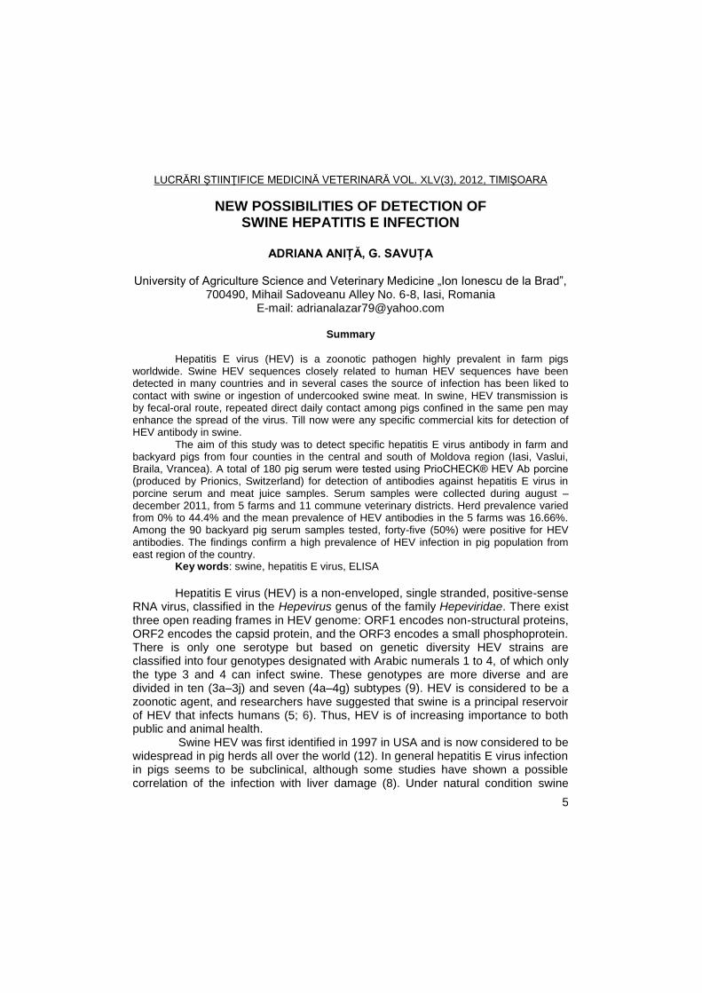
Table 1 Number of positive serum samples displayed by county
County No. of tested
samples No. of positive
samples % positivity
Iasi Farm 1 27 0 0
Vaslui Farm 2 20 0 0
Braila Farm 3 26 10 38.46
Vrancea
Farm 4 8 1 12.50
Farm 5 9 4 44.44
CVD Campuri 8 4 50
CVD Racoasa 5 3 60
CVD Maicanesti 6 0 0
CVD Golesti 8 0 0
CVD Garoafa 8 8 100
CVD Urechesti 8 5 62.50
CVD Tataranu 8 4 50
CVD Marasesti 8 8 100
CVD Ciorasti 16 9 56.25
CVD Gologaru 7 1 14.28
CVD Campenenca 8 3 37.50
The data indicate a high seroprevalence for anti-HEV antibodies in pig
herds, even if the percentage of seropositive animals varied widely among herds, being 0% in two farms and 44.44% on other.
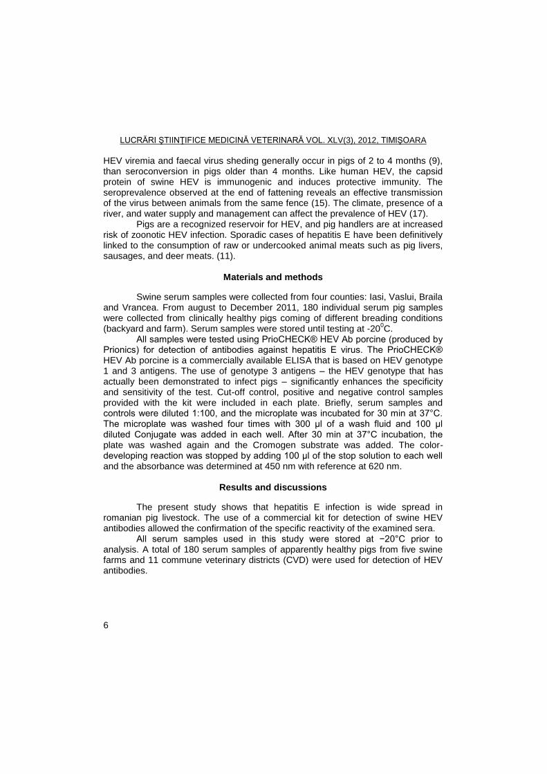
Our results point out that HEV may be circulating intensely among healthy backyard pigs in Vrancea County. A 50% (45/90) of the individual pig sera tested result positive by ELISA. Seroprevalence varied from 0% in Maicanesti and Golesti CMV, to 100% (8/8) in Garoafa and Marasesti CVD.
Fig. 1 Distribution of positive samples from Vrancea County
Two farms and 11 commune veterinary districts were included from the
same county, demonstrating that the swine HEV infection is spread over the region. Transmission of HEV among swine appears to occur through a fecal-oral route of
LUCRĂRI ŞTIINŢIFICE MEDICINĂ VETERINARĂ VOL. XLV(3), 2012, TIMIŞOARA
8
exposure at approximately 1-3 months of age, with a majority of animals exposed by 6-8 months of age (10, 15, 16). The IgM anti-HEV increase from 9 weeks of age, and pigs are IgG anti-HEV-positive at 22 weeks of age (4).
The seroprevalence found in our investigation is similar to that reported in pigs from commercial farms around European countries (3; 7; 14). Similar data describing a quite variable seroprevalence between relatively close regions have been reported in different provinces of Canada (38.3– 88.8%) (18).
In conclusion, these data support our previous results reporting a high prevalence of HEV infection among healthy pigs in Botosani (2) and Iasi (1) county.
Conclusions
Our serological data revealed that HEV is highly prevalent in swine commercial farms and backyard pigs in the east region of Romania.
Several issues rise from the discovery of swine hepatitis E infection, which seems to be ubiquitous all over the world. The main concern is about the possibility of its transmission to humans, since pigs are very common domestic animals and whose meat is also largely consumed.
Acknowledgements
This work was cofinanced from the European Social Fund through Sectoral Operational Programme Human Resources Development 2007-2013; project number POSDRU/I.89/1.5/S62371, Postdoctoral School in Agriculture and Veterinary Medicine area.
References
1. Aniţa, A., Hepatitis E – a new zoonosis present also in Romania, Scientific Works UASVM Iasi, Veterinary Medicine Series, 2009, 51 (10), 593-598.
2. Aniţa, A., Aniţă, D., Ludu, L., Savuţă, G., Seroepidemiological investigation of human and swine hepatitis E in Botoşani County, Bulletin UASVM Cluj-Napoca, Veterinary Medicine, 2010 67(2), 19-22.
3. Casas, M., Pujols, J., Rosell, R., de Deus, N., Peralta, B., Pina, S., Retrospective serological study of hepatitis E infection in pigs from 1985 to 1997 in Spain, Veterinary Microbiology, 2009, 135(3–4), 248–252.
4. De Deus, N., Casas, M., Peralta, B., Nofrarias, M., Pina, S., Martin, M., Segales, J, Hepatitis E virus infection dynamics and organic distribution in naturally infected pigs in a farrow-to-finish farm, Veterinary Microbiology, 2008, 132,19-28.
5. Huang, F.F., Haqshenas, G., Guenette, D.K., Halbur, P.G., Schommer, S.K., Pierson, F.W., Toth, T.E., Meng, X.J., Detection by reverse transcription-PCR and genetic characterization of field isolates of swine
LUCRĂRI ŞTIINŢIFICE MEDICINĂ VETERINARĂ VOL. XLV(3), 2012, TIMIŞOARA
9
hepatitis E virus from pigs in different geographic regions of the United States. Journal of Clinical Microbiology, 2002, 40(4),1326-1332.
6. Ijaz, S., Arnold, E., Banks, M., Bendall, R.P., Cramp, M.E., Cunningham, R., Dalton, H.R., Harrison, T.J., Non-travel-associated hepatitis E in England and Wales: demographic, clinical, and molecular epidemiological characteristics, J of Infectious Diseases, 2005, 192(7), 1166-1172.
7. Kaba, M., Davoust, B., Marie, J.L., Barthet, M., Henry, M., Tamalet, C., Frequent transmission of hepatitis E virus among piglets in farms in Southern France, Journal of Medical Virology, 2009, 81(10), 1750–1759.
8. Lee, S., Kang, S., Kim, D., Bae, J., Kim, J., Detection of swine hepatitis E virus in the porcine hepatic lesion in Jeju Island, Journal of Veterinary Science, 2007, 8, 51-55.
9. Lu, L., Li, C., Hagedorn, C.H., Phylogenetic analysis of global hepatitis E virus sequences: genetic diversity, subtypes and zoonoses, Review of Medical Virology, 2006, 16, 5-36.
10. Meng, X.J., Purcell, R.H., Halbur, P.G., Lehman, J.R., Webb, D.M., A novel virus in swine is closely related to the human hepatitis E virus. Proc Natl Acad Sci, 1997, 94, 9860-9865.
11. Meng, X.J., Hepatitis E virus: Animal reservoirs and zoonotic risk, Veterinary Microbiology, 2009, 140 (3-4), 256-265.
12. Panda, S.K., Thakral, D., Rehman, S., Hepatitis E virus, Review of Medical Virology, 2007, 17(3), 151-180.
13. Pavio, N., Meng, X.J., Renou, C., Zoonotic hepatitis E: animal reservoirs and emerging risks, Veterinary Research, 2010, 41(6), 46.
14. Reuter, G., Fodor, D., Forgach, P., Katai, A., Szucs, G., Characterization and zoonotic potential of endemic Hepatitis E virus (HEV) strains in humans and animals in Hungarym Journal of Clinical Virology, 2009, 44(4), 277-281.
15. Takahashi, M., Nishizawa, T., Miyajima, H., Gotanda, Y., Iita, T., Tsuda, F., Okamoto, H., Swine hepatitis E virus strains in Japan form four phylogenetic clusters comparable with those of Japanese isolates of human hepatitis E virus, Journal of General Virology, 2003, 84, 851-862.
16. Wu, J.C., Chen, C.M., Chiang, T.Y., Tsai, W.H., Jeng, W.J., Sheen, J., Lin, C.C., Meng, X.J., Spread of hepatitis E virus among different-aged pigs: two-year survey in Taiwan, Journal of Medical Virology, 2002, 66 (4), 488-492 .
17. Zhang, W., Shen, Q., Mou, J., Yang, Z.B., Yuan, C.L., Cui, L., Cross-species infection of hepatitis E virus in a zoo-like location, including birds, Epidemiology and Infection, 2008, 136(8),1020-1026.
18. Yoo, D., Wilson, P., Pei, Y., Hayes, M.A., Deckert, A., Dewey, C.E., Prevalence of Hepatitis E Virus antibodies in Canadian swine herds and identification of a novel variant of swine Hepatitis E virus, Clinical and Diagnostic Laboratory Immunology, 2001, 8(6), 1213–1219.
LUCRĂRI ŞTIINŢIFICE MEDICINĂ VETERINARĂ VOL. XLV(3), 2012, TIMIŞOARA
10
DETECTION OF BOVINE PAPILLOMAVIRUS E5 BY IMMUNOFLUORESCENCE IN BOVINE CUTANEOUS
FIBROPAPILLOMAS FROM EASTERN ROMANIA
FLORENTINA BOCANETI1, G. BORZACCHIELLO
2, G. ALTAMURA
2,
ANNUNZIATA CORTEGGIO2, F. ROPERTO
2, C. DARABAN
1, ELENA VELESCU
1
1Department of Public Health, Faculty of Veterinary Medicine, University of
Agriculture Sciences and Veterinary Medicine Ion Ionescu de la Brad, 700490, Mihail Sadoveanu Alley No. 6-8, Iasi, Romania
2Department of Pathology and Animal Health, University of Naples Federico II,
Napoli, Italy E-mail: [email protected]
Summary
Papillomaviruses are oncogenic DNA tumour viruses that infect cutaneous and
mucous epithelia in a variety of animals, including humans, inducing papillomas or warts, which generally regress, occasionally the lesions persist and progress to malignancy. In Romania, despite the high frequency of skin warts in bovines, studies involving the detection and the evaluation of BPV infection incidence are uncommon. E5 is the main oncoprotein of the BPV, being responsible for the neoplastic transformation of keratinocytes and fibroblasts. The aim of this study was to assess the presence of the BPV E5 protein by immunofluorescence in bovine skin fibropapillomas. Fifteen bovine samples were used in this study: eleven bovine cutaneous fibropapillomas and four normal skins. By indirect immunofluorescence, only the fibropapilloma samples expressed BPV E5, mostly within the cytoplasm of basal and differentiated keratinocytes layers. E5 was recorded intracytoplasmically, although some samples the neoplastic cells showed a very characteristic juxtanuclear and/or membranous staining pattern. This is the first report of BPV E5 expression in bovine fibropapillomas from Romania.
Key words: fibropapillomas, immunofluorescence, bovine papillomavirus, E5 Papillomaviruses are oncogenic DNA tumour viruses that infect
cutaneous and mucous epithelia in a variety of animals, including humans, through cuts and abrasions and induce papillomas or warts, which generally regress; occasionally the lesions persist and progress to malignancy; the immunosuppressed animals are unable to reject the infection and succumb to widespread cutaneous or mucosal involvement. This forms of papillomatosis are problematic and of economical significance (9). One important feature of the PVs is their strict species-specificity: even in experimental conditions, papillomaviruses do not infect any other host than the natural one. The only know case of cross-species infection is the BPV which infects equids (17). 12 BPV genotypes have been characterized so far; they are classified into three genera : Deltapapillomavirus (BPV 1 and 2), Epsilonpapillomavirus (BPV 5 and 8),
LUCRĂRI ŞTIINŢIFICE MEDICINĂ VETERINARĂ VOL. XLV(3), 2012, TIMIŞOARA
11
Xipapillomavirus (BPV 3, 4, 6, 9, 10, 11, 12) and an as yet unassigned PV genus (BPV 7) (10, 13, 14, 18, 19, 23). The BPV genome is divided into three main regions: a long control region (LCR), a region that encodes the early (non-structural) proteins and a late region encoding the capsid proteins (16). The PV infects the basal keratinocytes, expresses part of its genes in the basal and suprabasal layers, replicates its genome in the differentiating spinous and granular layers, express its structural genes and package its DNA in the squamous layers and new infectious virions are released with the keratinized squames. During its life cycle, the BPV genome persists in the infected cell as an episome (9). Delta-BPVs encode three oncoproteins, E5, E6 and E7, whereas the Xi- BPVs lack the E6 gene and encode only E5 and E7 (20). The BPV E5 is a small peptide (44 amino acids), being the major oncoprotein. It is very hydrophobic with high leucine content; it spontaneously localizes in the cell endomembrane compartments, particularly in the Golgi apparatus. It is expressed in the deep layers of the epithelium (22) and also in the basal and superficial differentiating keratinocytes of bovine fibropapillomas (4, 7, 5). High amounts of BPV E5 protein are also observed in bovine urinary bladder cancer (3, 5). E5 is present in papillomas at early stages of development but its expression is extinguished as the papilloma ages (1).
In Romania, despite the high frequency of skin warts in cows, studies involving the detection and the evaluation of BPV infection incidence are uncommon. Only one report (3) describes the presence of BPV-2 infection in bovine urinary bladder cancer.
The aim of this study was to assess the presence of the E5 protein by immunofluorescence in bovine skin fibropapillomas.
Materials and methods
Fifteen bovine samples were used in this study: eleven bovine cutaneous
fibropapillomas and four normal skins. The samples were collected from cows suffering of fibropapillomatosis, which were located in Moldova, Romania, the fibropapillomas were removed under local anesthesia. After the removal, the samples were immediately fixed in 10% neutral buffered formalin for histological examination. The fixed samples were embedded in paraffin by routine methods and the 5μm paraffin sections were stained with haematoxilin and eosin for histopathological assessment.
For immunofluorescence labeling, the sections were dewaxed in xylene for 20 minutes, then rehydrated by including for 10 minutes in absolute alcohol 1000, 10 minutes in denatured alcohol 1000, and for 5 minutes in each 950, 700 and 500 alcohols. The samples were washed for five minutes in distillated water. The antigen enhancement was made by pretreating the samples in citrate tampon solution, pH 6, with microwave heating at 525 W twice for five minutes each, then washed three times for five minutes in phosphate buffered saline, pH
LUCRĂRI ŞTIINŢIFICE MEDICINĂ VETERINARĂ VOL. XLV(3), 2012, TIMIŞOARA
12
7.4, 0.1 M (PBS). The sections were blocked with donkey antiserum diluted 1:20 in PBS for 30 minutes in humified chamber. The primary anti-E5 antibody (kindly provided by Prof. Maria Saveria Campo, University of Glasgow, Scotland) was applied overnight at 40C in a humified chamber at 1:50 dilution in PBS. The slides were washed for three times with PBS, and then incubated with the secondary antibody, Alexa Fluor 488 donkey anti-sheep diluted 1:100 in PBS at room temperature in a humified chamber. The slides were washed again three times for five minutes in PBS and mounted in PBS/Glycerol (1:1). Immunofluorescence staining was analyzed with a confocal laser scanning microscope LSM 510 (Carl Zeiss GmbH, Jena, Germany).
Results and discussions
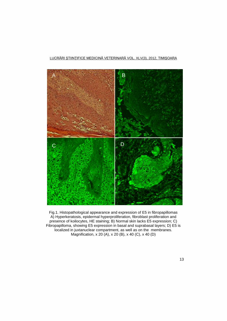
Macroscopically, the tumours were multiple, lobulated, grey-white, with cauliflower-like aspect. Histologically, the lesions were composed of connective tissue, which was covered by epidermal hyperplasia with acanthosis, and orthokeratotic hyperkeratosis. The spinous layer was hyperplastic, showing many koilocytes, arranged in small groups, indicating the presence of papillomavirus infection (Fig.1A).
We examined the localization of E5 protein expression in normal skin and in naturally induced fibropapillomas from different bovines by indirect immunofluorescence. Normal epithelium showed, as expected, negative staining for E5 (Fig.1B), while all fibropapillomas showed a positive staining for E5 oncoprotein. E5 was strongly expressed in the basal and granular cell layers and sporadically expressed in the spinous cell layer (Fig.1C). Within the neoplastic cells E5 was mostly recorded intracytoplasmically, often localized in a very characteristic juxtanuclear region, and/or membraneous staining pattern was also recorded (Fig.1D). The score of immunoreactivity is presented in table 1.
LUCRĂRI ŞTIINŢIFICE MEDICINĂ VETERINARĂ VOL. XLV(3), 2012, TIMIŞOARA
13
Fig.1. Histopathological appearance and expression of E5 in fibropapillomas A) Hyperkeratosis, epidermal hyperproliferation, fibroblast proliferation and presence of koilocytes, HE staining; B) Normal skin lacks E5 expression; C)
Fibropapilloma, showing E5 expression in basal and suprabasal layers; D) E5 is localized in juxtanuclear compartment, as well as on the membranes.
Magnification, x 20 (A), x 20 (B), x 40 (C), x 40 (D)
A B
C D
LUCRĂRI ŞTIINŢIFICE MEDICINĂ VETERINARĂ VOL. XLV(3), 2012, TIMIŞOARA
14
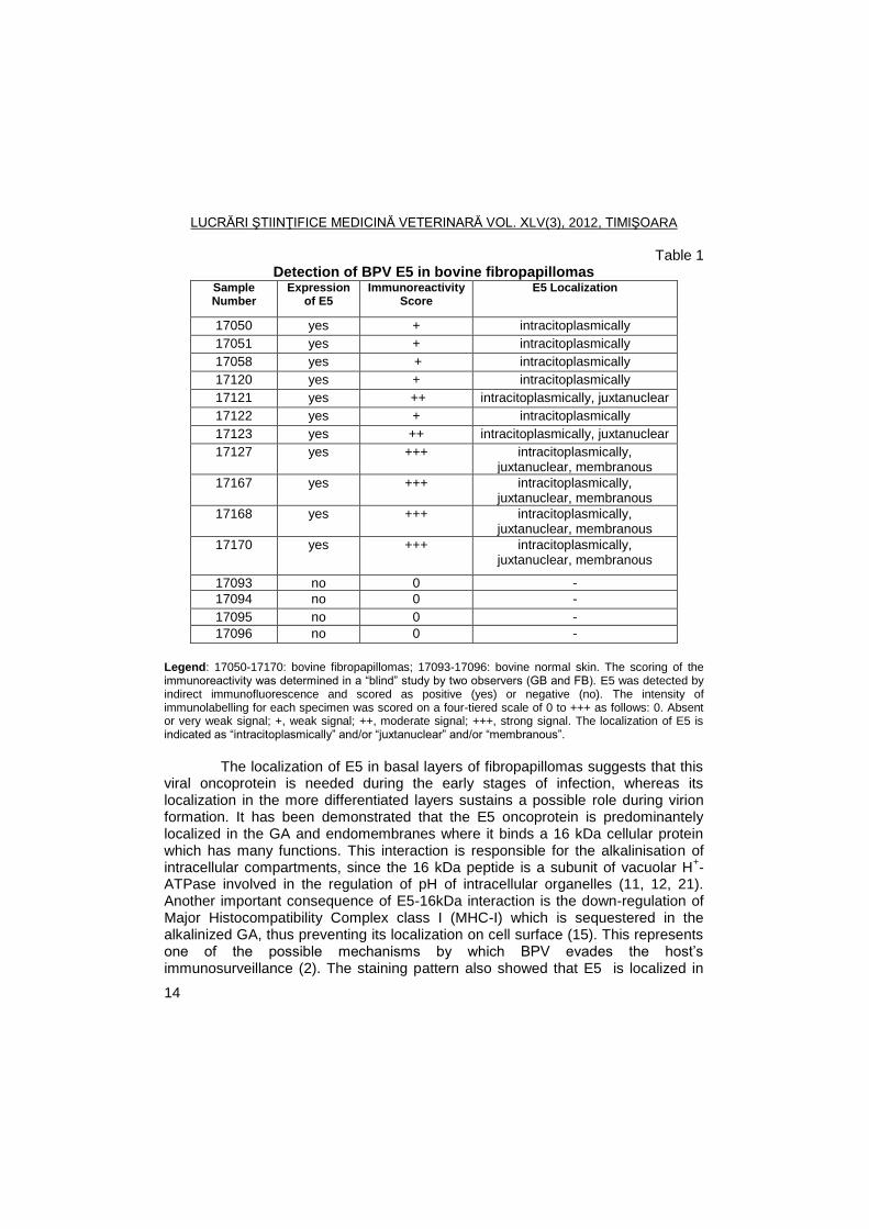
Table 1 Detection of BPV E5 in bovine fibropapillomas
Sample Number
Expression of E5
Immunoreactivity Score
E5 Localization
17050 yes + intracitoplasmically
17051 yes + intracitoplasmically
17058 yes + intracitoplasmically
17120 yes + intracitoplasmically
17121 yes ++ intracitoplasmically, juxtanuclear
17122 yes + intracitoplasmically
17123 yes ++ intracitoplasmically, juxtanuclear
17127 yes +++ intracitoplasmically, juxtanuclear, membranous
17167 yes +++ intracitoplasmically, juxtanuclear, membranous
17168 yes +++ intracitoplasmically, juxtanuclear, membranous
17170 yes +++ intracitoplasmically, juxtanuclear, membranous
17093 no 0 -
17094 no 0 -
17095 no 0 -
17096 no 0 -
Legend: 17050-17170: bovine fibropapillomas; 17093-17096: bovine normal skin. The scoring of the immunoreactivity was determined in a ―blind‖ study by two observers (GB and FB). E5 was detected by indirect immunofluorescence and scored as positive (yes) or negative (no). The intensity of immunolabelling for each specimen was scored on a four-tiered scale of 0 to +++ as follows: 0. Absent or very weak signal; +, weak signal; ++, moderate signal; +++, strong signal. The localization of E5 is indicated as ―intracitoplasmically‖ and/or ―juxtanuclear‖ and/or ―membranous‖.
The localization of E5 in basal layers of fibropapillomas suggests that this
viral oncoprotein is needed during the early stages of infection, whereas its localization in the more differentiated layers sustains a possible role during virion formation. It has been demonstrated that the E5 oncoprotein is predominantely localized in the GA and endomembranes where it binds a 16 kDa cellular protein which has many functions. This interaction is responsible for the alkalinisation of intracellular compartments, since the 16 kDa peptide is a subunit of vacuolar H
+-
ATPase involved in the regulation of pH of intracellular organelles (11, 12, 21). Another important consequence of E5-16kDa interaction is the down-regulation of Major Histocompatibility Complex class I (MHC-I) which is sequestered in the alkalinized GA, thus preventing its localization on cell surface (15). This represents one of the possible mechanisms by which BPV evades the host’s immunosurveillance (2). The staining pattern also showed that E5 is localized in
LUCRĂRI ŞTIINŢIFICE MEDICINĂ VETERINARĂ VOL. XLV(3), 2012, TIMIŞOARA
15
the cell membrane where it is known to bind to platelet-derived growth factor beta receptor (PDGFβ-R) leading to a stable activation of the receptor, thus inducing cell transformation (6). We have showed that the major BPV oncoprotein E5 is expressed only in the tumours, not in the normal skin, suggesting a causal role of the virus in the neoplastic transformation.
Conclusions
To the best of authors’ knowledge, this study demonstrates for the first time
the expression of BPV E5 oncoprotein in bovine fibropapillomas from cows bred in Romania. These results are important since demonstrate the widespread of the BPV in Eastern Europe.
Finally, the immunofluorescence can be a useful method to detect the presence of BPV in fibropapillomas and E5 oncoprotein can be used as a marker for a BPV productive infection.
References
1. Anderson, R.A., Scobie, L., O’Neil, B.W., Grindlay, G.J., Campo, Maria
Saveria, Viral proteins of bovine papillomavirus type 4 during the development of alimentary tumours, Vet J, 1997, 154, 69-78.
2. Ashrafi, G.H, Tsirimonaki, E., Marchetti, B., Down-regulation of MHC class I by bovine papillomavirus E5 oncoproteins. Oncogene, 2002, 21, 248-259.
3. Balcos, L.G., Borzacchiello, P., Russo, V., Popescu, O., Roperto, S., Roperto, S., Association of Bovine papillomavirus type-2 and urinary bladder tumours in cattle from Romania, Res Vet Sci, 2008, 85, 145-148.
4. Bohl, J., Hull, B., Vande Pol, S.B., Cooperative transformation and coexpression of bovine papillomavirus type 1 E5 and E7 proteins, J Virol, 2001, 75, 513-521.
5. Borzacchiello, G., Iovane, G., Marcante, M.L., Poggiali, F., Roperto, F., Venuti, A., Presence of bovine papillomavirus type 2 DNA and expression of the viral oncoprotein E5 in naturally occurring urinary bladder tumours in cows, J Gen Virol, 2003, 84, 2921-2926.
6. Borzacchiello, G., Russo, V., Gentile, F., Roperto, F., Venuti, A., Nitsch, L., Campo, Maria Saveria, Roperto, S., Bovine papillomavirus E5 oncoprotein binds to the activated form of the plateled-derived growth factor β receptor in naturally occurring bovine urinary bladder tumours. Oncogene, 2006, 25, 1251-1260.
7. Burnett, S., Jareborg, N., DiMaio, D., Localization of bovine papillomavirus type 1 E5 protein to transformed basal keratinocytes and permissive differentiated cells in fibropapilloma tissue, Proc Natl Acad Sci U S A, 1992, 89, 5665-5669.
LUCRĂRI ŞTIINŢIFICE MEDICINĂ VETERINARĂ VOL. XLV(3), 2012, TIMIŞOARA
16
8. Campo, Maria Saveria, Animal model of papillomavirus pathogenesis, Virus Res, 2002, 89, 249-261.
9. Campo, Maria Saveria, Papillomavirus research from natural history to vaccines and beyond, Ed. Caister Academic Press, Norflok, 2006.
10. Claus, M.P., Lunardi, M., Alfieri, A.F., Ferracin, L.M., Fungaro, M.H.P., Alfieri, A.A., Identification of unreported putative new bovine papillomavirus types in Brazilian cattle herds, Vet Microbiol, 2008, 132, 369-401.
11. Faccini, A.M., Cairney, M., Ashrafi, G.H., Finbow, M.E., Campo, Maria Saveria, Pitts, J.D., The bovine papillomavirus type 4 E8 protein binds to ductin and causes loss of gap junctional intercellular communication in primary fibroblasts, J Virol, 1996, 70, 9041-9045.
12. Goldstein, D.J., Finbow, M.E., Andresson, T., McLean, P., Smith, K., Bubb, V., Schlegel, R., Bovine papillomavirus E5 oncoprotein binds to the 16K component of vacuolar H(+)-ATPases, Nature, 1991, 352, 347-349.
13. Hatama, S., Nobumato, K., Kanno, T., Genomic and phylogenetic analysis of two novel bovine papillomaviruses, BPV-9 and BPV-10, J Gen Virol, 2008, 89, 158-163.
14. Hatama, S., Ishihara, R., Ueda, Y., Kanno, T., Uchida, I., Detection of a novel bovine papillomavirus type 11 (BPV-11) using xipapillomavirus consensus polymerase chain reaction primers, Arch Virol, 2011, 156, 1281-1285.
15. Marchetti B., Ashrafi G.H., Tsirimonaki E., O'Brien PM, Campo MS. The bovine papillomavirus oncoprotein E5 retains MHC class I molecules in the Golgi apparatus and prevents their transport to the cell surface. Oncogene, 2002, 21, 7808-16.
16. Morgan, I.M., Campo, Maria Saveria, Recent developments in Bovine Papillomaviruses, Papillomavirus Report, 2000, 11, 127-132.
17. Nasir, Lubna, Gault, E., Morgan, I. M., Chambers, G., Ellsmore, V., Campo, Maria Saveria, Identification and functional analysis of sequence variants in the long control region and the E2 open reading frame of bovine papillomavirus type 1 isolated from equine sarcoids, Virology, 2007, 364, 355-361.
18. Ogawa, T., Tomita, Y., Okada, M., Shinozaki, K., Kubonoya, H., Kaiho, I., Shirasawa, H., Broad-spectrum detection of papillomaviruses in bovine teat papillomas and healthy teat skin, J Gen Virol, 2004, 85, 2191-2197.
19. Ogawa, T., Tomita, Y., Okada, M., Shirasawa, H., Complete genome and phylogenetic position of bovine papillomavirus type 7, J Gen Virol, 2007, 88, 1935-1938.
20. Pennie, W.D., Grindlay, G.J., Cairney, M., Campo, Maria Saveria, Analysis of the transforming functions of bovine papillomavirus type 4, Virol, 1993, 193, 614-620.
LUCRĂRI ŞTIINŢIFICE MEDICINĂ VETERINARĂ VOL. XLV(3), 2012, TIMIŞOARA
17
21. Schapiro, F., Sparkowski, J., Adduci, A., Suprynowicz, F., Schlegel, R., Grinstein, S., Golgi alkalinization by papillomavirus E5 oncoprotein, J Cell Biol, 2000, 148, 305-315.
22. Venuti, A., Campo, Maria Saveria, The E5 protein of papillomavirus, in: McCance, D., Human Papillomavirus, Perspectives in Medical Virology, 2002, Vol.8, Elsevier Science, Amsterdam, 143-164.
23. Zhu, W., Dong, J., Shimizu, E., Hatama, S., Kadota, K., Goto, Y., Haga, T., Characterization of novel bovine papillomavirus type 12 (BPV-12) causing epithelial papilloma, Arch Virol, 2012, 157, 85-91.
LUCRĂRI ŞTIINŢIFICE MEDICINĂ VETERINARĂ VOL. XLV(3), 2012, TIMIŞOARA
18
TUBERCULOSIS OF THE HIGH PRODUCTING DAIRY COWS – CASE REPORT –
J. BOJKOVSKI
1, B.SAVIŠ
2, R. RELIŠ
3, I. PAVLOVIŠ
2, D. ROGOŢARSKI
4,
T. PETRUJKIŠ1
1 Faculty of Veterinary Medicine, Belgrade, Bulevar Oslobodenja 18, Serbia
2 Scientific Veterinary Institute, Belgrade, Serbia 3 Agricultural Faculty, Belgrade-Zemun, Serbia
4 Veterinary Specialistc Institute, Poţarevac, Serbia
E-mail: [email protected]
Summary
Many species of domestic animals are affected under natural conditions by spontaneous tuberculosis caused by Mycobacterium tuberculosis. The most common affected species are cattle, pigs, and poultry. Bovine tuberculosis is caused by Mycobacterium bovis and sometimes Bovine tuberculosis. Disease is regularly chronic nature. It can be localized in any part of their body; most often localized in lymphoid tissues, lung, and mammary glands of cows, less commonly localized in the liver, kidney, and other organs. Infected animals usually do not show any clinical symptoms manifest. Depending on the localization of these processes in various ways and various secretions and excretions are excreted causes tuberculosis. For expansion tuberculosis among the most significant is the cause of secretion of milk over cow tuberculosis.
In this paper we show tuberculosis of lungs and intestines of high productive dairy cow, which are sent to slaughter as positive animals. Postive cows were coming from one private mini cattle farm with confined breeding system.
Key words: bovine tuberculosis, high dairy productive cows
Tuberculosis is a chronic disease of old people and domestic animals.
Widespread is worldwide and is the main problem people in less developed countries, and noted an increase in some industrialized countries, usually because of immunosuppressive effect of AIDS and migration transport of infected animals (6). Among the people is passed Mycobacterium tuberculosis, as a source of infection is an important Mycobacterium bovis, which is transmitted unpasteurized milk cows with tuberculosis of the udder tuberculosis (1, 11). It is widely known that many species of domestic animals suffer spontaneously under natural conditions with tuberculosis caused by Mycobacterium tuberculosis. Usually affects cattle, pigs and poultry. Bovine tuberculosis caused by Mycobacterium bovis and Mycobacterium tuberculosis sometimes. The disease is chronic nature of most regularly are localized in the lymphoid tissues, lungs and mammary glands. Depending on the localization process of the animals in various ways and various secretions and excretions are excreted causes tuberculosis. For the spread of tuberculosis is the most important causes secretion of milk by cows tuberculosis.
LUCRĂRI ŞTIINŢIFICE MEDICINĂ VETERINARĂ VOL. XLV(3), 2012, TIMIŞOARA
19
The aim of this study was to give a contribution to the knowledge of tuberculosis of the lungs and intestines of high-producing dairy cows.
Materials and methods
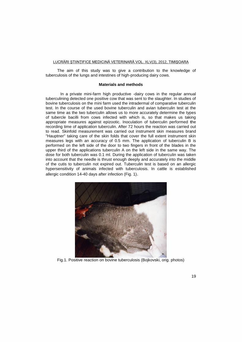
In a private mini-farm high productive -dairy cows in the regular annual
tuberculining detected one positive cow that was sent to the slaughter. In studies of bovine tuberculosis on the mini farm used the intradermal of comparative tuberculin test. In the course of the used bovine tuberculin and avian tuberculin test at the same time as the two tuberculin allows us to more accurately determine the types of tubercle bacilli from cows infected with which is, so that makes us taking appropriate measures against epizootic. Inoculation of tuberculin performed the recording time of application tuberculin. After 72 hours the reaction was carried out to read. Skinfold measurement was carried out instrument skin measures brand "Hauptner" taking care of the skin folds that cover the full extent instrument skin measures legs with an accuracy of 0.5 mm. The application of tuberculin B is performed on the left side of the door to two fingers in front of the blades in the upper third of the applications tuberculin A on the left side in the same way. The dose for both tuberculin was 0.1 ml. During the application of tuberculin was taken into account that the needle is thrust enough deeply and accurately into the middle of the cutis to tuberculin not expired out. Tuberculin test is based on an allergic hypersensitivity of animals infected with tuberculosis. In cattle is established
allergic condition 14-40 days after infection (Fig. 1).
Fig.1. Positive reaction on bovine tuberculosis (Bojkovski, orig. photos)
LUCRĂRI ŞTIINŢIFICE MEDICINĂ VETERINARĂ VOL. XLV(3), 2012, TIMIŞOARA
20
Results and discussions
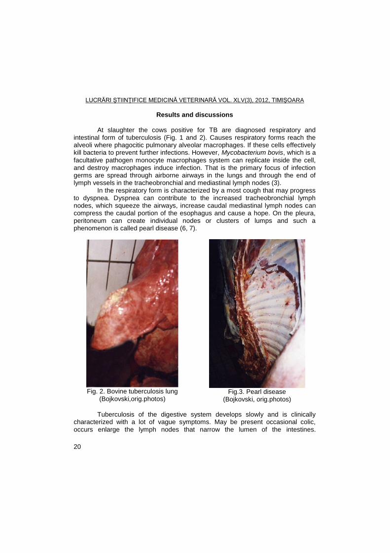
At slaughter the cows positive for TB are diagnosed respiratory and intestinal form of tuberculosis (Fig. 1 and 2). Causes respiratory forms reach the alveoli where phagocitic pulmonary alveolar macrophages. If these cells effectively kill bacteria to prevent further infections. However, Mycobacterium bovis, which is a facultative pathogen monocyte macrophages system can replicate inside the cell, and destroy macrophages induce infection. That is the primary focus of infection germs are spread through airborne airways in the lungs and through the end of lymph vessels in the tracheobronchial and mediastinal lymph nodes (3).
In the respiratory form is characterized by a most cough that may progress to dyspnea. Dyspnea can contribute to the increased tracheobronchial lymph nodes, which squeeze the airways, increase caudal mediastinal lymph nodes can compress the caudal portion of the esophagus and cause a hope. On the pleura, peritoneum can create individual nodes or clusters of lumps and such a phenomenon is called pearl disease (6, 7).
Fig. 2. Bovine tuberculosis lung
(Bojkovski,orig.photos)
Fig.3. Pearl disease
(Bojkovski, orig.photos)
Tuberculosis of the digestive system develops slowly and is clinically characterized with a lot of vague symptoms. May be present occasional colic, occurs enlarge the lymph nodes that narrow the lumen of the intestines.
LUCRĂRI ŞTIINŢIFICE MEDICINĂ VETERINARĂ VOL. XLV(3), 2012, TIMIŞOARA
21
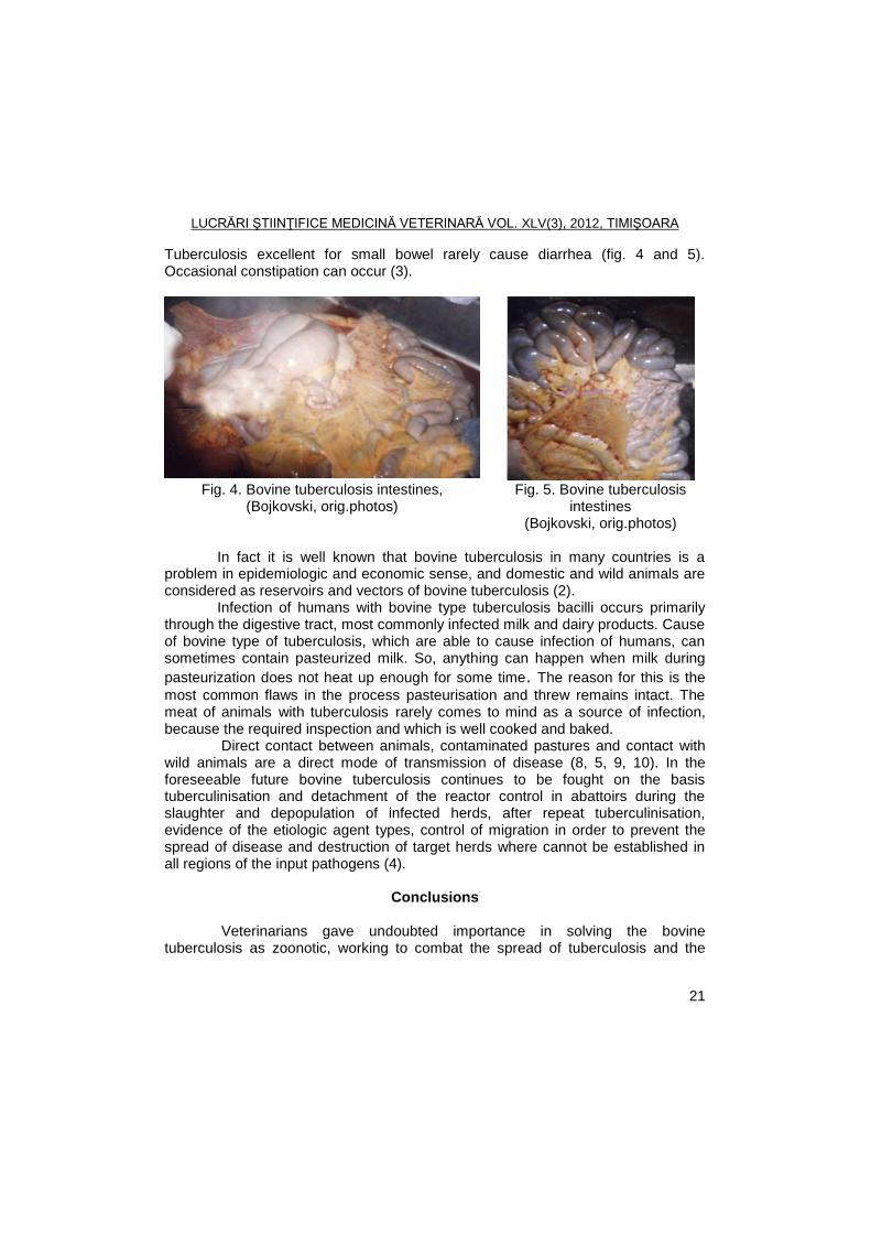
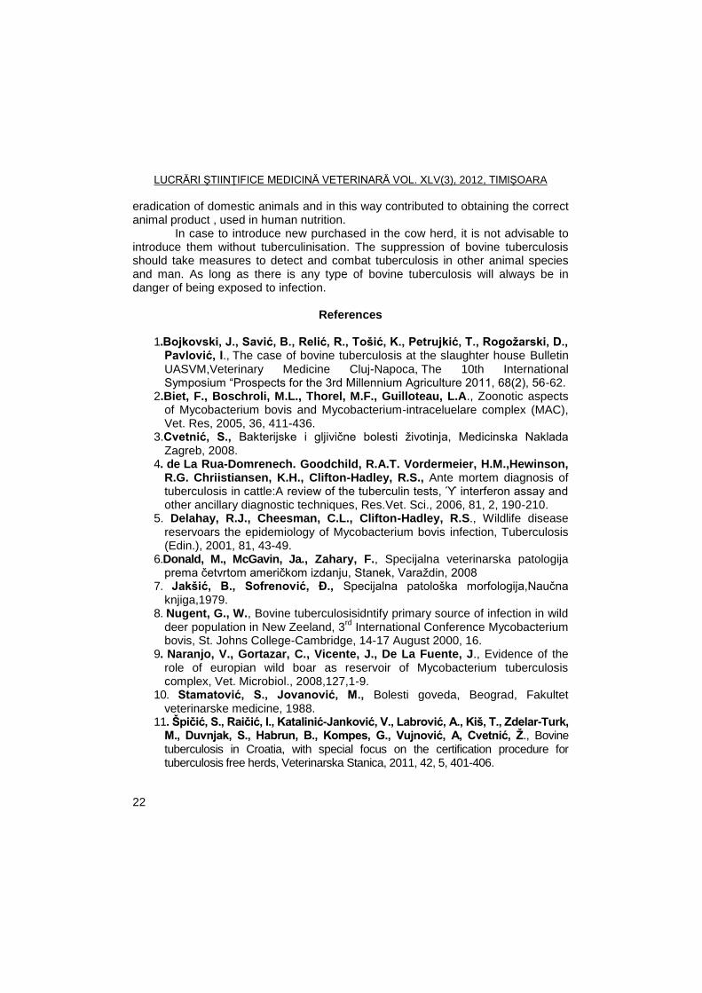
Tuberculosis excellent for small bowel rarely cause diarrhea (fig. 4 and 5). Occasional constipation can occur (3).
Fig. 4. Bovine tuberculosis intestines, (Bojkovski, orig.photos)
Fig. 5. Bovine tuberculosis intestines
(Bojkovski, orig.photos) In fact it is well known that bovine tuberculosis in many countries is a
problem in epidemiologic and economic sense, and domestic and wild animals are considered as reservoirs and vectors of bovine tuberculosis (2).
Infection of humans with bovine type tuberculosis bacilli occurs primarily through the digestive tract, most commonly infected milk and dairy products. Cause of bovine type of tuberculosis, which are able to cause infection of humans, can sometimes contain pasteurized milk. So, anything can happen when milk during
pasteurization does not heat up enough for some time. The reason for this is the
most common flaws in the process pasteurisation and threw remains intact. The meat of animals with tuberculosis rarely comes to mind as a source of infection, because the required inspection and which is well cooked and baked.
Direct contact between animals, contaminated pastures and contact with wild animals are a direct mode of transmission of disease (8, 5, 9, 10). In the foreseeable future bovine tuberculosis continues to be fought on the basis tuberculinisation and detachment of the reactor control in abattoirs during the slaughter and depopulation of infected herds, after repeat tuberculinisation, evidence of the etiologic agent types, control of migration in order to prevent the spread of disease and destruction of target herds where cannot be established in all regions of the input pathogens (4).
Conclusions
Veterinarians gave undoubted importance in solving the bovine
tuberculosis as zoonotic, working to combat the spread of tuberculosis and the
LUCRĂRI ŞTIINŢIFICE MEDICINĂ VETERINARĂ VOL. XLV(3), 2012, TIMIŞOARA
22
eradication of domestic animals and in this way contributed to obtaining the correct animal product , used in human nutrition.
In case to introduce new purchased in the cow herd, it is not advisable to introduce them without tuberculinisation. The suppression of bovine tuberculosis should take measures to detect and combat tuberculosis in other animal species and man. As long as there is any type of bovine tuberculosis will always be in danger of being exposed to infection.
References
1.Bojkovski, J., Saviš, B., Reliš, R., Tošiš, K., Petrujkiš, T., Rogoţarski, D.,
Pavloviš, I., The case of bovine tuberculosis at the slaughter house Bulletin
UASVM,Veterinary Medicine Cluj-Napoca, The 10th International Symposium ―Prospects for the 3rd Millennium Agriculture 2011, 68(2), 56-62.
2.Biet, F., Boschroli, M.L., Thorel, M.F., Guilloteau, L.A., Zoonotic aspects of Mycobacterium bovis and Mycobacterium-intraceluelare complex (MAC), Vet. Res, 2005, 36, 411-436.
3.Cvetniš, S., Bakterijske i gljivične bolesti ţivotinja, Medicinska Naklada Zagreb, 2008.
4. de La Rua-Domrenech. Goodchild, R.A.T. Vordermeier, H.M.,Hewinson, R.G. Chriistiansen, K.H., Clifton-Hadley, R.S., Ante mortem diagnosis of tuberculosis in cattle:A review of the tuberculin tests, ϓ interferon assay and other ancillary diagnostic techniques, Res.Vet. Sci., 2006, 81, 2, 190-210.
5. Delahay, R.J., Cheesman, C.L., Clifton-Hadley, R.S., Wildlife disease reservoars the epidemiology of Mycobacterium bovis infection, Tuberculosis (Edin.), 2001, 81, 43-49.
6.Donald, M., McGavin, Ja., Zahary, F., Specijalna veterinarska patologija prema četvrtom američkom izdanju, Stanek, Varaţdin, 2008
7. Jakšiš, B., Sofrenoviš, Đ., Specijalna patološka morfologija,Naučna knjiga,1979.
8. Nugent, G., W., Bovine tuberculosisidntify primary source of infection in wild deer population in New Zeeland, 3
rd International Conference Mycobacterium
bovis, St. Johns College-Cambridge, 14-17 August 2000, 16. 9. Naranjo, V., Gortazar, C., Vicente, J., De La Fuente, J., Evidence of the
role of europian wild boar as reservoir of Mycobacterium tuberculosis complex, Vet. Microbiol., 2008,127,1-9.
10. Stamatoviš, S., Jovanoviš, M., Bolesti goveda, Beograd, Fakultet veterinarske medicine, 1988.
11. Špiţiš, S., Raiţiš, I., Kataliniš-Jankoviš, V., Labroviš, A., Kiš, T., Zdelar-Turk, M., Duvnjak, S., Habrun, B., Kompes, G., Vujnoviš, A, Cvetniš, Ţ., Bovine tuberculosis in Croatia, with special focus on the certification procedure for tuberculosis free herds, Veterinarska Stanica, 2011, 42, 5, 401-406.
LUCRĂRI ŞTIINŢIFICE MEDICINĂ VETERINARĂ VOL. XLV(3), 2012, TIMIŞOARA
23
RESEARCH ON THE ROLE OF AVIAN REOVIRUS IN TRANSMISSIBLE VIRAL ETIOLOGY PROVENTRICULITIS
N. CĂTANĂ, ALINA COMAN,
IONICA FODOR
Department of Infectious Diseases and Preventive Medicine, Banat's University of
Agricultural Sciences and Veterinary Medicine Timisoara, Faculty of Veterinary Medicine, 300645, Aradului Street No. 119, Timisoara, Romania
E-mail: [email protected]
Summary
Transmissible viral proventriculitis (TVP) is a poorly understood disease of broiler
chickens. The major economic problem caused by TVP it is ability to affect overall flock performance in broiler complexes. TVP is considered to be a disease of unknown etiology, several infectious agents, nutritional factors, and toxic factors have been associated as causes.
In this study avian reovirus was identified in all age groups, followed in frequency
by: ChPV, IBV, CAstV and AvRV viruses, and IBDV, CAV, ANV and CPNV were not identified.
Key words: broiler, proventriculitis, PCR
Transmissible viral proventriculitis (TVP) is an infectious disease that
develops in broilers, was first reported in the Netherlands, then in the U.S.A. and Australia. In intensive poultry farming is associated with runting stunding syndrome causing significant economic damage.
In 1996, Goodwin et al., identified by electron microscopy, positive contrast, a virus, subsequently by other researchers called adenovirus-like. In subsequent years, particularly after 2000, many researchers have studied the disease etiology and according to the results obtained at present it is considered that the disease is caused by a combination of the following viruses: ARV, IBV, IBDV, CAstV, AvRV, CAV, ChPV, ANV and CPNV. Viral association was demonstrated by the fact that TVP reproduction by experimental infection is possible only with the proventriculi triturat with specific injuries and attempts to reproduce only one or two viruses were not possible (6, 8, 9).
In subsequent years, the disease has spread in more countries, including Romania, both imported broilers at the age of one day, and the broiler breeding flocks coming from local farms (1, 2, 3, 4, 5).
Research has been conducted in a broiler farm, raised on litter permanent. In that farm, the disease was suspected clinically at more effective.
LUCRĂRI ŞTIINŢIFICE MEDICINĂ VETERINARĂ VOL. XLV(3), 2012, TIMIŞOARA
24
Materials and methods This study was made in a broiler flock of 6500 chickens, where TVP
evolved to confirm the etiology, being made molecular biology tests and serological examinations.
Proventriculus samples were harvested from cadavers in the following age groups: 7 days, 14 days, 28 days and 35 days.
From cadaver with TVP lesions, proventriculi were sampled for examination of molecular biology, respectively, by RT-PCR techniques. Isolations of viral nucleic acid was made using QIAamp viral RNA Mini Kit (Qiagen, Germany) and High Pure Viral Nucleic Acid Kit (Roche, Switzerland). Revers transcription and amplification were made using Qiagen OneStep RT-PCR Kit (Roche, Switzerland).
Serological examination was performed in order to identify specific antibodies against avian reovirus, because it had the highest frequency determined by RT-PCR technique. For this purpose blood samples were taken from chickens, randomized as follows:
- R 1 - at 21 days old (48 blood samples); - R 2 - at 35 days old (45 blood samples).
Specific antibodies were detected by ELISA, using FlockChek® Avian
Reovirus Antibody Test Kit, made by IDEXX Laboratories, Inc.
Results and discussions
The cadavers from outbreak were collected from four different age categories to highlight the possible viruses responsible for disease etiology. Analyzing data is observed that avian reovirus was identified in all age groups.
The results are identical with the results communicated by most research teams dealing with TVP study (7, 8, 9).
Serological examination was performed in order to confirm avian reovirus infection because the virus was identified by molecular biology tests, samples proventriculi at all age categories.
The results of this examination, performed by ELISA, are shown in Table 1. After reading the responses and process the results, according to the interpretation of the kit software FlockChek
® Avian Reovirus Antibody Test Kit, made by IDEXX
Laboratories, Inc., were established for each harvest: titer groups, minimum titer, maximum titer and geometric mean. Titres are expressed in optical density (O.D.).
First harvest, that the age of 21 days, six groups were identified titers (0-5), the minimum titer of 12 OD and 1022 OD maximum titer.
The secound harvest, that the age of 35 days, eight groups were identified titers (0-7), the minimum titer of 63 OD and 1453 OD maximum titer.
Analyzing these results, we see that at the age of 21 days (R 1) 29 sera were positive, respectively 60.42% and the age of 35 days 35 sera were positive, 77.77% respectively. These data show that, within 14 days, the proportion of
LUCRĂRI ŞTIINŢIFICE MEDICINĂ VETERINARĂ VOL. XLV(3), 2012, TIMIŞOARA
25
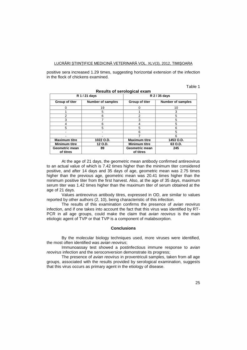
positive sera increased 1.29 times, suggesting horizontal extension of the infection in the flock of chickens examined.
Table 1 Results of serological exam
R 1 / 21 days R 2 / 35 days
Group of titer Number of samples Group of titer Number of samples
0 19 0 10
1 5 1 3
2 6 2 5
3 7 3 5
4 6 4 5
5 5 5 6
- - 6 5
- - 7 6
Maximum titre 1022 O.D. Maximum titre 1453 O.D.
Minimum titre 12 O.D. Minimum titre 63 O.D.
Geometric mean of titres
89 Geometric mean of titres
245
At the age of 21 days, the geometric mean antibody confirmed antireovirus
to an actual value of which is 7.42 times higher than the minimum titer considered positive, and after 14 days and 35 days of age, geometric mean was 2.75 times higher than the previous age, geometric mean was 20.41 times higher than the minimum positive titer from the first harvest. Also, at the age of 35 days, maximum serum titer was 1.42 times higher than the maximum titer of serum obtained at the age of 21 days.
Values antireovirus antibody titres, expressed in OD, are similar to values reported by other authors (2, 10), being characteristic of this infection.
The results of this examination confirms the presence of avian reovirus infection, and if one takes into account the fact that this virus was identified by RT-PCR in all age groups, could make the claim that avian reovirus is the main etiologic agent of TVP or that TVP is a component of malabsorption.
Conclusions
By the molecular biology techniques used, more viruses were identified, the most often identified was avian reovirus;
Immunoassay test showed a postinfectious immune response to avian reovirus infection and the seroconversion demonstrate its progress;
The presence of avian reovirus in proventriculi samples, taken from all age groups, associated with the results provided by serological examination, suggests that this virus occurs as primary agent in the etiology of disease.
LUCRĂRI ŞTIINŢIFICE MEDICINĂ VETERINARĂ VOL. XLV(3), 2012, TIMIŞOARA
26
References
1. Cătană, N., Fodor, Ionica, Coman, Alina, Epidemiologic, clinic and anatomopathologic research in an outbreak of chickens with transmissible viral proventriculitis, Lucrări ştiinţifice medicină veterinară Timişoara, 2011,, vol. XLIV (1), 122-125.
2. Cătană, N., Popa, Virgilia, Herman, V., Fodor, Ionica, Cercetări anatomoclinice şi serologice într-un focar de reoviroză la puii de carne, Lucr. Şt. Med. Vet. Iaşi, 2008, 51 (10), 263-266.
3. Cătană, N., Proventriculita virală transmisibilă, Magazin Avicol, 2011, 8, 32, 19-20.
4. Coman, Alina, Fodor, Ionica, Cătană, N., Study on Outbreak of Transmissible Viral Proventriculis in Broiler Chickens - The 10th International Symposium Prospects for 3rd millennium agriculture 29 September – 1 October, Cluj-Napoca, Romania, 2011, 96-99.
5. Coman, Alina, Rusvai, M., Demeter, Z., Palade, Elena Alina, Cătană, N., Histopathological changes of Transmissible Viral Proventriculitis in Broiler Flocks, The 10th International Symposium Prospects for 3rd millennium agriculture 29 September – 1 October, Cluj-Napoca, Romania, 2011, 93-95.
6. Goodwin, M.A., Hafner, S., Viral proventriculitis. In: Diseases Of Poultry, 11th ed. H. J. Barnes, J. R. Glisson, A. M. Fadly, L. R. McDougald, Y. M. Saif, and D. E. Swayne, eds. Iowa State Press, Ames, IA. 2003, 383-388.
7. Hafner, S., Goodwin, M. A., Guy, J. S., Pantin-Jackwood, M., Proventriculitis and Proventricular Dilatation of Broiler Chickens. In: Diseases Of Poultry, 12th ed. Y. M. Saif, A. M. Fadly, J. R. Glisson, L. R. McDougald, L. K. Nolan and D. E. Swayne, eds. Blackwell Publishing, State Avenue, Ames, Iowa, USA. 2008, 1272-1277.
8. Pantin-Jackwood, M.J., Brown, T.P., Huff, G.R., Reproduction of proventriculitis in commercial and specific-pathogen-free broiler chickens, Avian Diseases, 2005, 49, 352-360.
9. Pantin-Jackwood, M. J., Brown, T.P., Infectious bursal disease virus and proventriculitis in broiler Chickens, Avian Diseases, 2003, 47, 681-690.
10. Van Der Heide, L., The history of avian reovirus, Avian Diseases, 2000, 44, 638-641.
LUCRĂRI ŞTIINŢIFICE MEDICINĂ VETERINARĂ VOL. XLV(3), 2012, TIMIŞOARA
27
USING A SIMPLIFIED SCHEME FOR ISOLATION AND TYPING STAPHYLOCOCCI ISOLATED FROM ANIMALS
N. CĂTANĂ, FODOR IONICA, J. DÉGI
Department of Infectious Diseases and Preventive Medicine, Banat's University of
Agricultural Sciences and Veterinary Medicine Timisoara, Faculty of Veterinary Medicine, 300645, Aradului Street No. 119, Timisoara, Romania
E-mail: [email protected]
Summary
In routine diagnosis, staphylococcal infections, it is necessary to use a simplified
working scheme to differentiate pathogens such as Staphylococcus or other nonpathogenic
and hull, which is based on some characteristic phenotypic characters. Methicillin-resistant staphylococcus bacteria are considered with zoonotic risk,
causing nosocomial infections in humans. Methicillin-resistance is present in several species coagulase positive or negative, in laboratories, its presence being compulsorily tested.
In this study have been identified 27 of methicillin-resistant strains, from all animal species, is shown as epidemiological cycle of intra-and interspecific these strains.
Bacterial performed according to the methodology used allowed the isolation and identification of three species of staphylococci (S. aureus subps. aureus, S. hycus, S. xylosus) from animals of rent and the Group intermedius from dogs and cats.
Key words: staphilococi, methicillin-resistance, animals
Staphylococcal infections in animals are quite common, have a variable
clinical course, being produced by several species of staphylococci, with an intra-and interspecific epidemiological circuit that includes people (3).
In recent years methicillin-resistant Staphylococcus bacteria are considered particularly zoonotic risk, causing nosocomial infections in humans. Methicillin-resistance is present in several species coagulase positive or negative, in laboratories, its presence being compulsorily tested (1, 2, 4, 5).
In routine diagnosis, staphylococcal infections, it is necessary to use a simplified working scheme to differentiate pathogens such as Staphylococcus or other nonpathogenic and hull, which is based on some characteristic phenotypic characters (3).
In this context research covered by this paper were performed in order: -to characterize the phenotypic strains of staphylococci isolated from
animals, based on a simplified scheme; -to identify staphylococci coagulase positive and negative rapid test; -to identify methicillin-resistant strains of staphylococci present in animals.
LUCRĂRI ŞTIINŢIFICE MEDICINĂ VETERINARĂ VOL. XLV(3), 2012, TIMIŞOARA
28
Materials and methods
Pathological material samples were taken from animals (sheep, cattle, horses, pigs, dogs and cats), with different clinical disease or healthy. Pathological samples were taken from a number of rent and 50 animals from 55 different pets with skin diseases or healthy (Table 1).
Of pathological material were made early sowing on agar sheep blood defibrinated 5%, the plates were incubated at 37˚C in normal atmosphere for 18 -20 hours. Early sowings were made in this way to obtain isolated colonies, in order to assess cultural character. Strains of staphylococci were isolated and purified on biochemical tests and pathogenicity of characters.
Controlled biochemical properties were manita the environment Chapmann fermentation and fermentation sugars present in several API Staph system. Control pathogenicity factors were the presence of coagulase-related (clumping factor) and hemolisins. Haemolytic activity was assessed on sheep blood defibrinated agar 5%.
Highlighting related coagulase was highlighted Staph Latex Kit KIT PROLEX quickly. Related strains possessing coagulase (clumping factor) or protein A, placed in contact with latex particles sensitised with fibrinogen and IgG, producing mixed agglutination, indicating the emergence of coagulase-related small clot.
For susceptibility testing of staphylococci at novobiocine, strains of staphylococci isolated was used Kirby-Bauer difusimetric method were used the following ingredients: Mueller-Hinton broth and agar, Petri plates and impregnated biodiscs with meticilin and novobiocine by Oxoid products.
Results and discussions
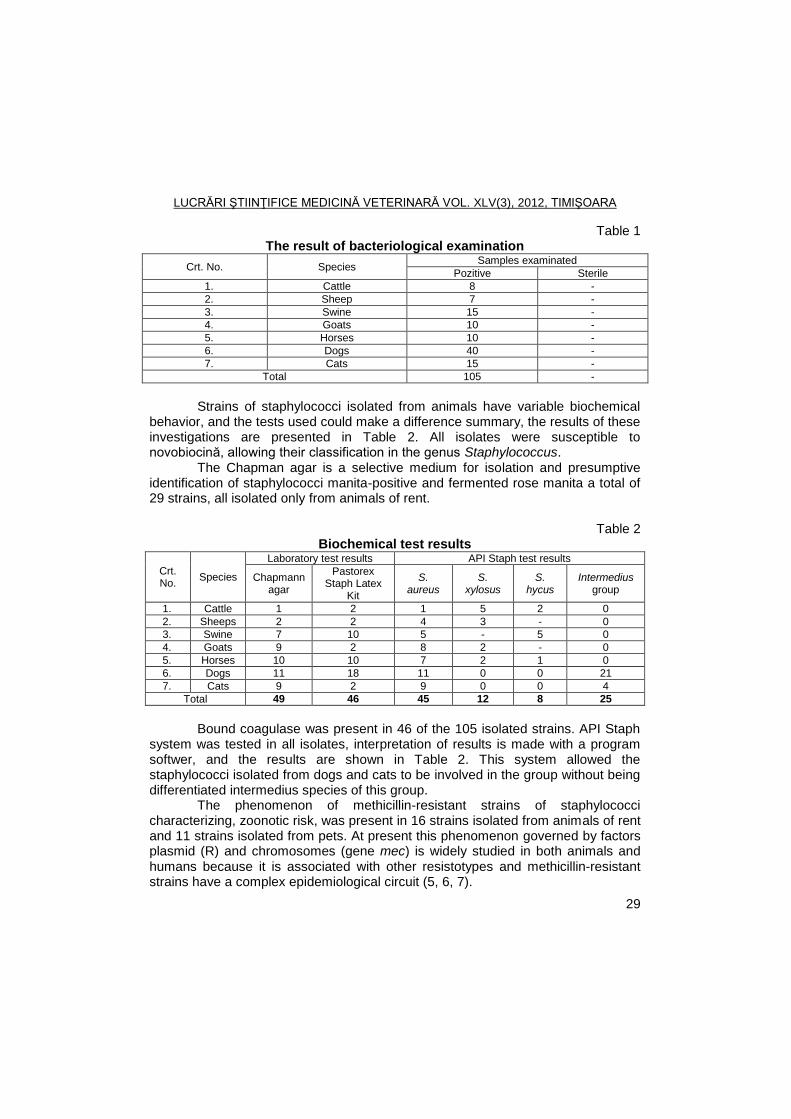
Bacteriological examination of samples submitted were isolated 105 strains
of staphylococci, in October 2010 - May 2011, in the Bacterial Infectious Diseases Research Laboratory - Faculty of Veterinary Medicine Timisoara.
The 105 strains studied, the Gram stain were assigned to Gram-positive group and were presented in the form of cocci, with size between 0.8-1.5 mm in diameter, and how was the predominant group cluster and occasionally in pairs or solitary bacterial cells.
Both in primary cultures and subcultures isolates produced yellow pigment is either white pigment beta haemolysis type, more commonly, as well as alpha (incomplete).
LUCRĂRI ŞTIINŢIFICE MEDICINĂ VETERINARĂ VOL. XLV(3), 2012, TIMIŞOARA
29
Table 1 The result of bacteriological examination
Crt. No. Species Samples examinated
Pozitive Sterile
1. Cattle 8 -
2. Sheep 7 -
3. Swine 15 -
4. Goats 10 -
5. Horses 10 -
6. Dogs 40 -
7. Cats 15 -
Total 105 -
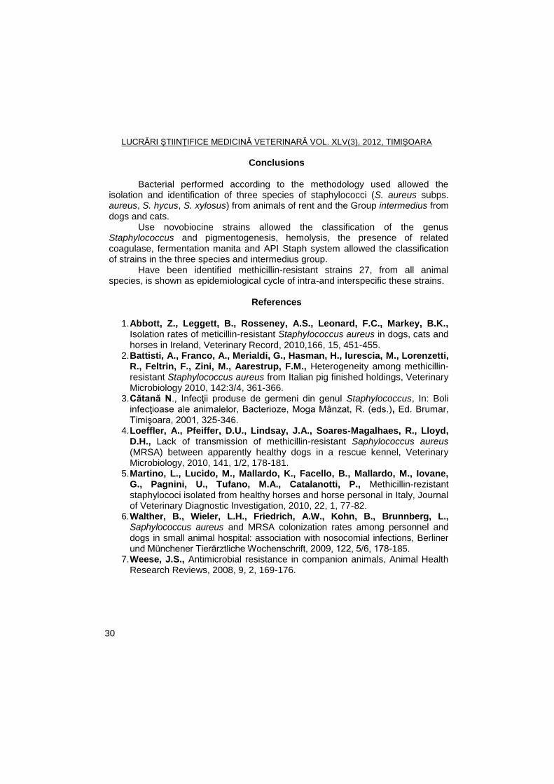
Strains of staphylococci isolated from animals have variable biochemical
behavior, and the tests used could make a difference summary, the results of these investigations are presented in Table 2. All isolates were susceptible to novobiocină, allowing their classification in the genus Staphylococcus.
The Chapman agar is a selective medium for isolation and presumptive identification of staphylococci manita-positive and fermented rose manita a total of 29 strains, all isolated only from animals of rent.
Table 2
Biochemical test results
Crt. No.
Species
Laboratory test results API Staph test results
Chapmann agar
Pastorex Staph Latex
Kit
S. aureus
S. xylosus
S. hycus
Intermedius group
1. Cattle 1 2 1 5 2 0
2. Sheeps 2 2 4 3 - 0
3. Swine 7 10 5 - 5 0
4. Goats 9 2 8 2 - 0
5. Horses 10 10 7 2 1 0
6. Dogs 11 18 11 0 0 21
7. Cats 9 2 9 0 0 4
Total 49 46 45 12 8 25
Bound coagulase was present in 46 of the 105 isolated strains. API Staph
system was tested in all isolates, interpretation of results is made with a program softwer, and the results are shown in Table 2. This system allowed the staphylococci isolated from dogs and cats to be involved in the group without being differentiated intermedius species of this group.
The phenomenon of methicillin-resistant strains of staphylococci characterizing, zoonotic risk, was present in 16 strains isolated from animals of rent and 11 strains isolated from pets. At present this phenomenon governed by factors plasmid (R) and chromosomes (gene mec) is widely studied in both animals and humans because it is associated with other resistotypes and methicillin-resistant strains have a complex epidemiological circuit (5, 6, 7).
LUCRĂRI ŞTIINŢIFICE MEDICINĂ VETERINARĂ VOL. XLV(3), 2012, TIMIŞOARA
30
Conclusions
Bacterial performed according to the methodology used allowed the isolation and identification of three species of staphylococci (S. aureus subps. aureus, S. hycus, S. xylosus) from animals of rent and the Group intermedius from dogs and cats.
Use novobiocine strains allowed the classification of the genus Staphylococcus and pigmentogenesis, hemolysis, the presence of related coagulase, fermentation manita and API Staph system allowed the classification of strains in the three species and intermedius group.
Have been identified methicillin-resistant strains 27, from all animal species, is shown as epidemiological cycle of intra-and interspecific these strains.
References
1. Abbott, Z., Leggett, B., Rosseney, A.S., Leonard, F.C., Markey, B.K.,
Isolation rates of meticillin-resistant Staphylococcus aureus in dogs, cats and horses in Ireland, Veterinary Record, 2010,166, 15, 451-455.
2. Battisti, A., Franco, A., Merialdi, G., Hasman, H., Iurescia, M., Lorenzetti, R., Feltrin, F., Zini, M., Aarestrup, F.M., Heterogeneity among methicillin-resistant Staphylococcus aureus from Italian pig finished holdings, Veterinary Microbiology 2010, 142:3/4, 361-366.
3. Cătană N., Infecţii produse de germeni din genul Staphylococcus, In: Boli infecţioase ale animalelor, Bacterioze, Moga Mânzat, R. (eds.), Ed. Brumar, Timişoara, 2001, 325-346.
4. Loeffler, A., Pfeiffer, D.U., Lindsay, J.A., Soares-Magalhaes, R., Lloyd, D.H., Lack of transmission of methicillin-resistant Saphylococcus aureus (MRSA) between apparently healthy dogs in a rescue kennel, Veterinary Microbiology, 2010, 141, 1/2, 178-181.
5. Martino, L., Lucido, M., Mallardo, K., Facello, B., Mallardo, M., Iovane, G., Pagnini, U., Tufano, M.A., Catalanotti, P., Methicillin-rezistant staphylococi isolated from healthy horses and horse personal in Italy, Journal of Veterinary Diagnostic Investigation, 2010, 22, 1, 77-82.
6. Walther, B., Wieler, L.H., Friedrich, A.W., Kohn, B., Brunnberg, L., Saphylococcus aureus and MRSA colonization rates among personnel and dogs in small animal hospital: association with nosocomial infections, Berliner und Münchener Tierärztliche Wochenschrift, 2009, 122, 5/6, 178-185.
7. Weese, J.S., Antimicrobial resistance in companion animals, Animal Health Research Reviews, 2008, 9, 2, 169-176.
LUCRĂRI ŞTIINŢIFICE MEDICINĂ VETERINARĂ VOL. XLV(3), 2012, TIMIŞOARA
31
CHANGES IN HUMORAL COMPARTMENT OF THE IMMUNE SYSTEM ASSOCIATED WITH ENZOOTIC BOVINE LEUKOSIS
CĂTĂLINA CENUȘE
1, E. TÎRZIU
1, ILEANA NICHITA
1, C. CUMPĂNĂŞOIU
1,
R.V. GROS1, R. LAZĂR
2, MONICA ȘEREȘ
1
1Banat's University of Agricultural Sciences and Veterinary Medicine, Faculty of
Veterinary Medicine Timisoara, 300645, Aradului Street No. 119, Timisoara, Romania
2DSVSA Constanţa
E-mail: [email protected]
Summary
Enzootic bovine leukosis (EBL), disease of adult cattle caused by bovine leukaemia virus (BLV), a retrovirus, is accompanied by a series of changes in cellular and humoral compartments of the immune system. If cellular changes, especially those of B cells, target of the viral infection, have been extensively studied and described, humoral changes, particularly those involving cytokines are subject only in few studies.
Knowing how cytokines, due their changes, influence the evolution EBL is extremely important both for understanding the viral biology and pathogenesis of infection and to identify viable therapeutic measures, and this review describe in detail all their humoral and cellular implications.
Key words: enzootic bovine leukosis, antibodies against BLV, cytokines alteration
Enzootic bovine leukosis (EBL) is a disease of adult cattle caused by a retrovirus, bovine leukaemia virus (BLV). Cattle may be infected at any age, including the embryonic stage. Most infections are subclinical, but a proportion of cattle (30-70%) over 3 years old develop persistent lymphocytosis, and a smaller proportion (0.1-10%) develop lymphosarcomas (tumors) in various internal organs. Natural infection has also been recorded in buffaloes, sheep and capybaras (18).
Infection with the virus in cattle is lifelong and gives rise to a persistent antibody response, which can be detected as early as 3 weeks after infection (18). Changes in the humoral compartment of the immune system are not limited to the synthesis of specific antibodies against BLV, but also include the alteration of cytokine secretion, such as interleukins (ILs), interferons (IFNs) and tumor necrosis factors (TNFs), which shows also an alteration of T cells and macrophages.
Humoral changes, taken together, are important for elucidating the pathogenic mechanisms underlying EBL, for early and certain diagnosis of this viral disease and for therapeutic strategies.
Antibody-mediate immune response against BLV Natural or iatrogenic transmission BLV mainly involves transfer of infected
cells through the blood or milk. Phenomena which follow the primary infections are not yet fully understood, but one of the first signs of disease, detected in
LUCRĂRI ŞTIINŢIFICE MEDICINĂ VETERINARĂ VOL. XLV(3), 2012, TIMIŞOARA
32
approximately 1-8 weeks after experimental infection, is the antiviral antibody response (14). In natural infection in cattle, the antibodies against various viral antigens can be first detected between 3 and 16 weeks after exposure (18).
Maternally derived antibodies may take up to 6 or 7 months to disappear, and there is no way of distinguishing passively transferred antibodies from those resulting from active infection (18).
For these reasons, most studies of the antibody-mediated immune response made so far used the model of experimental infection in sheep. They have shown that antibodies against BLV belong to IgG1 class (9) and have the ability to recognize structural proteins (gp51 and p24 from the envelope, respectively capsid) (10) and regulatory proteins (Tax and Rex) (11). Also, antibodies directed against infected B cells were detected (9).
The anti-gp51 and p24 antibodies level can be directly correlated with replication VLB, lowered titers were associated with low proviral loads, and the higher ones with intense replication (4). Thus, one can distinguish two phases (14):
- The early phase - low viral replication associated with the synthesis of neutralizing antibodies directed specifically against the capsid components;
- The late phase - intense replication accompanied by a massive increase in antiviral antibodies, which proceeds the release of many infected cells into the blood.
Subsequently, although the number of cells expressing viral proteins significantly decreases, neutralizing antibody titer is kept stable even eight months after experimental infection (14). This persistence of anti-BLV antibodies suggest continued antigenic stimulation in vivo.
Alteration in cytokines expression Types 1 and 2 cytokine imbalance is generally considered to cause
disease progression in chronic retroviral infections, the typical example being human immunodeficiency virus type 1 (HIV-1) infection. Type 1 cytokines such as IFN-γ, IL-2, and IL-12 are involved in cellular immunity, whereas type 2 cytokines including IL-4, IL-5, IL-6, IL-10 and IL-13 deal with the humoral immunity (16).
Knowing how cytokines, due their changes, influence the EBL evolution is extremely important both for understanding the viral biology and pathogenesis of infection and to identify viable therapeutic measures. Until now, there are only several reports about implication of some cytokines (IL-2, IL-6, IL-10, IL-12, TNF-α, and IFN-γ) in the evolution of infection, and this review describe in detail all their humoral and cellular implications.
Interleukins 2, 6, 10, and 12 The first evidence regarding the importance of cytokines in EBL
interconnects IL-10, viral expression and proliferation of B cells. These initial studies showed a significantly higher expression of messenger RNA (mRNA)
LUCRĂRI ŞTIINŢIFICE MEDICINĂ VETERINARĂ VOL. XLV(3), 2012, TIMIŞOARA
33
specific IL-10 in cattle with persistent lymphocytosis, in late stage of infection (2, 12, 13).
Combination BLV - IL-10 plays an important role both in neoplasms development and in the evolution of the disease to one of their forms. First, IL-10 inhibits expression of other cytokines, particularly those of type 1 (i.e. IL-2 and IFN-γ), and class II major histocompatibility complex molecules (MHC) on the macrophages surface, inhibiting antigen presentation. Inability of T cells to respond to foreign peptides may also lead to decline in IL-2, IL-6, and IFN-γ (12).
Second, IL-10 is able to induce programmed cell death of Th1 cells. Th1 cells express high levels of Fas ligand and will disappear in the process of programmed death, while Th2 lymphocytes (which are the cells that synthesize large amounts of IL-10), whose expression of Fas ligand is reduced, do not become apoptotic. Fas mediate Th1 cell activation in early immune response, in its late stages being an apoptotic factor. Thus, in animals infected with BLV, Th1 cells activated in the first phase will become apoptotic, with a subsequent decrease of the IL-2 and IFN-γ (12).
Peripheral blood B cells do not express detectable amounts of IL-10 in vivo, suggesting that autocrine secretion of this interleukin is not essential to maintain CD5
+ B cell subpopulation. It should be noted however that the high level
of mRNA for IL-10 detected in peripheral blood mononuclear cells (PBMCs) cultures may be associated with to progress to persistent lymphocytosis (2).
Although there is a debate, it is considered that IL-2 is synthesized at high levels in cultured PBMCs mitogenic stimulated obtained from asymptomatic cattle or with persistent lymphocytosis (1, 5).
In vivo expression of IL-2 can be detected from 2-3 weeks after in cattle, its level decreased significantly at 12 weeks post infection (17).
In cultures of unstimulated PBMCs mRNA expression of IL-2 is significantly lower in cattle with persistent lymphocytosis compared with the healthy animals. Also, CD4
+ T cells isolated from the first category of animals have a small
amount of mRNA for IL-2 (1). Regarding the effect on B cells isolated from cattle with persistent
lymphocytosis, IL-2 increases viral CA protein synthesis and expression of IL-2 receptors, being also a trigger of cell proliferation (3).
T lymphocytes isolated from lymph nodes and peripheral blood of cattle infected with BLV contains IL-2 mRNA, but the amount of genetic material is significantly less than in CD4
+ T cells of cattle with persistent lymphocytosis (5).
Although IL-6 mRNA is almost undetectable in B cells processed immediately after sampling from cattle with persistent lymphocytosis, the nucleic acid becomes evident after cultivation (2).
In addition, compared with cattle VLB+ and aleukemic, serum levels of IL-6 is significantly higher in those with persistent lymphocytosis; the same phenomenon was reported in cultures of PBMCs exposed to BLV antigens. Viral
LUCRĂRI ŞTIINŢIFICE MEDICINĂ VETERINARĂ VOL. XLV(3), 2012, TIMIŞOARA
34
expression is significantly inhibited if exogenous IL-6 is added to cultures infected in vitro, suggesting that IL-6 plays an important role in inducing viral latency (3).
Regarding IL-12, some researchers found that asymptomatic cattle with persistent lymphocytosis exhibit high levels of this cytokine, aspect confirmed also in vitro, on PBMCs cultures (5). In vivo, IL-12 can be detected in PBMCs from the first 1-3 weeks after infection in cattle, but this presence is temporary (17).
However, compared with aleukemic cattle, IL-12 mRNA expression is significantly lower in animals with persistent lymphocytosis, this phenomenon being observed also in experimentally infected sheep (3).
TNF-α In sheep inoculated with VLB it was observed that mRNA for type I
receptor of the TNF-α (TNF-R1) is reduced, while mRNA of TNF-R2 remains constant. PBMCs maintained in cultures expressed TNF-α associated to membrane and proliferate in the presence of this cytokine. Also, TNF-α expression is higher in sheep resistant to infection following vaccination against EBL (15).
In BLV+ cattle, average level of TNF-α mRNA is higher in cultures of
PBMCs proliferate spontaneously (without antigens and mitogens) or after antigenic challenge (8).
Addition of exogenous TNF-α to cell cultures in vitro infected with BLV significantly inhibit viral expression. Cells isolated from cattle with persistent lymphocytosis show an intense proliferative response in the presence of TNF-α, expressing high levels of recombinant bovine mRNA for TNF-R2, maintaining a constant level of mRNA for TNF-R1 (7). Most cells expressing TNF-R2 are CD5
+
and IgM+ and are less prone to apoptosis induced by TNF-α (15).
Thus, we can consider that TNF-α coupled to the surface of B cells may promote their growth in animals infected with BLV by autocrine effects, manifested through TNF-R2, and possibly by modulating interaction with other cells, such as T helper cells (15). Also, TNF-R2 may act in tandem with IL-2 to stimulate cell proliferation by inhibiting apoptosis, thus contributing to the development of lymphocytosis and lymphosarcomas in infected cattle (7).
It can be concluded that TNF-α is involved in BLV elimination in the early phase of infection, but in the late phase (persistent lymphocytosis or lymphosarcomatosis), this cytokine contributes to cell proliferation and progression of infection.
IFN-γ As suggested by antiviral activity, IFN-γ suppresses viral replication in
bovine cell cultures infected with BLV in vitro. However, in sheep, which have high levels of mRNA for IFN-γ, there was a lower proviral load than in cattle (16). Intraperitoneal inoculation of IFN-γ in cattle infected with BLV resulted in an increase of γδ T cells and infected B cells remained at low levels for a week after administration (3).
LUCRĂRI ŞTIINŢIFICE MEDICINĂ VETERINARĂ VOL. XLV(3), 2012, TIMIŞOARA
35
Messenger RNA for IFN-γ can be detected in T cells isolated from lymph nodes of infected cattle (5). In cultures of PBMCs, mRNA expression for IFN-γ rises four weeks after in vitro infection, but antiviral activity remains constant (3).
In experimental infection in cattle, IFN-γ is detectable in 2-3 weeks after exposure to BLV, its level decreases significantly by week 12, then remains constant (17).
Comparative study conducted on aleukemic and with persistent lymphocytosis cattle showed that the first category of animals expressed significantly higher levels of IFN-γ (6). In addition, IFN-γ reached higher concentrations in cell cultures from asymptomatic cattle (5).
From the above it can be concluded that administration of type 1 cytokines,
such as IFN-γ, TNF-α, IL-2, and IL-12, in the first phase of infection could be a viable therapeutic solution for animals of economic value. Of course, their therapeutic action is conditioned by early detection of EBL, which can be made on the basis of the antibody-mediated immune response presence and level.
References
1. Amills, M., Ramiya, V., Norimine, J., Olmstead, C.A., Lewin, H.A., Reduced
IL-2 and IL-4 mRNA expression in CD4+ T cells from bovine leukemia virus-infecte cows with persistent lymphocytosis, Virology, 2002, 304, 1-9.
2. Amills, M., Norimine, J., Olmstead, C.A., Lewin, H.A., Cytokine mRNA expression in B cells from bovine leukemia virus-infected cattle with persistent lymphocytosis, Cytokine, 2004, 28, 25-28.
3. Gillet, N., Florins A,. Boxus, M, Burteau, Catherine, Nigro, Annamaria, Vandermeers, F., Balon, H., Bouzar, A.-B., Defoiche, Mechanisms of leukemogenesis induced by bovine leukemia virus: prospects for novel anti-retroviral therapies in human, Retrovirology, 2007, 4, 1-32.
4. Juliarena, M.A., Poli, M., Ceriani, C., Sala, L., Rodríguez, E., Gutierrez, S., Dolcini, G., Odeon, A., Esteban, E.N., Antibody response against three widespread bovine viruses is not impaired in Holstein cattle carrying bovine leukocyte antigen DRB3.2 alleles associated with bovine leukemia virus resistance, J. Dairy Sci., 2009, 92, 375-381.
5. Keefe, R.G., Choi, Y., Ferrick, D.A., Stott, J.L., Bovine cytokine expression during different phases of bovine leukemia virus infection, Vet Immunol Immunopathol, 1997, 56, 39-51.
6. Konnai, S., Usui, T., Ohashi, K., Onuma, M., The rapid quantitative analysis of bovine cytokine genes by real-time RT-PCR, Vet Microbiol, 2003, 94, 283.
7. Konnai, S., Usui, T., Ikeda, M., Kohara, J., Hirata, T., Okada, K., Ohashi, K., Onuma, M., Imbalance of tumor necrosis factor receptors during progression in bovine leukemia virus infection, Virology, 2005, 339, 239-248.
LUCRĂRI ŞTIINŢIFICE MEDICINĂ VETERINARĂ VOL. XLV(3), 2012, TIMIŞOARA
36
8. Konnai, S., Usui, T., Ikeda, M., Kohara, J., Hirata, T., Okada, K., Tumor necrosis factor-alpha up-regulation in spontaneously proliferating cells derived from bovine leukemia virus-infected cattle, Arch Virol., 2006, 151, 347-360.
9. Portetelle, D., Bruck, C., Burny, A., Dekegel, D., Mammerickx, M., Urbain, J., Detection of complement-dependent lytic antibodies in sera from bovine leukemia virus-infected animals, Ann Rech Vet, 1978, 9, 667-674.
10. Portetelle, D., Mammerickx, M., Burny, A., Use of two monoclonal antibodies in an ELISA test for the detection of antibodies to bovine leukaemia virus envelope protein gp51, J Virol Methods, 1989, 23, 211-222.
11. Powers, Maureen A., Grossman, Deborah, Kidd, Lynn C., Radke, Kathryn, Episodic occurrence of antibodies against the bovine leukemia virus rex protein during the Course of Infection in Sheep, J Virol, 1991, 65(9), 4959-4965.
12. Pyeon, D., O'Reilly, K.L., Splitter, G.A., Increased interleukin-10 mRNA expression in tumor-bearing or persistently lymphocytotic animals infected with bovine leukemia virus, J Virol, 1996, 70, 5706-5710.
13. Pyeon, D., Diaz, F.J., Splitter, G.A., Prostaglandin E(2) increases bovine leukemia virus tax and pol mRNA levels via cyclooxygenase 2: regulation by IL-2, IL-10, and bovine leukemia virus, J Virol, 2000, 74, 5740-5745.
14. Radke, K., Grossman, D., Kidd, L.C., Humoral immune response of experimentally infected sheep defines two early periods of bovine leukemia virus replication, Microb Pathog., 1990, 9, 159.
15. Usui, T., Konnai, S., Ohashi, K., Onuma, M., Expression of tumor necrosis factor-alpha in IgM+ B-cells from bovine leukemia virusinfected lymphocytotic sheep, Vet Immunol Immunopathol, 2006, 112, 296-301.
16. Usui, T., Konnai, S., Ohashi, K., Onuma, M., Interferon-gamma expression associated with suppression of bovine leukemia virus at the early phase of infection in sheep, Vet Immunol Immunopathol, 2007, 15, 17-23.
17. Yakobson, B., Brenner, J., Ungar, H., Short-termed expression of interleukin-12 during experimental BLV infection may direct disease progression to persistent lymphocytosis, Vet Immunol Immunopathol, 1998, 64(3), 207-218.
18. Enzootic Bovine Leukosis, In: Manual of Diagnostic Tests and Vaccines for Terrestrial Animals 2011, vol. 2, p. 729-738, http://www.oie.int/fileadmin/ Home/eng/Health_standards/tahm/2.04.11_EBL.pdf (11.02.2012)
LUCRĂRI ŞTIINŢIFICE MEDICINĂ VETERINARĂ VOL. XLV(3), 2012, TIMIŞOARA
37
SEROPREVALENCE OF BOVINE LEUKEMIA VIRUS INFECTION IN CATTLE FROM TIMIS COUNTY BETWEEN 2006 AND 2010
CĂTĂLINA CENUȘE, E. TÎRZIU, ILEANA NICHITA, C. CUMPĂNĂȘOIU,
R.V. GROS, MONICA ȘEREȘ
Banat's University of Agricultural Sciences and Veterinary Medicine Timisoara, Faculty of Veterinary Medicine Timisoara, 300645, Aradului Street No. 119,
Timisoara, Romania E-mail: [email protected]
Summary
The purpose of this study was to determine the seroprevalence of bovine leukemia virus infection in Timis County in 2006-2010. So, serum samples were collected from cattle population and tested according to Programs of surveillance, prevention and control of animal diseases, those transmitted from animals to humans, animals and environmental protection of National Sanitary Veterinary and Food Safety Authority. Samples were
processed using ELISA and AGID techniques, and the following seroprevalence values were obtained: 0.39% in 2006, 0.41% in 2007 and 2008, 0.2% in 2009, and 0.24% in 2010. We notice there is a gradual decrease of BLV positive cattle number during the years studied because of sanitary veterinary measured applied to control enzootic bovine leukosis.
Key words: bovine leukemia virus, seroprevalence, cattle, Timis County Enzootic bovine leukosis (EBL) is the most common neoplastic disease of
cattle and is caused by bovine leukemia virus (BLV), which belong to Deltaretrovirus genus, from Retroviridae family, Orthoretroviridae subfamily (7).
Cattle, the natural hosts, may be infected at any age, including the embryonic stage. Most infections are subclinical, but a proportion of cattle (30-70%) over 3 years old develop persistent lymphocytosis, and a smaller proportion (0.1-10%) develop lymphosarcomas in various internal organs (14). Infected animals develop, after several weeks, a persistent antibody response, mainly against gp51membrane glycoprotein and p24 major protein (3).
The evolution of EBL is associated with significant economic losses due to morbidity, mortality, carcass depreciation, decreased milk production, costs of prevention and control measures, and perhaps most important, export restrictions (1, 12). Despite integrated control measures applied globally, the level of BLV infections remains high both in EU and other regions (North America, South America, Africa, Australia, Asia) (2, 5, 6, 10, 12).
Given the importance of EBL for cattle herds, in the last 20-30 years it has been tested and applied a series of laboratory diagnostic methods. Currently, worldwide, two techniques are preferred - the Agar-gel immunodiffusion (AGID) and serum or milk ELISA - which helped the viroses eradication in many countries (14).
LUCRĂRI ŞTIINŢIFICE MEDICINĂ VETERINARĂ VOL. XLV(3), 2012, TIMIŞOARA
38
The purpose of this study was to determine the seroprevalence of bovine leukemia virus (BLV) infection in Timis County in 2006-2010.
Materials and methods
Cattle population and blood sampling. Epidemiological study was
conducted between 2006 and 2010 on existing cattle population in private farms and households in Timis County. In this period, a variable number of cattle was evaluated, respectively: 34,644 cattle in 2006, 31,252 in 2007, 27,673 in 2008, 27,645 in 2009, and 22,856 cattle in 2010.
Investigations were conducted on serum samples collected from cattle in Timis County, according to the Programs of surveillance, prevention and control of animal diseases, those transmitted from animals to humans, animals and environmental protection of National Sanitary Veterinary and Food Safety Authority (NSVFSA), established for 2006-2010.
Biological samples were obtained from the animals subjected to annual LEB active surveillance and sampling frequency was acording to NSVFSA methodological rules – for bulls: twice a year (in 2006 and 2007) or once a year (2008-2010); for all cattle over 24 months from EBL-free herds: once a year, and for cattle over 12 months from private farms and households in which LEB was previously detected: twice a year.
Serological methods. Serum samples were evaluated by ELISA technique, using Pourquier
®ELISA Bovine Leukosis Screening Kit (Institute
Pourquier, France), confirmation of positivity being achieved by AGID test, using the Kit for Serological Diagnosis of EBL (SN Pasteur Institute SA, Romania).
Blocking ELISA technique has followed the principles recommended by the diagnostic kit. Briefly, sera (controls and samples) were diluted to 1/20 and incubated for 1 hour at 37
oC; after washing, an anti-bovine antibody
immunoglobulin linked to an enzyme was added and incubated 30 minutes at 37oC;
after another washing, the enzyme substrate (TMB) was added to the wells. If the conjugate is fixed to the immune-complex, the enzyme transforms the substrate into a blue compound becoming yellow after blocking (after the addition of stop solution). The intensity of the colour is a function of the level of antibodies present in the sample to be tested.
The optical densities were read at 450 nm (OD 450), the photometer being first blanked on air. The results were considered reliable if: the positive control had a minimal mean OD 450 value of 0.350 and if the ratio between the mean OD 450 value of the positive control and the OD 450 value of the negative control was greater than or equal to 3.
For each sample %E/P was calculated using the formula:
% E/P = (OD 450 value of sample / OD 450 value of positive control) × 100
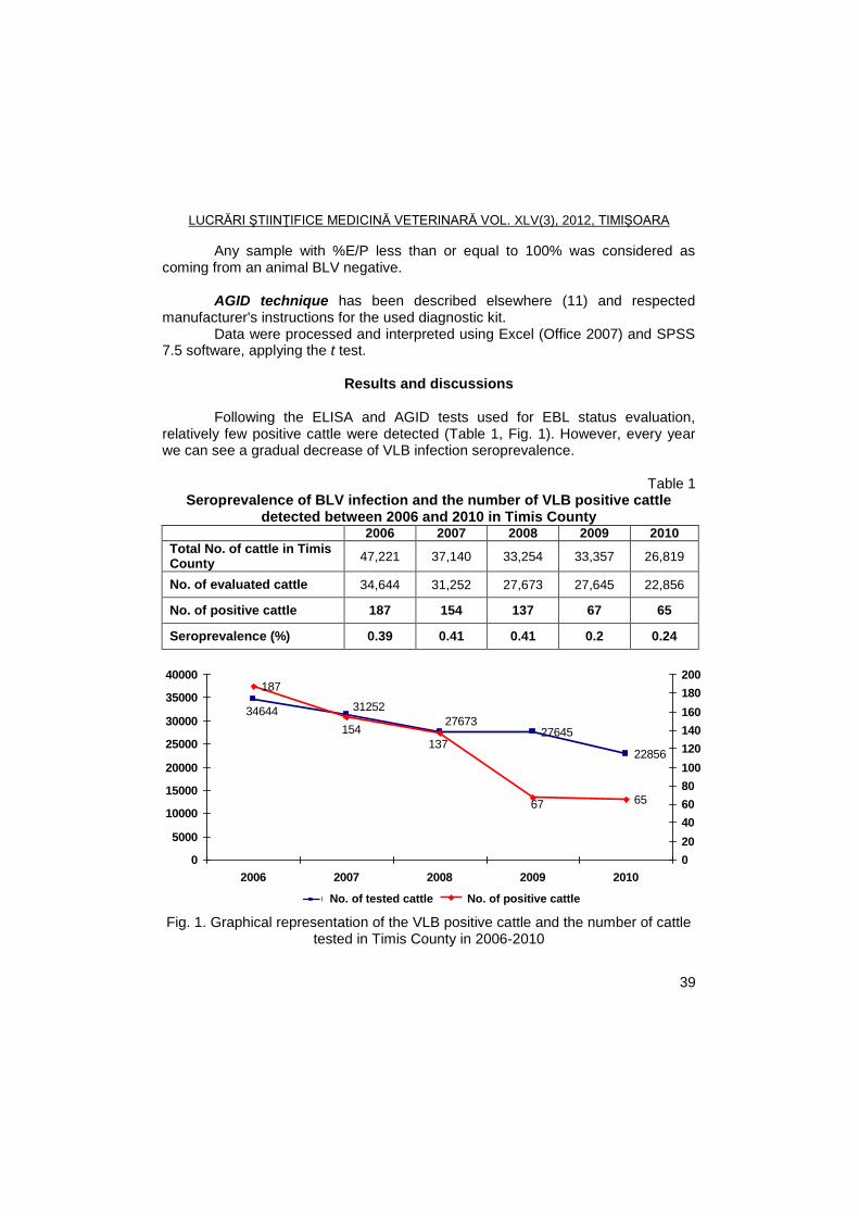
LUCRĂRI ŞTIINŢIFICE MEDICINĂ VETERINARĂ VOL. XLV(3), 2012, TIMIŞOARA
39
Any sample with %E/P less than or equal to 100% was considered as coming from an animal BLV negative.
AGID technique has been described elsewhere (11) and respected
manufacturer's instructions for the used diagnostic kit. Data were processed and interpreted using Excel (Office 2007) and SPSS
7.5 software, applying the t test.
Results and discussions Following the ELISA and AGID tests used for EBL status evaluation,
relatively few positive cattle were detected (Table 1, Fig. 1). However, every year we can see a gradual decrease of VLB infection seroprevalence.
Table 1
Seroprevalence of BLV infection and the number of VLB positive cattle detected between 2006 and 2010 in Timis County
2006 2007 2008 2009 2010
Total No. of cattle in Timis County
47,221 37,140 33,254 33,357 26,819
No. of evaluated cattle 34,644 31,252 27,673 27,645 22,856
No. of positive cattle 187 154 137 67 65
Seroprevalence (%) 0.39 0.41 0.41 0.2 0.24
27645
22856
2767334644 31252
187
6567
137
154
0
5000
10000
15000
20000
25000
30000
35000
40000
2006 2007 2008 2009 2010
0
20
40
60
80
100
120
140
160
180
200
Bovine controlate Bovine pozitive
27645
22856
2767334644 31252
187
6567
137
154
0
5000
10000
15000
20000
25000
30000
35000
40000
2006 2007 2008 2009 2010
0
20
40
60
80
100
120
140
160
180
200
Bovine controlate Bovine pozitive No. of tested cattle
27645
22856
2767334644 31252
187
6567
137
154
0
5000
10000
15000
20000
25000
30000
35000
40000
2006 2007 2008 2009 2010
0
20
40
60
80
100
120
140
160
180
200
Bovine controlate Bovine pozitive No. of positive cattle
Fig. 1. Graphical representation of the VLB positive cattle and the number of cattle tested in Timis County in 2006-2010
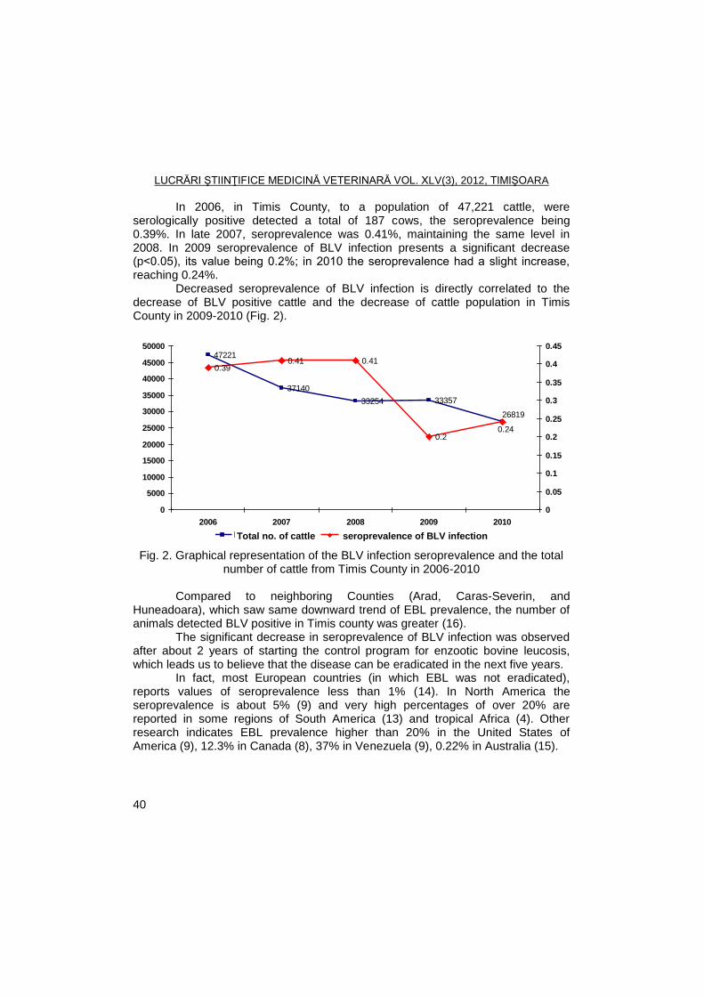
LUCRĂRI ŞTIINŢIFICE MEDICINĂ VETERINARĂ VOL. XLV(3), 2012, TIMIŞOARA
40
In 2006, in Timis County, to a population of 47,221 cattle, were serologically positive detected a total of 187 cows, the seroprevalence being 0.39%. In late 2007, seroprevalence was 0.41%, maintaining the same level in 2008. In 2009 seroprevalence of BLV infection presents a significant decrease (p˂0.05), its value being 0.2%; in 2010 the seroprevalence had a slight increase, reaching 0.24%.
Decreased seroprevalence of BLV infection is directly correlated to the decrease of BLV positive cattle and the decrease of cattle population in Timis County in 2009-2010 (Fig. 2).
26819
33254
47221
37140
33357
0.390.41 0.41
0.20.24
0
5000
10000
15000
20000
25000
30000
35000
40000
45000
50000
2006 2007 2008 2009 2010
nr.
cap
ete
0
0.05
0.1
0.15
0.2
0.25
0.3
0.35
0.4
0.45
% i
nfe
cţi
e
Total bovine %infecţie
27645
22856
2767334644 31252
187
6567
137
154
0
5000
10000
15000
20000
25000
30000
35000
40000
2006 2007 2008 2009 2010
0
20
40
60
80
100
120
140
160
180
200
Bovine controlate Bovine pozitive Total no. of cattle
27645
22856
2767334644 31252
187
6567
137
154
0
5000
10000
15000
20000
25000
30000
35000
40000
2006 2007 2008 2009 2010
0
20
40
60
80
100
120
140
160
180
200
Bovine controlate Bovine pozitive seroprevalence of BLV infection
Fig. 2. Graphical representation of the BLV infection seroprevalence and the total number of cattle from Timis County in 2006-2010
Compared to neighboring Counties (Arad, Caras-Severin, and
Huneadoara), which saw same downward trend of EBL prevalence, the number of animals detected BLV positive in Timis county was greater (16).
The significant decrease in seroprevalence of BLV infection was observed after about 2 years of starting the control program for enzootic bovine leucosis, which leads us to believe that the disease can be eradicated in the next five years.
In fact, most European countries (in which EBL was not eradicated), reports values of seroprevalence less than 1% (14). In North America the seroprevalence is about 5% (9) and very high percentages of over 20% are reported in some regions of South America (13) and tropical Africa (4). Other research indicates EBL prevalence higher than 20% in the United States of America (9), 12.3% in Canada (8), 37% in Venezuela (9), 0.22% in Australia (15).
LUCRĂRI ŞTIINŢIFICE MEDICINĂ VETERINARĂ VOL. XLV(3), 2012, TIMIŞOARA
41
Conclusions
During the five years of study, the seroprevalence of BLV infection decreased to about half the value initially recorded which shows that control measures applied are effective.
ELB seroprevalence downward trend shows that the disease can be eradicated in Romania in the near future.
Acknowledgments
This work was supported by the grant POSDRU/21/1.5/G/38347.
References
1. Abd El-Haifez, Y.G.M., Metias, K.N., Ibrahim, I.G.A., Comparative serological detection of enzootic bovine leucosis virus (EBLV) in cattle sera, Global Veterinaria, 2010, 4, 3, 267-270.
2. Acaite, J., Tamosiunas, V., Lukauskas, K., Milius, J., Pieskus, J., The eradication experience of enzootic bovine leukosis from Lithuania, Preventive Veterinary Medicine, 2007, 82, 83-89.
3. De Giuseppe, A., Feliziani, F., Rutili, D., De Mia, G.M., Expression of the Bovine Leukemia Virus Envelope Glycoprotein (gp51) by Recombinant Baculovirus and Its Use in an ELISA, Clin Diagn Lab Immunol., 2004, 11, 147.
4. Hesterberg, Uta, A serological prevalence study of important infectious diseases of cattle in rural areas of Kwa Zulu Natal, South Africa, Thesis, Faculty of Veterinary Science, University of Pretoria, 2007.
5. Monti, G.E., Frankena, K., De Jong, M.C.M., Evaluation of natural transmission of bovine leukaemia virus within dairy herds of Argentina, Epidemiol. Infect., 2007, 135, 228-237.
6. Murakami, K., Kobayashi, S., Konishi, M., Kameyama, K., Yamamoto, T., Tsutsui, T., The recent prevalence of bovine leukemia virus (BLV) infection among Japanese cattle, Veterinary Microbiology, 2011, 148, 1, 80-84.
7. Murphy, F.A., Gibbs, E.P., Horzinek, Marian C., Studdert, M.J., Veterinary Virology, 3
rd Edition, Academic Press, San Diego, California, 1999.
8. Olaloku, O.A., The prevalence of antibodies to bovine leukemia virus, neospora caninum and risk factors, and biosecurity practices in beef cow-calf herds in Canada, Thesis, Western College of Veterinary Medicine, University of Saskatchewan, 2010.
9. Radostits, O.M., Gay, C.C., Hinchcliff, K.W., Constable, P.D., Veterinary Medicine: A Textbook of the Diseases of Cattle, Sheep, Pigs and Goats, Saunders Elsevier Press, London, 2007.
LUCRĂRI ŞTIINŢIFICE MEDICINĂ VETERINARĂ VOL. XLV(3), 2012, TIMIŞOARA
42
10. Rukkwamsuk, Theera, Rungruang, S., Seroprevalence of Bovine Leukemia Virus (BLV) Infection in Pregnant Replacement Dairy Heifers in Saraburi Province, Thailand, Kasetsart J. (Nat. Sci.), 2008, 42 , 278-281.
11. Tîrziu, E., Trif., R., Vior, C., Cumpanasoiu, C., Metode de diagnostic imunologic, Ed. Waldpress, Timisoara, 2002.
12. Trainin, Z., Brenner, J., The direct and indirect economic impacts of bovine leukemia virus infection on dairy cattle, Israel Journal of Veterinary Medicine, 2005, 60, 4, 121-130.
13. Trono, K.G., Perez-Filgueira, D.M., Duffy, S., Broca, M.V., Seroprevalence of bovine leukemia virus in dairy cattle in Argentina: comparison of sensitivity and specificity of different detection methods, Vet. Microbiol., 2001, 83, 253-248.
14. ***Enzootic Bovine Leukosis, In: OIE Terrestrial Manual 2008, vol. 2, pp. 729-738, http://www.oie.int/eng/normes/mmanual/2008/pdf/2.04.11_EBL.pdf (10.03.2012).
15. ***Enzootic Bovine Leukosis, Australian Government, Department of Agriculture, Fisheries and Forestry Biosecurity, http://www.daff.gov.au/animal-plant-health/pests-diseases-weeds/animal/ebl (10.03.2012).
16. ***Rapoarte de activitate DSVSA, http://www.ansvsa.ro/?pag=759 (10.03.2012).
LUCRĂRI ŞTIINŢIFICE MEDICINĂ VETERINARĂ VOL. XLV(3), 2012, TIMIŞOARA
43
PREVALENCE AND ANTIMICROBIAL SUSCEPTIBILITY OF STAPHYLOCOCCI ISOLATED FROM OTITIS EXTERNA OF CATS
J. DÉGI
1, R.T.CRISTINA
2, IONICA FODOR
1, V. HERMAN
1, N. CĂTANĂ
1
1 Banat’s University of Agricultural Science and Veterinary MedicineTimisoara
Department of Infectious Diseases and Preventive Medicine, Faculty of Veterinary Medicine, 300645, 119 Calea Aradului Street, Timişoara, Romania
2Banat’s University of Agricultural Science and Veterinary MedicineTimisoara,
Faculty of Veterinary Medicine, Department of Pharmacology E-mail: [email protected]
Summary
Were collected 45 samples from adult cats with clinical otitis externa, processed
from isolation Staphylococcus species and tested for susceptibility for ciprofloxacin, pristinamicin, ampicillin / sulbactan, gentamicin, kanamycin, novobiocin, methicillin, vancomycin, ceftriaxon, cefoxitin, rifampicin, lincomycin, cefaclor, amoxicillin / clavulanic acid, eritromycin, polymyxin B, tetracyclyn and doxycyclin. Was also determined methicilin resistance. Processed samples were isolated from thirty-two strains of staphylococci (71.11% growth positivity). Coagulase-positive staphylococci were obtained from 21 cats, and the species recovered was Staphylococcus Intermedius Group (Staphylococcus intermedius), S. aureus and S. hycus. Were isolated coagulase-negative staphylococci also like S. felis, S. haemolyticus and S. epidermidis. Resistance of antibiotics was frequently observed, with 71.875% of the isolates showing resistance to at least one drug. Resistance were observed to polymyxin B, in 23 in the 32 isolated (71.875%), 20 to doxycycline (62.5%), 15 to erythromycin (46.87%), 8 to amoxicillin / clavulanic acid (25%) and 5 to methicillin (15.62%). Most active antimicrobial substances were ciprofloxacin, ampicillin / sulbactan and cephalosporin group. These microorganisms have an important role in the pathogenesis of ear infections in cats.
Key words: staphylococci, antimicrobials, otitis externa, cat
In cats, Staphylococci are commonly and opportunistic bacteria, associated
with skin infection and otitis. Coagulase-positive and coagulase negative Staphylococci are found in cats, but although both groups have been isolated from cats with skin lesions and otitis, reports of pathogenic coagulase-negative staphylococci in cats remains rare (1).
S. intermedius and S. aureus are important pathogens of cats. S. intermedius causes bacterial skin infections, otitis externa and others diseases. S. intermedius are now classified as the Staphylococcus intermedius Group (SIG), which contains three genetically distinct species; S. intermedius, S. pseudointermedius and S. delphinii, each of which occupy distinct ecological niches. It is currently though that S. pseudointermedius is the primary feline pathogen in this group (2). Currently, all staphylococcus species that infect cats exhibit, some degree of antimicrobial resistance. Cross resistance to classes of
LUCRĂRI ŞTIINŢIFICE MEDICINĂ VETERINARĂ VOL. XLV(3), 2012, TIMIŞOARA
44
antimicrobials were common. It has been shown that methicillin resistance in this species may express coresistance to any combination of other drug classes, including aminoglycosides, flouroquinolone and macrolides (3).
Materials and methods
Cats
Samples were obtained from 45 unmedicated adult cats of both sex wich were presented otitis externa, judged following a detailed physical examination and otoscopy. Animals with recent history of antimicrobial agents or systemic drug administration, exclude from the study. Specimens were collected from trhe cats in University Veterinary clinics of the Faculty of Veterinary Medicine of Timisoara and in their owners home.
Samples collecting and processing
For collection ear exudate from the ear canal used a sterile cotton swabs and inoculated immediately if this possibility in 5% Sheep Blood agar broth and incubated at 37ºC or within 24 hours, keeping the sample in a refrigerator. If growth occurred after 24 or 48 h, smear were made, Gram stained and examined microscopically for microorganism. Samples with morphologies compatible with Staphylococcus spp. were transferred to Mannitol Salt agar.
Identification of isolates
After growth, staphylococci were identified on the basis of colony characteristics, Gram stain, pigment production, hemolysis on 5% Sheep Blood agar, and the following biochemical reactions using for this purpose API Staph multitest system.
Isolates of other bacterial genera were not considerated in this study.
Susceptibility test All the staphylococci strains were tested for susceptibility to antimicrobial
agents by Kirby Bauer agar disc difussion method on Muller Hinton agar, incubated at 37ºC. Discs of methicillin (30μg), gentamicin (10μg), tetracycline (30μg), ciprofloxacin (30μg), kanamycin (30μg), novobiocin (30μg), doxycycline (30μg), erythromycin - 15 mg, vancomycin - 30 mg, ceftriaxone - 30 mg, cefoxotin - 10 mg, polymyxin B (50UI), rifampicin (30μg), lincomycin (30μg), cefaclor (30μg), pristamicină (15μg), ampicillin / sulbactan (30μg) and amoxicillin / clavulanic acid (30μg), were tested. Interpretation of antibiotic sensitivity test was performed by measuring the growth inhibition zone diameter, in accordance with standards provided by the company producing the disc (Bioanalyse ®).
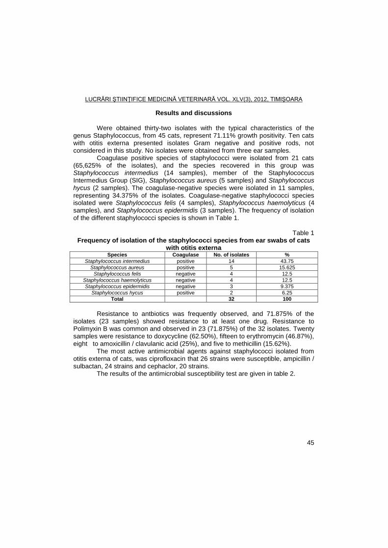
LUCRĂRI ŞTIINŢIFICE MEDICINĂ VETERINARĂ VOL. XLV(3), 2012, TIMIŞOARA
45
Results and discussions
Were obtained thirty-two isolates with the typical characteristics of the genus Staphylococcus, from 45 cats, represent 71.11% growth positivity. Ten cats with otitis externa presented isolates Gram negative and positive rods, not considered in this study. No isolates were obtained from three ear samples.
Coagulase positive species of staphylococci were isolated from 21 cats (65,625% of the isolates), and the species recovered in this group was Staphylococcus intermedius (14 samples), member of the Staphylococcus Intermedius Group (SIG), Staphylococcus aureus (5 samples) and Staphylococcus hycus (2 samples). The coagulase-negative species were isolated in 11 samples, representing 34.375% of the isolates. Coagulase-negative staphylococci species isolated were Staphylococcus felis (4 samples), Staphylococcus haemolyticus (4 samples), and Staphylococcus epidermidis (3 samples). The frequency of isolation of the different staphylococci species is shown in Table 1.
Table 1
Frequency of isolation of the staphylococci species from ear swabs of cats with otitis externa
Species Coagulase No. of isolates %
Staphylococcus intermedius positive 14 43.75
Staphylococcus aureus positive 5 15.625
Staphylococcus felis negative 4 12.5
Staphylococcus haemolyticus negative 4 12.5
Staphylococcus epidermidis negative 3 9.375
Staphylococcus hycus positive 2 6.25
Total 32 100
Resistance to antbiotics was frequently observed, and 71.875% of the
isolates (23 samples) showed resistance to at least one drug. Resistance to Polimyxin B was common and observed in 23 (71.875%) of the 32 isolates. Twenty samples were resistance to doxycycline (62.50%), fifteen to erythromycin (46.87%), eight to amoxicillin / clavulanic acid (25%), and five to methicillin (15.62%).
The most active antimicrobial agents against staphylococci isolated from otitis externa of cats, was ciprofloxacin that 26 strains were susceptible, ampicillin / sulbactan, 24 strains and cephaclor, 20 strains.
The results of the antimicrobial susceptibility test are given in table 2.
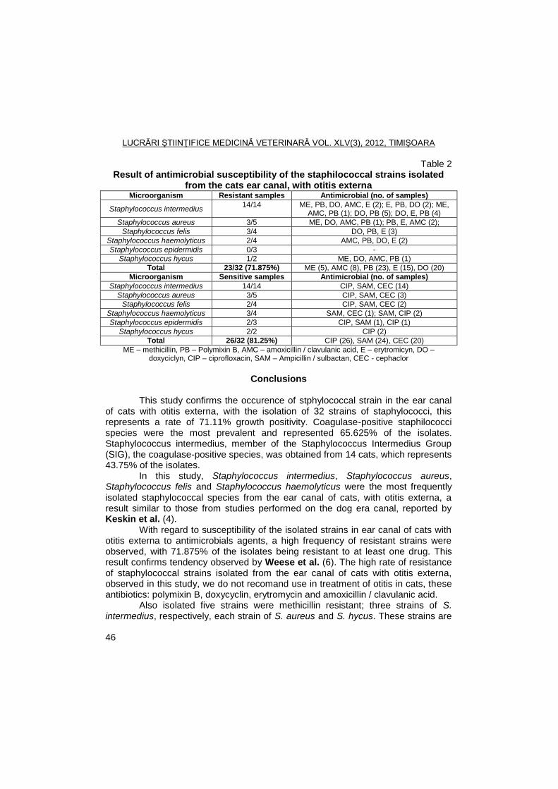
LUCRĂRI ŞTIINŢIFICE MEDICINĂ VETERINARĂ VOL. XLV(3), 2012, TIMIŞOARA
46
Table 2 Result of antimicrobial susceptibility of the staphilococcal strains isolated
from the cats ear canal, with otitis externa Microorganism Resistant samples Antimicrobial (no. of samples)
Staphylococcus intermedius 14/14 ME, PB, DO, AMC, E (2); E, PB, DO (2); ME,
AMC, PB (1); DO, PB (5); DO, E, PB (4)
Staphylococcus aureus 3/5 ME, DO, AMC, PB (1); PB, E, AMC (2);
Staphylococcus felis 3/4 DO, PB, E (3)
Staphylococcus haemolyticus 2/4 AMC, PB, DO, E (2)
Staphylococcus epidermidis 0/3 -
Staphylococcus hycus 1/2 ME, DO, AMC, PB (1)
Total 23/32 (71.875%) ME (5), AMC (8), PB (23), E (15), DO (20)
Microorganism Sensitive samples Antimicrobial (no. of samples)
Staphylococcus intermedius 14/14 CIP, SAM, CEC (14)
Staphylococcus aureus 3/5 CIP, SAM, CEC (3)
Staphylococcus felis 2/4 CIP, SAM, CEC (2)
Staphylococcus haemolyticus 3/4 SAM, CEC (1); SAM, CIP (2)
Staphylococcus epidermidis 2/3 CIP, SAM (1), CIP (1)
Staphylococcus hycus 2/2 CIP (2)
Total 26/32 (81.25%) CIP (26), SAM (24), CEC (20)
ME – methicillin, PB – Polymixin B, AMC – amoxicillin / clavulanic acid, E – erytromicyn, DO – doxyciclyn, CIP – ciprofloxacin, SAM – Ampicillin / sulbactan, CEC - cephaclor
Conclusions
This study confirms the occurence of stphylococcal strain in the ear canal
of cats with otitis externa, with the isolation of 32 strains of staphylococci, this represents a rate of 71.11% growth positivity. Coagulase-positive staphilococci species were the most prevalent and represented 65.625% of the isolates. Staphylococcus intermedius, member of the Staphylococcus Intermedius Group (SIG), the coagulase-positive species, was obtained from 14 cats, which represents 43.75% of the isolates.
In this study, Staphylococcus intermedius, Staphylococcus aureus, Staphylococcus felis and Staphylococcus haemolyticus were the most frequently isolated staphylococcal species from the ear canal of cats, with otitis externa, a result similar to those from studies performed on the dog era canal, reported by Keskin et al. (4).
With regard to susceptibility of the isolated strains in ear canal of cats with otitis externa to antimicrobials agents, a high frequency of resistant strains were observed, with 71.875% of the isolates being resistant to at least one drug. This result confirms tendency observed by Weese et al. (6). The high rate of resistance of staphylococcal strains isolated from the ear canal of cats with otitis externa, observed in this study, we do not recomand use in treatment of otitis in cats, these antibiotics: polymixin B, doxycyclin, erytromycin and amoxicillin / clavulanic acid.
Also isolated five strains were methicillin resistant; three strains of S. intermedius, respectively, each strain of S. aureus and S. hycus. These strains are
LUCRĂRI ŞTIINŢIFICE MEDICINĂ VETERINARĂ VOL. XLV(3), 2012, TIMIŞOARA
47
considered zoonotic risk, is very important since human contact ever more closely with these pets.
The most active antibiotic agents against staphylococci isolated from ear canal of cats with otitis externa, was ciprofloxacin, ampicillin / sulbactan and cephaclor. This finding agreement with others studies (1, 4, 5, 6) and was also observed with staphylococci isolated from the skin of cats (1).
As staphylococcal strains from the ear canal of the cats with otitis externa may develop an important role in pathogenesis of otitis in cats.
Also by determining sensitivity to antibiotics, we ensure a more accurate treatment of this disease in cats.
Acknowledgements
This work was published during the project "POSTDOCTORAL SCHOOL
OF AGRICULTURE AND VETERINARY MEDICINE", POSDRU/89/1.5/S/62371, co-financed by the European Social Fund through the Sectorial Operational Programme for the Human Resources Development 2007-2013.
References
1. Abraham, J.L., Morris, D.O., Griffeth, G.C., Shofer, F.S., Rankin, Sh.C.,
Surveillance of healthy cats and cats with inflammatory skin disease for colonization of the skin by methicillin-resistant coagulase-positive staphylococci and Staphylococcus schleiferi ssp. schleiferi, Veterinary Dermatology, 2007, 18(4), 252-259.
2. Bannoehr, Jeanette, Zakour Ben, N.L., Waller, A.S., Guardabassi, L., Thoday, K.L., Van den Broek, A.H.M., Fitzgerald, J.R., Population Genetic Structure of the Staphylococcus intermedius Group: Insights into Diversification and the Emergence of Methicillin-Resistant Strains, Journal of Bacteriology, 2007, 189(23), 8685-8692.
3. Berger-Bachi, Brigitte, Rohner, Susane, Factors influencing methicillin resistance in staphylococci, Archives of Microbiology, 2002, 178(3), 165-171.
4. Keskin, O., Tel, O.Y., Kaya Arserim, N.B., Aerobic Bacteria and Fungi Isolated from External Ear Canal of Healthy Dogs and the Antibiotic Susceptibility of Staphylococci, Journal of Animal and Veterinary Advances, 2010, 9(3), 496-500.
5. Linek, Monika, Otitis externa and media in the dog and cat, Tierarztl Prax Ausg K Kleintiere Heimtiere, 2011, 39(6), 451-63.
6. Weese, J.S., Antimicrobial resistance: time for action, Veterinary Record, 2011, 169(5), 122-130.
LUCRĂRI ŞTIINŢIFICE MEDICINĂ VETERINARĂ VOL. XLV(3), 2012, TIMIŞOARA
48
RESEARCH ON THE FREQUENCY OF ISOLATION STRAINS OF APEC IN BROILERS
IONICA FODOR, IOANA GROZA, NICOLAE CĂTANĂ
Banat's University of Agricultural Sciences and Veterinary Medicine Timisoara, Faculty
of Veterinary Medicine, Department of Infectious Diseases and Preventive Medicine,300645, Aradului Street No. 119, Timisoara, Romania
E-mail: [email protected]
Summary
In the poultry industry, avian colibacillosis is considered a major bacterial disease,
one of the most common diseases of chickens and some strains are with zoonotic risk. This study was carried out over several years to determine the frequency of APEC strains isolated from broilers with colibacillosis in the Western side of Romania. Were autopsied 3276 cadavers with colibacillosis lesions and were isolated a total of 203 strains of E. coli and 60 were isolated from the air. Following PCR examination established that, of the 108 strains tested, 84 strains (77.7%) were employed in APEC pathotype having at least one virulence gene and fixed Congo red.
Key words: poultry, colibacillosis, avian pathogenic Escherichia coli.
Avian colibacillosis is an infectious disease common in the poultry sector,
having a major impact on poultry production worldwide. Economic losses are caused by high mortality and decrease in weight.
E. coli strains are frequently isolated from internal organs and cause systemic colibacillosis, but strains belonging to the APEC pathotype can be isolated from respiratory mucosa of broilers and growing halls.
Frequent respiratory virosis in broilers can be complicated with infections caused by APEC strains increasing the poultry industry losses.
Some APEC strains are able to fix Congo red, an acid dye. This feature of APEC strains is used as an epidemiological marker in the study of these strains.
Materials and methods
The investigations were carried out broiler farms in western side of Romania, studied herds ranged from 8000 to 14,450 chickens per house.
Cadavers were autopsied by the classical method on the farm and in the Bacteriology Laboratory of Infectious Diseases Department of the Faculty of Veterinary Medicine Timisoara. Since 3276 a number of bodies of broilers of different ages were sampled from the internal organs, long bone, brain, and in the air.
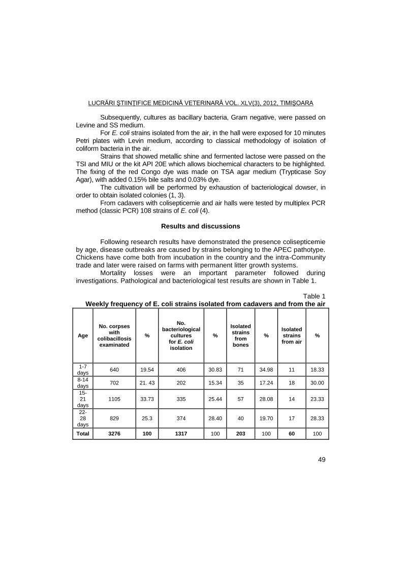
LUCRĂRI ŞTIINŢIFICE MEDICINĂ VETERINARĂ VOL. XLV(3), 2012, TIMIŞOARA
49
Subsequently, cultures as bacillary bacteria, Gram negative, were passed on Levine and SS medium.
For E. coli strains isolated from the air, in the hall were exposed for 10 minutes Petri plates with Levin medium, according to classical methodology of isolation of coliform bacteria in the air.
Strains that showed metallic shine and fermented lactose were passed on the TSI and MIU or the kit API 20E which allows biochemical characters to be highlighted. The fixing of the red Congo dye was made on TSA agar medium (Trypticase Soy Agar), with added 0.15% bile salts and 0.03% dye.
The cultivation will be performed by exhaustion of bacteriological dowser, in order to obtain isolated colonies (1, 3).
From cadavers with colisepticemie and air halls were tested by multiplex PCR method (classic PCR) 108 strains of E. coli (4).
Results and discussions
Following research results have demonstrated the presence colisepticemie
by age, disease outbreaks are caused by strains belonging to the APEC pathotype. Chickens have come both from incubation in the country and the intra-Community trade and later were raised on farms with permanent litter growth systems.
Mortality losses were an important parameter followed during investigations. Pathological and bacteriological test results are shown in Table 1.
Table 1 Weekly frequency of E. coli strains isolated from cadavers and from the air
Age
No. corpses with
colibacillosis examinated
%
No. bacteriological
cultures for E. coli isolation
%
Isolated strains from
bones
% Isolated strains from air
%
1-7 days
640 19.54 406 30.83 71 34.98 11 18.33
8-14 days
702 21. 43 202 15.34 35 17.24 18 30.00
15-21
days 1105 33.73 335 25.44 57 28.08 14 23.33
22-28
days 829 25.3 374 28.40 40 19.70 17 28.33
Total 3276 100 1317 100 203 100 60 100
LUCRĂRI ŞTIINŢIFICE MEDICINĂ VETERINARĂ VOL. XLV(3), 2012, TIMIŞOARA
50
Following the weekly mortality is observed that the largest number of cadavers have been between 15-21 days, respectively in 1105 cadavers (33.73%), were isolated from a total of 57 strains of E. coli.
It also notes that the greatest number of E. coli strains were isolated from broilers in the first week of life (71 strains).
The air were a total of 60 isolated strains of E. coli, of which several strains were isolated in the second week of life (18 strains) and the last week of life (17 strains), and less E. coli strains were isolated in the first week of life (11 strains).
Bacterial provided the following results: - E. coli cultures were obtained from bacteriologycal cultivation
made by the common methodology in nutritive broth and agar; - Smears made from these cultures revealed the presence of Gram
negative coccobacili; - The SS medium, isolates were lactose positive, forming red
colonies, while the Levine medium, colonies were blackish and showed metallic shine, characteristic for strains of E. coli;
- E. coli strains isolated from broilers with colisepticemie and from air had properties biochemical characteristic of APEC strains;
- On the red Congo agar the colonies were identified by their dark red color and dry, wrinkled appearence. The results are similar to the bacteriological examination of the literature,
indicating that E. coli strains isolated from outbreaks of colisepticemie ferment lactose on culture media, this biochemical feature is constant in APEC strains (1, 3).
Following PCR examination established that, of the 108 strains tested 84 strains (77.7%) were employed in APEC pathotype having at least one virulence gene.
It also confirm that isolates from the air were identical to those isolated from cadavers, at 98%. Framing isolates the APEC pathotype was based on fixation of Congo red on the detection of virulence genes (iss, fim H, omp A).
Research conducted shows that, in broilers, APEC strains can be transmitted through direct contact or indirectly through various sources of shelter, as confirmed by the identification of identical APEC strains from both the air and from the chicks with colisepticemie.
Increasing the number of germs in the shelter above the permissible limits, has led to increased mortality and the number of strains of E. coli isolated, these data are according with the literature (1, 2, 5).
The large number of E. coli strains isolated from air in the second week of life, then led to increased mortality in the third week of life and that the increase of APEC strains isolated from outbreaks with colisepticemie.
LUCRĂRI ŞTIINŢIFICE MEDICINĂ VETERINARĂ VOL. XLV(3), 2012, TIMIŞOARA
51
Conclusions
The highest number of dead was in the third week of life (1105 dead) and the lowest number of dead was in the first week of life (640 dead).
The highest number of E. coli strains were isolated from cadavers in the first week of life (71 strains) and the lowest number recorded in the second week of life (35 strains).
From the air, the large number of strains were isolated in the second week of life (18 strains), and fewest were isolated first week of life (11 strains).
E. coli strains isolated had biochemical properties characteristic of APEC. PCR examination established that 84 strains (77.7%) of the 108 tested had
at least one virulence gene. Strains isolated from the air were identical to those isolated from cadavers,
at 98%
Acknowledgments
This study has been financed by UEFISCU - Human Resources Program Projects Doctoral Research - PD 111/28.07.2010.
References
1. Barnes, H. J., Lisa K. Nolan, Vaillantcourt, J.P. Colibacillosis, in Diseases of Poultry, 12 th edition, editor in Chief, Saif, Y.M., Blackwell Publishing, Ames, Iowa, 2008, 691-732.
2. Decun, M., Nichita, Ileana, Țibru, I., Lăzărescu, C., Îndrumător practic pentru igiena animalelor și a mediului, Ed. Mirton, Timișoara, România, 2005.
3. Ewers, Christa, Traute, Ja., Wieler, L.H. Aviare pathogene Escherichia coli (APEQ), Munch. Tierarztl. Wschr., 2003, 116, 381-395.
4. Popa, Virgilia, Stanica, Andreea, Botus, Daniela, Mihailescu, Ramona, Iss, ompA and fimH genes detection by multiplex PCR in Escherichia coli of avian origin strains, Scientific Papers Veterinary Medicine Timisoara, 2004, 37, 522-528.
5. Zanella, A., Alborali, G.L., Bardotti, M., Candotti, P., Guadagnini, P.F., Martino, P.A., Stonfer, M., Severe Escherichia coli O111 septicaemia and polyserositis in hens at start of lay, Avain Pathol., 2000, 29, 311-317.
LUCRĂRI ŞTIINŢIFICE MEDICINĂ VETERINARĂ VOL. XLV(3), 2012, TIMIŞOARA
52
RABIES CASES IN ROMANIA DURING 2001 AND 2010
V. HERMAN, N. CĂTANĂ, CORINA PASCU
Banat's University of Agricultural Sciences and Veterinary Medicine Timisoara, Faculty of Veterinary Medicine, 300645, Aradului Street No. 119, Timisoara, Romania
E-mail: [email protected]
Summary
In the paper are presented in tables, the data of rabies development in Romania
comparatively with Europe, between 2001 and 2010. In Romania the number of rabies cases was decrease in 2001 and 2003, but in the
next years the number of cases was significant increased. In Europe the rabies cases were decrease from 10,208 in 2001, to 7,589 in 2010.
Increasing cases of rabies in Romania and Europe has occurred due to increasing cases of rabies in wild animals, in foxes particularly.
Key words: rabies, Romania, Europe, wild animals Rabies continues to be a severe infectious disease that is important due
looses, in animal and human population, too. This paper wants to be an alarm signal about presence of rabies in
Romania, at high share, yet, which means high vigilance from both veterinarians and physicians.
Materials and methods
In the paper are presented and comparatively analyzed the epidemiological data about rabies evolution during 2001-2010 in Romania and epidemiological situation of rabies in whole Europe in the same period (1, 2, 3, 5).
Data from this paper were taken over Internet address ―http://www.who-rabies-bulletin.org/Queries/Default.aspx‖ (5). The primary data were remaking and interpretation the paper contain those information that are suggestive and important for realize the purpose (2, 3, 4).
Results and discussions
In table 1 are present the rabies cases register in Romania during 2001-2010 and the distribution of affected species.
It’s observe the total number of rabies cases was decrease, year by year in the first three years and the total number of rabies cases was reduced at 115 in 2002 and at 95 in 2003. The total number of rabies cases was increase at 187 in 2004 and at 530, in 2005. In 2006, the total number of rabies cases was 293, but in
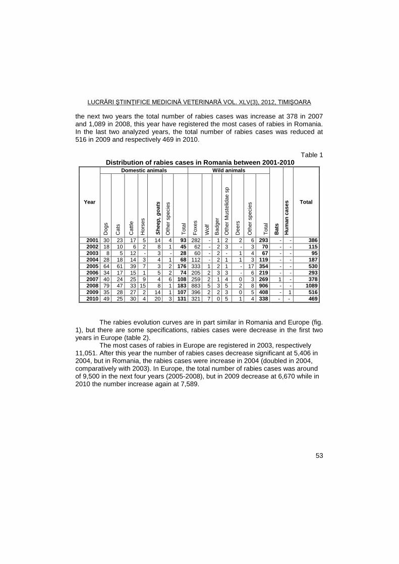
LUCRĂRI ŞTIINŢIFICE MEDICINĂ VETERINARĂ VOL. XLV(3), 2012, TIMIŞOARA
53
the next two years the total number of rabies cases was increase at 378 in 2007 and 1,089 in 2008, this year have registered the most cases of rabies in Romania. In the last two analyzed years, the total number of rabies cases was reduced at 516 in 2009 and respectively 469 in 2010.
Table 1
Distribution of rabies cases in Romania between 2001-2010
Year
Domestic animals Wild animals
Bats
Hu
man
cases
Total
Dogs
Cats
Catt
le
Hors
es
Sh
eep
, g
oa
ts
Oth
er
specie
s
To
tal
Fo
xes
Wolf
Badger
Oth
er
Muste
lidae s
p
Deers
Oth
er
specie
s
To
tal
2001 30 23 17 5 14 4 93 282 - 1 2 2 6 293 - - 386
2002 18 10 6 2 8 1 45 62 - 2 3 - 3 70 - - 115
2003 8 5 12 - 3 - 28 60 - 2 - 1 4 67 - - 95
2004 28 18 14 3 4 1 68 112 - 2 1 1 3 119 - - 187
2005 64 61 39 7 3 2 176 333 1 2 1 - 17 354 - - 530
2006 34 17 15 1 5 2 74 205 2 3 3 - 6 219 - - 293
2007 40 24 25 9 4 6 108 259 2 1 4 0 3 269 1 - 378
2008 79 47 33 15 8 1 183 883 5 3 5 2 8 906 - - 1089
2009 35 28 27 2 14 1 107 396 2 2 3 0 5 408 - 1 516
2010 49 25 30 4 20 3 131 321 7 0 5 1 4 338 - - 469
The rabies evolution curves are in part similar in Romania and Europe (fig. 1), but there are some specifications, rabies cases were decrease in the first two years in Europe (table 2).
The most cases of rabies in Europe are registered in 2003, respectively 11,051. After this year the number of rabies cases decrease significant at 5,406 in 2004, but in Romania, the rabies cases were increase in 2004 (doubled in 2004, comparatively with 2003). In Europe, the total number of rabies cases was around of 9,500 in the next four years (2005-2008), but in 2009 decrease at 6,670 while in 2010 the number increase again at 7,589.
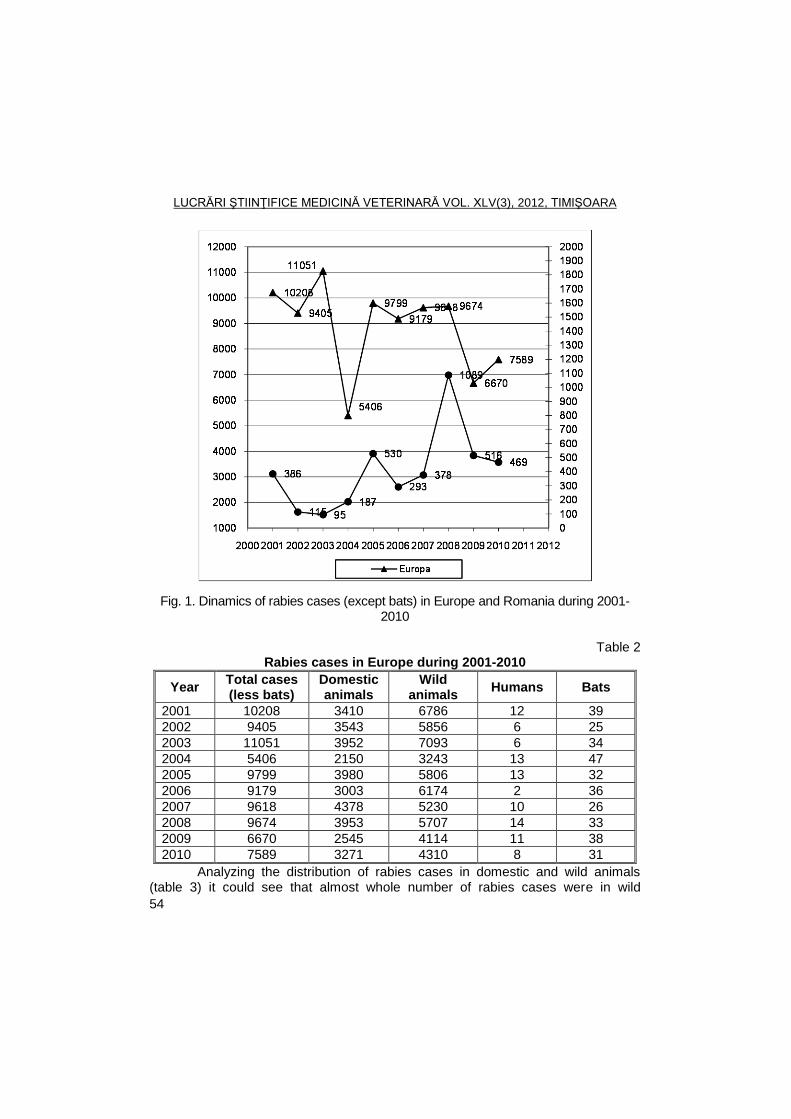
LUCRĂRI ŞTIINŢIFICE MEDICINĂ VETERINARĂ VOL. XLV(3), 2012, TIMIŞOARA
54
Fig. 1. Dinamics of rabies cases (except bats) in Europe and Romania during 2001-2010
Table 2
Rabies cases in Europe during 2001-2010
Year Total cases (less bats)
Domestic animals
Wild animals
Humans Bats
2001 10208 3410 6786 12 39
2002 9405 3543 5856 6 25
2003 11051 3952 7093 6 34
2004 5406 2150 3243 13 47
2005 9799 3980 5806 13 32
2006 9179 3003 6174 2 36
2007 9618 4378 5230 10 26
2008 9674 3953 5707 14 33
2009 6670 2545 4114 11 38
2010 7589 3271 4310 8 31
Analyzing the distribution of rabies cases in domestic and wild animals (table 3) it could see that almost whole number of rabies cases were in wild
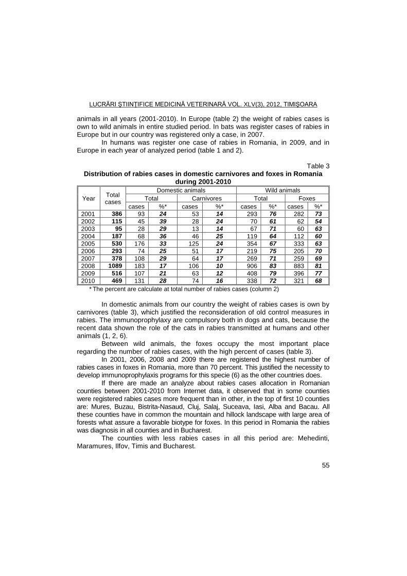
LUCRĂRI ŞTIINŢIFICE MEDICINĂ VETERINARĂ VOL. XLV(3), 2012, TIMIŞOARA
55
animals in all years (2001-2010). In Europe (table 2) the weight of rabies cases is own to wild animals in entire studied period. In bats was register cases of rabies in Europe but in our country was registered only a case, in 2007.
In humans was register one case of rabies in Romania, in 2009, and in Europe in each year of analyzed period (table 1 and 2).
Table 3 Distribution of rabies cases in domestic carnivores and foxes in Romania
during 2001-2010
Year Total cases
Domestic animals Wild animals
Total Carnivores Total Foxes
cases %* cases %* cases %* cases %*
2001 386 93 24 53 14 293 76 282 73
2002 115 45 39 28 24 70 61 62 54
2003 95 28 29 13 14 67 71 60 63
2004 187 68 36 46 25 119 64 112 60
2005 530 176 33 125 24 354 67 333 63
2006 293 74 25 51 17 219 75 205 70
2007 378 108 29 64 17 269 71 259 69
2008 1089 183 17 106 10 906 83 883 81
2009 516 107 21 63 12 408 79 396 77
2010 469 131 28 74 16 338 72 321 68
* The percent are calculate at total number of rabies cases (column 2)
In domestic animals from our country the weight of rabies cases is own by
carnivores (table 3), which justified the reconsideration of old control measures in rabies. The immunoprophylaxy are compulsory both in dogs and cats, because the recent data shown the role of the cats in rabies transmitted at humans and other animals (1, 2, 6).
Between wild animals, the foxes occupy the most important place regarding the number of rabies cases, with the high percent of cases (table 3).
In 2001, 2006, 2008 and 2009 there are registered the highest number of rabies cases in foxes in Romania, more than 70 percent. This justified the necessity to develop immunoprophylaxis programs for this specie (6) as the other countries does.
If there are made an analyze about rabies cases allocation in Romanian counties between 2001-2010 from Internet data, it observed that in some counties were registered rabies cases more frequent than in other, in the top of first 10 counties are: Mures, Buzau, Bistrita-Nasaud, Cluj, Salaj, Suceava, Iasi, Alba and Bacau. All these counties have in common the mountain and hillock landscape with large area of forests what assure a favorable biotype for foxes. In this period in Romania the rabies was diagnosis in all counties and in Bucharest.
The counties with less rabies cases in all this period are: Mehedinti, Maramures, Ilfov, Timis and Bucharest.
LUCRĂRI ŞTIINŢIFICE MEDICINĂ VETERINARĂ VOL. XLV(3), 2012, TIMIŞOARA
56
Conclusions
The obtained results about rabies evolution in Romania during 2001-2010 allow the next conclusions:
In the studied period (2001-2010) the rabies cases were decrease in Romania in the first three years, but in the next years the numbers of cases were increased. In Europe the rabies cases were decrease from 10,208 in 2001, to 7,589 in 2010. In Europe in 2004 were recorded at fewer cases of rabies, the number of rabies cases (except bats) decrease at 5,406, the higher cases of rabies in Europe were registered in 2003.
The increase of rabies cases in Romania and Europe is due to increase of rabies cases in wild animals.
In the studied period the Romanian counties with the number of cases exceeds the 150, are Mures, Buzau, Bistrita-Nasaud, Cluj, Salaj, Suceava, Iasi, Alba and Bacau county.
In Romania, in wild animals the highest number of rabies cases was registered in foxes and in carnivores for domestic animals. Because of this, it is necessary to maintain and develop the actual program for rabies immunophrophylaxis in dogs, cats and foxes. For cats and foxes the vaccination against rabies was not put into practice systematically.
References
1. Herman, V., Aspecte epidemiologice ale evoluţiei turbării în România în perioada 1977-2001, Lucr. şt. Med. Vet. Iaşi 2002, 45, (4), p.618-623.
2. Herman, V., Cătană, N., Pascu, Corina, Văduva, Ioana, Faur, B., Evolution of rabies in Romania between 2000 and 2007, Lucr. şt. Med. Vet. Timişoara, 2009, 42 (1), p.212-215.
3. Perianu, T., Savuta, Ghe., Ghiţă, S., Burtan, E., Epidemiologie de la rage en Roumanie, Epidémiol. santé anim., 1997, 31-32, 04.A.52
4. Văduva, Ioana, Herman, V., Cătană, N., Pascu, Corina, Faur, B., Evolution of rabies in Romania between 2000 and 2008, The 16
th Scient. Conf. with
International Participation - Animal Protection and Welfare, 22. - 23. September 2009, Faculty of Veterinary Hygiene and Ecology, BRNO, Czeck Republic, p. 283-286
5. ****http://www.who-rabies-bulletin.org/. 6. ****Programul de supraveghere, profilaxie şi combatere a bolilor la animale,
de prevenire a transmiterii de boli de la animale la om şi de protecţie a mediului pentru anul 2011.
LUCRĂRI ŞTIINŢIFICE MEDICINĂ VETERINARĂ VOL. XLV(3), 2012, TIMIŞOARA
57
EQUINE INFECTIOUS ANEMIA EVOLUTION IN WESTERN AREA
OF ROMANIA DURING PERIOD 2007 AND 2011
AL. LAZĂU, I. OLARIU-JURCA, M. UROŞ, J. DÉGI
Banat’s University of Agricultural Science and Veterinary Medicine Timisoara, Department of Infectious Diseases and Preventive Medicine, Faculty of Veterinary
Medicine, 300645, Aradului Street No. 119, Romania E-mail: [email protected]
Summary
In this study we followed evolution of equine infectious anemia (EIA), in three
counties in western Romania (Timis, Arad, Bihor respectively), in durring period 2007 and 2011. The basis of this study formed the large number of cases (subclinical or clinical) in this area, where the number of horses is very high. Monitoring the disease, considered both private sector and the state. It also aimed to: recreation programs of the EIA; prevention and control measures, their implementation and their effectiveness and number of horses diagnosed with EIA serologically using AGID assay (which is mandatory).
Key words: equine infectious anemia, epidemiology, horse
Equine infectious anemia is one of the most common diseases of horses, being diagnosed every continent except Antarctica (1, 4, 5). Highest prevalence in the continent of Europe we find in northern and central regions. Ideally the occurrence of disease incidence of infection can approach 100% in a period of several weeks. Incidence varies considerably depending on the virus and hematophagous insects (2, 3, 7).
The disease causes serious damage equine mortality, slaughter necessary, veterinary restrictions and prevention and control costs (3, 7).
Materials and methods
The work is an epidemiological study and statistical character, made in
Timis, Arad and Bihor counties, between 2007 and 2011. To make its reports were used and results of the Veterinary and Food Safety Department, highlighting the evolution of serological reactions of horses belonging to state and private sector. According to epidemiological papers on eradication EIA in Romania, developed by ANSVSA (Veterinary National Agency and Food Safety), Arad and Bihor counties fall into the category high epidemiological risk, while Timis County in moderate risk category.
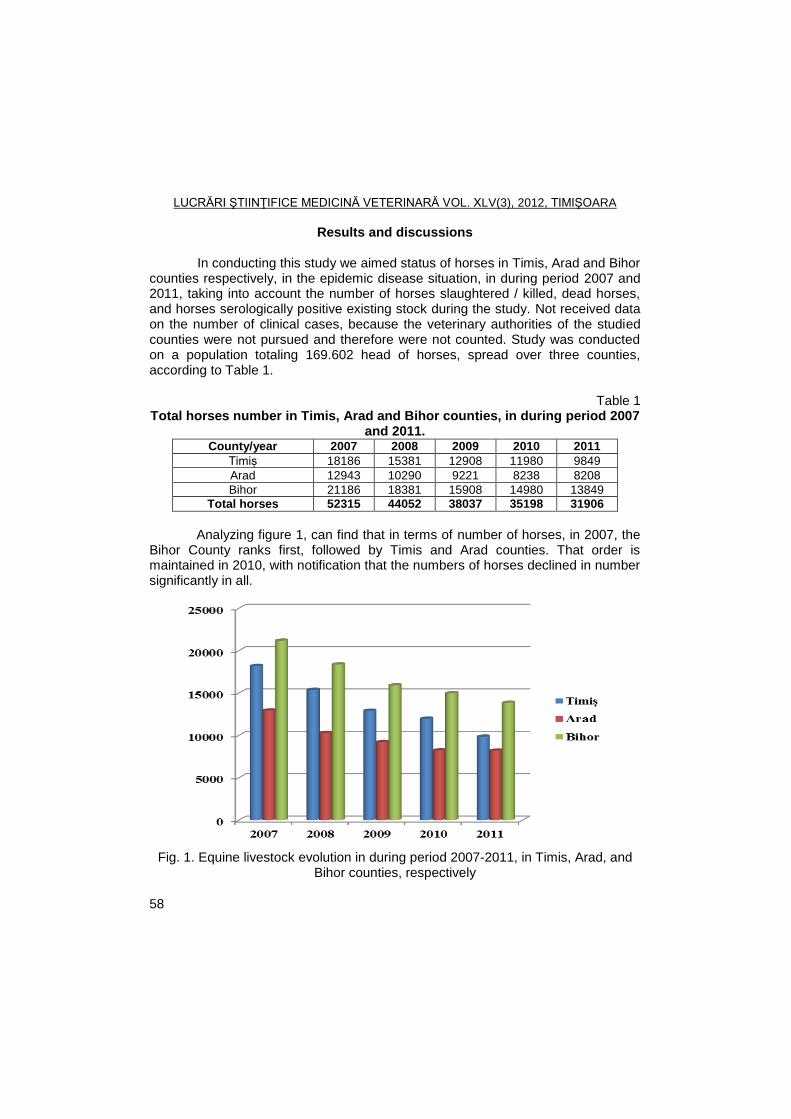
LUCRĂRI ŞTIINŢIFICE MEDICINĂ VETERINARĂ VOL. XLV(3), 2012, TIMIŞOARA
58
Results and discussions
In conducting this study we aimed status of horses in Timis, Arad and Bihor counties respectively, in the epidemic disease situation, in during period 2007 and 2011, taking into account the number of horses slaughtered / killed, dead horses, and horses serologically positive existing stock during the study. Not received data on the number of clinical cases, because the veterinary authorities of the studied counties were not pursued and therefore were not counted. Study was conducted on a population totaling 169.602 head of horses, spread over three counties, according to Table 1.
Table 1
Total horses number in Timis, Arad and Bihor counties, in during period 2007 and 2011.
County/year 2007 2008 2009 2010 2011
Timiș 18186 15381 12908 11980 9849
Arad 12943 10290 9221 8238 8208
Bihor 21186 18381 15908 14980 13849
Total horses 52315 44052 38037 35198 31906
Analyzing figure 1, can find that in terms of number of horses, in 2007, the
Bihor County ranks first, followed by Timis and Arad counties. That order is maintained in 2010, with notification that the numbers of horses declined in number significantly in all.
Fig. 1. Equine livestock evolution in during period 2007-2011, in Timis, Arad, and
Bihor counties, respectively
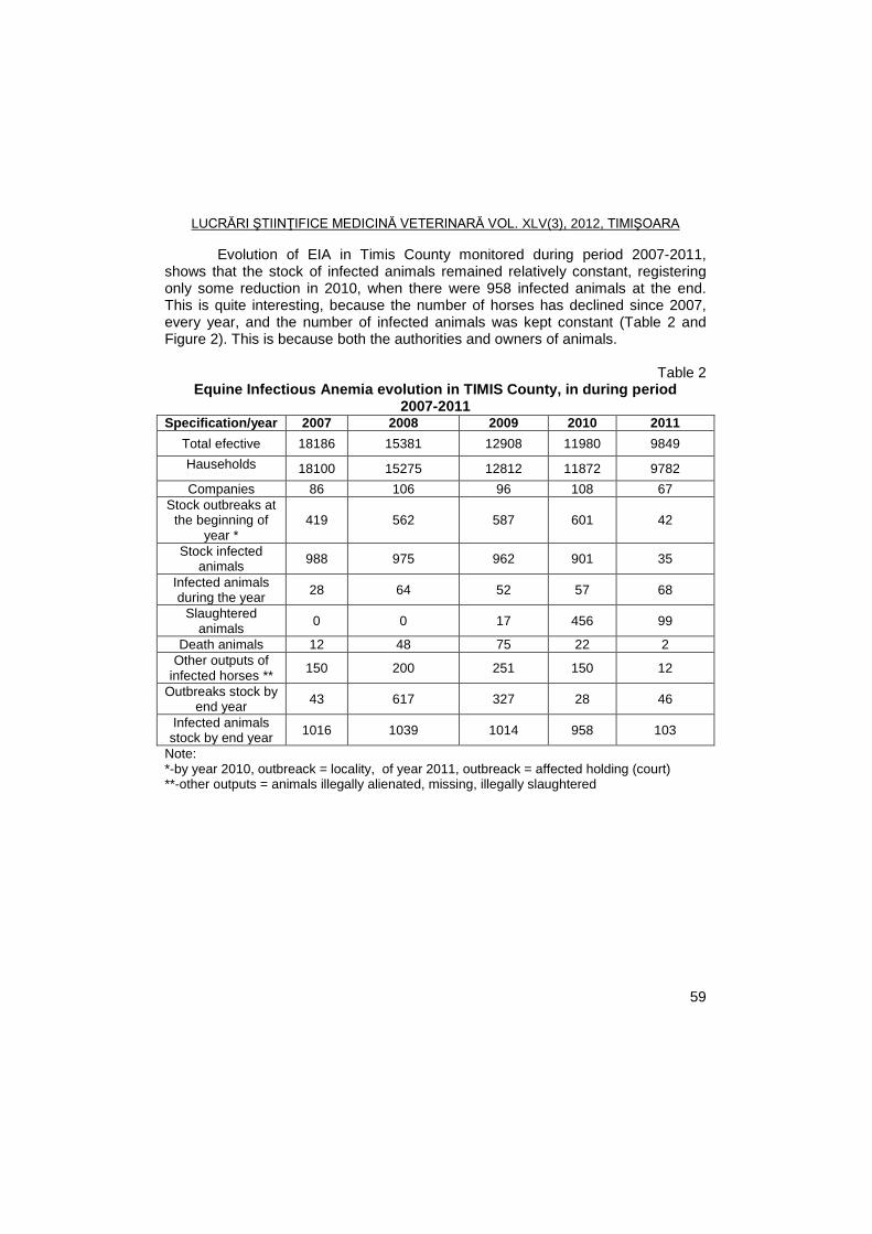
LUCRĂRI ŞTIINŢIFICE MEDICINĂ VETERINARĂ VOL. XLV(3), 2012, TIMIŞOARA
59
Evolution of EIA in Timis County monitored during period 2007-2011, shows that the stock of infected animals remained relatively constant, registering only some reduction in 2010, when there were 958 infected animals at the end. This is quite interesting, because the number of horses has declined since 2007, every year, and the number of infected animals was kept constant (Table 2 and Figure 2). This is because both the authorities and owners of animals.
Table 2
Equine Infectious Anemia evolution in TIMIS County, in during period 2007-2011
Specification/year 2007 2008 2009 2010 2011
Total efective 18186 15381 12908 11980 9849
Hauseholds 18100 15275 12812 11872 9782
Companies 86 106 96 108 67
Stock outbreaks at the beginning of
year * 419 562 587 601 42
Stock infected animals
988 975 962 901 35
Infected animals during the year
28 64 52 57 68
Slaughtered animals
0 0 17 456 99
Death animals 12 48 75 22 2
Other outputs of infected horses **
150 200 251 150 12
Outbreaks stock by end year
43 617 327 28 46
Infected animals stock by end year
1016 1039 1014 958 103
Note: *-by year 2010, outbreack = locality, of year 2011, outbreack = affected holding (court) **-other outputs = animals illegally alienated, missing, illegally slaughtered
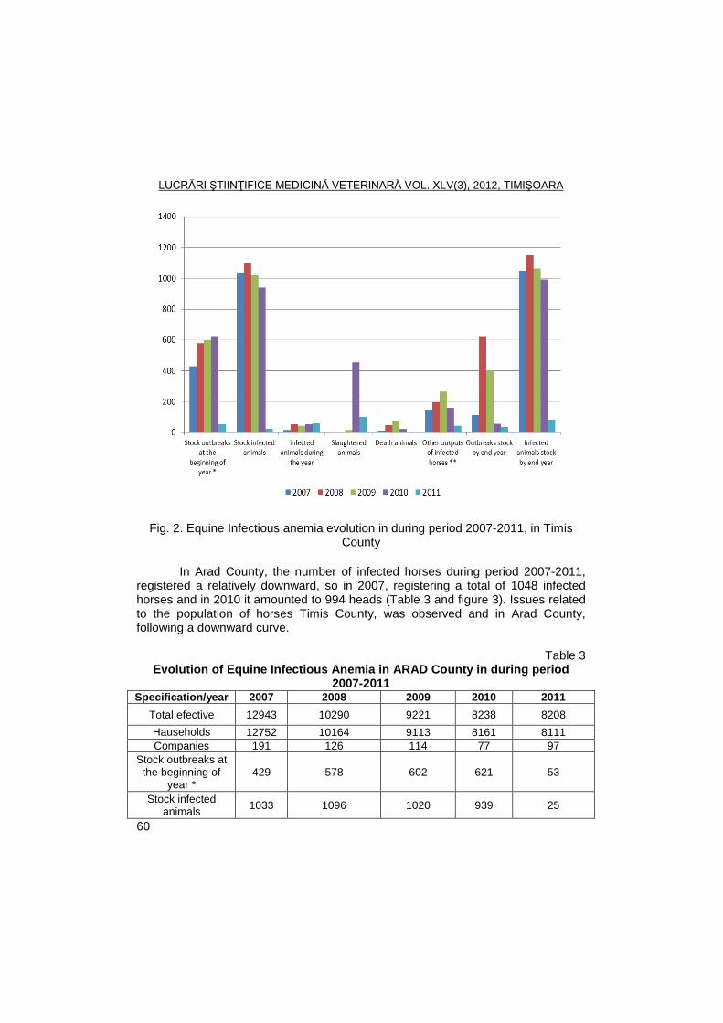
LUCRĂRI ŞTIINŢIFICE MEDICINĂ VETERINARĂ VOL. XLV(3), 2012, TIMIŞOARA
60
Fig. 2. Equine Infectious anemia evolution in during period 2007-2011, in Timis County
In Arad County, the number of infected horses during period 2007-2011,
registered a relatively downward, so in 2007, registering a total of 1048 infected horses and in 2010 it amounted to 994 heads (Table 3 and figure 3). Issues related to the population of horses Timis County, was observed and in Arad County, following a downward curve.
Table 3
Evolution of Equine Infectious Anemia in ARAD County in during period 2007-2011
Specification/year 2007 2008 2009 2010 2011
Total efective 12943 10290 9221 8238 8208
Hauseholds 12752 10164 9113 8161 8111
Companies 191 126 114 77 97
Stock outbreaks at the beginning of
year * 429 578 602 621 53
Stock infected animals
1033 1096 1020 939 25
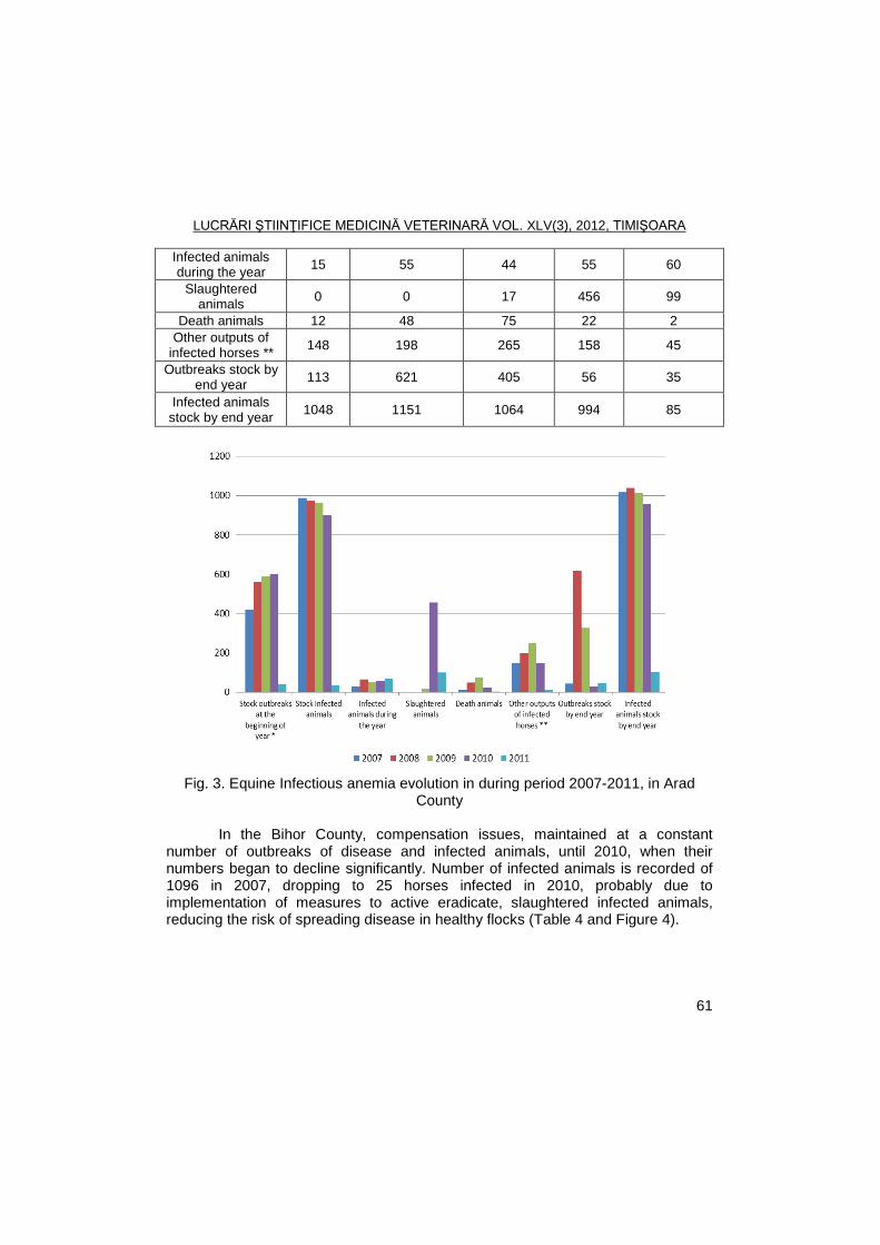
LUCRĂRI ŞTIINŢIFICE MEDICINĂ VETERINARĂ VOL. XLV(3), 2012, TIMIŞOARA
61
Infected animals during the year
15 55 44 55 60
Slaughtered animals
0 0 17 456 99
Death animals 12 48 75 22 2
Other outputs of infected horses **
148 198 265 158 45
Outbreaks stock by end year
113 621 405 56 35
Infected animals stock by end year
1048 1151 1064 994 85
Fig. 3. Equine Infectious anemia evolution in during period 2007-2011, in Arad
County
In the Bihor County, compensation issues, maintained at a constant number of outbreaks of disease and infected animals, until 2010, when their numbers began to decline significantly. Number of infected animals is recorded of 1096 in 2007, dropping to 25 horses infected in 2010, probably due to implementation of measures to active eradicate, slaughtered infected animals, reducing the risk of spreading disease in healthy flocks (Table 4 and Figure 4).
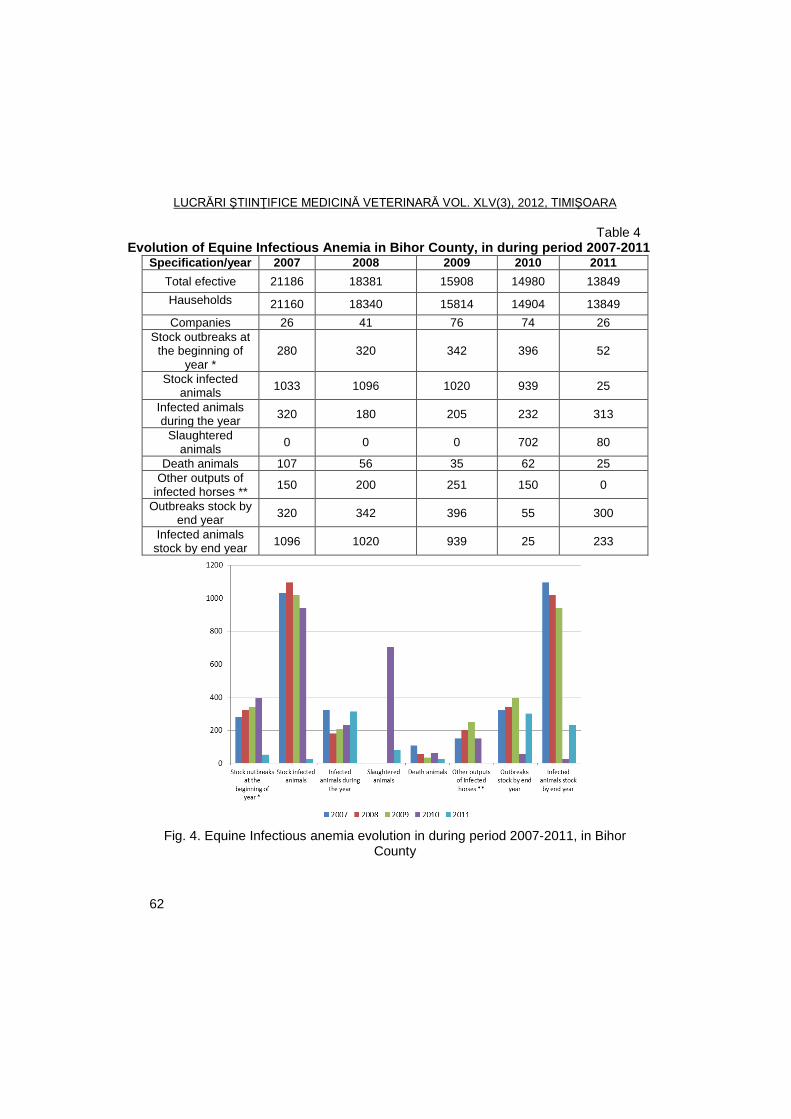
LUCRĂRI ŞTIINŢIFICE MEDICINĂ VETERINARĂ VOL. XLV(3), 2012, TIMIŞOARA
62
Table 4 Evolution of Equine Infectious Anemia in Bihor County, in during period 2007-2011
Specification/year 2007 2008 2009 2010 2011
Total efective 21186 18381 15908 14980 13849
Hauseholds 21160 18340 15814 14904 13849
Companies 26 41 76 74 26
Stock outbreaks at the beginning of
year * 280 320 342 396 52
Stock infected animals
1033 1096 1020 939 25
Infected animals during the year
320 180 205 232 313
Slaughtered animals
0 0 0 702 80
Death animals 107 56 35 62 25
Other outputs of infected horses **
150 200 251 150 0
Outbreaks stock by end year
320 342 396 55 300
Infected animals stock by end year
1096 1020 939 25 233
Fig. 4. Equine Infectious anemia evolution in during period 2007-2011, in Bihor
County
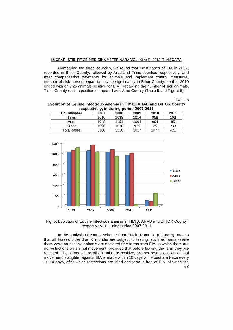
LUCRĂRI ŞTIINŢIFICE MEDICINĂ VETERINARĂ VOL. XLV(3), 2012, TIMIŞOARA
63
Comparing the three counties, we found that most cases of EIA in 2007, recorded in Bihor County, followed by Arad and Timis counties respectively, and after compensation payments for animals and implement control measures, number of sick horses began to decline significantly in Bihor County, so that 2010 ended with only 25 animals positive for EIA. Regarding the number of sick animals, Timis County retains position compared with Arad County (Table 5 and Figure 5).
Table 5
Evolution of Equine Infectious Anemia in TIMIŞ, ARAD and BIHOR County respectively, in during period 2007-2011
Countie/year 2007 2008 2009 2010 2011
Timiș 1016 1039 1014 958 103
Arad 1048 1151 1064 994 85
Bihor 1096 1020 939 25 233
Total cases 3160 3210 3017 1977 421
Fig. 5. Evolution of Equine infectious anemia in TIMIŞ, ARAD and BIHOR County respectively, in during period 2007-2011
In the analysis of control scheme from EIA in Romania (Figure 6), means
that all horses older than 6 months are subject to testing, such as farms where there were no positive animals are declared free farms from EIA, in which there are no restrictions on animal movement, provided that before leaving the farm they are retested. The farms where all animals are positive, are set restrictions on animal movement, slaughter against EIA is made within 10 days while pest are twice every 10-14 days, after which restrictions are lifted and farm is free of EIA, allowing the
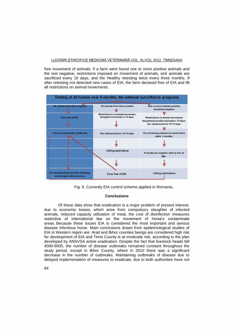
LUCRĂRI ŞTIINŢIFICE MEDICINĂ VETERINARĂ VOL. XLV(3), 2012, TIMIŞOARA
64
free movement of animals. If a farm were found one or more positive animals and the rest negative, restrictions imposed on movement of animals, sick animals are sacrificed every 10 days, and the Healthy retesting twice every three months. If after retesting not detected new cases of EIA, the farm declared free of EIA and lift all restrictions on animal movements.
Fig. 6. Currently EIA control scheme applied in Romania.
Conclusions
Of these data show that eradication is a major problem of present interest, due to economic losses, which arise from compulsory slaughter of infected animals, reduced capacity utilization of meat, the cost of disinfection measures restrictive of international law on the movement of horse’s contaminate areas. Because these issues EIA is considered the most important and serious disease infectious horse. Main conclusions drawn from epidemiological studies of EIA in Western region are: Arad and Bihor counties beings are considered high risk for development of EIA and Timis County is at moderate risk, according to the plan developed by ANSVSA active eradication. Despite the fact that livestock heads fell 4000-6000, the number of disease outbreaks remained constant throughout the study period, except in Bihor County, where in 2010 there was a significant decrease in the number of outbreaks. Maintaining outbreaks of disease due to delayed implementation of measures to eradicate, due to both authorities have not
LUCRĂRI ŞTIINŢIFICE MEDICINĂ VETERINARĂ VOL. XLV(3), 2012, TIMIŞOARA
65
provided the necessary funds to compensate owners of sick animals and horse owners who have lost confidence in the measures proposed by the authorities. Compensate owners of animals in Bihor county led to the possibility of implementing some control measures have had the purpose of decreasing the number of infected animals, from 1096 ends in 2007, to 25 heads in 2010.
References
1. Brangan, P., Bailey, D.C., Larkin, J.F., Myers, T., More, S.J., Management
of the national programme to eradicate equine infectious anaemia from Ireland during 2006: a review, Equine Vet J., 2008, 40(7), 702-704.
2. Cappelli, K., Capomaccio, S., Cook, F.R., FelicettI, M., Marenzoni, M.L., Coppola, G., Verini-Supplizi, A., Coletti, M., Passamonti, F., Molecular detection, epidemiology, and genetic characterization of novel European field isolates of equine infectious anemia virus, J Clin Microbiol., 2011, 49(1), 27-33.
3. Kaiser, A., Meier, H.P., Straub, R., Gerber, V., Equine Infectious Anemia (EIA), Schweiz Arch Tierheilkd., 2009, 151(4), 159-164.
4. Lazău, A., Legislatie si Deontologie Veterinara, Ed Mirton, 2003, Timisoara. 5. Moga-Manzat, R., Boli virotice si prionice ale animalelor, Ed. Brumar, 2005
Timisoara. 6. Vasiu, C., Viroze și boli prionice la animale, Ed. Nereamia Napra, 2003, Cluj-
Napoca. 7. Zielonka, J., Bravo, I.G., Marino, D., Conrad, E., Perkoviš, M.,
Battenberg, M., Cichutek, K., Münk, C., Restriction of Equine Infectious Anemia Virus by Equine APOBEC3 Cytidine Deaminases, J. Virol., 2009, 83(15), 7547-7559.
LUCRĂRI ŞTIINŢIFICE MEDICINĂ VETERINARĂ VOL. XLV(3), 2012, TIMIŞOARA
66
HISTOPATHOLOGICAL AND ULTRASTRUCTURAL ASPECTS OF THE GUMBORO DISEASE VIRUS FARAGHER 52/70 STRAIN
EXPERIMENTAL INFECTION IN SPF CHICKENS
A. POPOVICI1, CRISTINA DIACONU
1, DANIELA STAMATE
1, ŞTEFANIA
RAITA2, M. TURCITU
1, B. GEORGESCU
2, C. BELU
2, G. PREDOI
2, FLORICA
BARBUCEANU1
1Institute for Diagnosis and Animal Health, Dr. Staicovici No. 63, District 5, 050557,
Bucharest, Romania 2Faculty of Veterinary Medicine Bucharest
E-mail: [email protected]
Summary
The present work is a complex study of histological, immunohistochemical and electronomycroscopical investigation performed on samples of thymus, spleen and bursa of Fabricius, collected from poultry SPF as a result of experimental infection with the virus disease of Gumboro, classical reference strain Faragher 52\/70.
As a result of carrying out of different histological staining methods and examination of histological preparations were seen lesions, characterized by intense lymphocyte depletion accompanied by the proliferation of reticular dendritic cells and macrophages exhibiting the highest intensity in the thimus and Fabricius bursa. Intense phenomena of necrosis were observed in the white pulp of the spleen.
Ultrastructural exams have revealed in the cytoplasm of the bursitis cells icosahedrale virus particles with characteristic morphology of Gumboro disease.
Comparative study of the results of histological examinations, immunohistochemical and electronomycroscopical examinations have confirmed the evolution of acute infection with the virus disease of Gumboro and the installation of a severe immunodeficiency to 72 hours after infection.
Key words: avian infectious bursitis, SPF chickens, cytology,
immunohistopathology, electronomicroscopy
Avian Infectious Bursitis Disease - AIBD is an acute contagious disease of young chickens, which was initially described by Crosgove in 1962, the disease being reported for the first time in 1957 in the village of Gumboro in the US (1).The increased incidence and severity of the disease is dependent on the age of the infected birds, virus strain pathogenicity and the presence or absence of immunity.The ethiology is represented by a virus, belonging to the genus Avibirnavirus, family Birnaviridae, from here and the name of the Birnavirus (bi-TS). Its genome is represented by two molecules of double-stranded lined dsRNA segments, designated Aand B wich are enclosed within a nonenveloped icosahedral, diameter 60 nm, capsid (4). The target is B-cell expressing IgM (B-IgM +) but specific receptor is not yet specified. After virus infection, severe injuries occur in Fabricius bursal cells, resulting in pronounced immunosuppression and in other lymphoid
LUCRĂRI ŞTIINŢIFICE MEDICINĂ VETERINARĂ VOL. XLV(3), 2012, TIMIŞOARA
67
tissues (1, 4). Diagnosis involves the use of indirect serological reactions and especially of ELISA - viral antigen capture. In viral antigen detection, sensitivity of serological reactions was significantly increased by using polyclonal instead of virus monoclonal antibodies (3). Detection of the virus by these methods create premises the feasibility for further cultivation and isolation of virus in embryonated eggs or cell cultures. Currently has been described and used Reverse Transcriptase PCR method (RT-PCR, RNA-PCR) in epidemiological surveillance of infection with the virus and discrimination of the type IBDV classic different variants of serotype 1 (4, 7). Due to the high antigenity diversity, it is necessary to further improvement and development of new methods used in diagnosis, identification and isolation of virus.
The objective of this study consisted in develop and improv methods and techniques for optimization diagnostic microscopy in birds infected with infectious bursitis virus (IBV).
Materials and methods
a) Experimental protocol. Examinations were performed anatomo-
clinical, cytological, histopathological, immunohistochemical (IHC) and electrononomycroscopical (EM) in a group of 10 specific-pathogen free (SPF) chickens, Leghorn breed, aged 21 days, experimentally infected with classical virulent strain reference, Faragher 52/70. The infection was achieved by administration of viral suspension (dilution 1: 10 MEM) in the form of drops in the eyes, nose, oral and drinking water. To compare the results was established a control group (n = 4) of uninfected SPF chickens. After establishing the IBDV model of chicken, the animals were euthanatized for welfare resons. From birds slaughtered at 72 hours post infection, were collected fragments of spleen, thymus and bursa of Fabricius.
b) Cytology. Cytological exams were made on the smears obtained by the method of touchprint and/or homogenate of the three categories of lymphoid organs mentioned and routinely stained with May-Grűnwald-Giemsa method (5).
c) Histopathology. For the detection of histopathological lesions, sampling fragments of thymus, spleen and bursa of Fabricius were immediately fixed in Baker solution, embedded in Paraplast Plus (Sigma) embedding media. Parafin sections (5 µm), were de-waxed, routinely stained with trichromic Masson method (HEA).Tissue lymphoid lesions were determined and compared between groups.
d) Immunohistochemistry. For immunolabelling of viral antigen has been used Indirect Immunofluorescent assay (IFI) on lymphoid tissues.The bursal, spllen and thymus tissue samples taken both from experimental infected chickens and control group, were cutting at cryotom (4 µm) and fixed in acetone during 30 minutes at 4°C. The endogenous peroxidase in sections was eliminated by 3% H2O2. Nonspecific reactions were blocked by normal rabbit serum. Slides have been coated with polyclonal serum control (positive or negative) as primary antibody, diluted (1/10, 1/20, 1/30, 1/401/50) in PBS with 4% bovine albumin serum
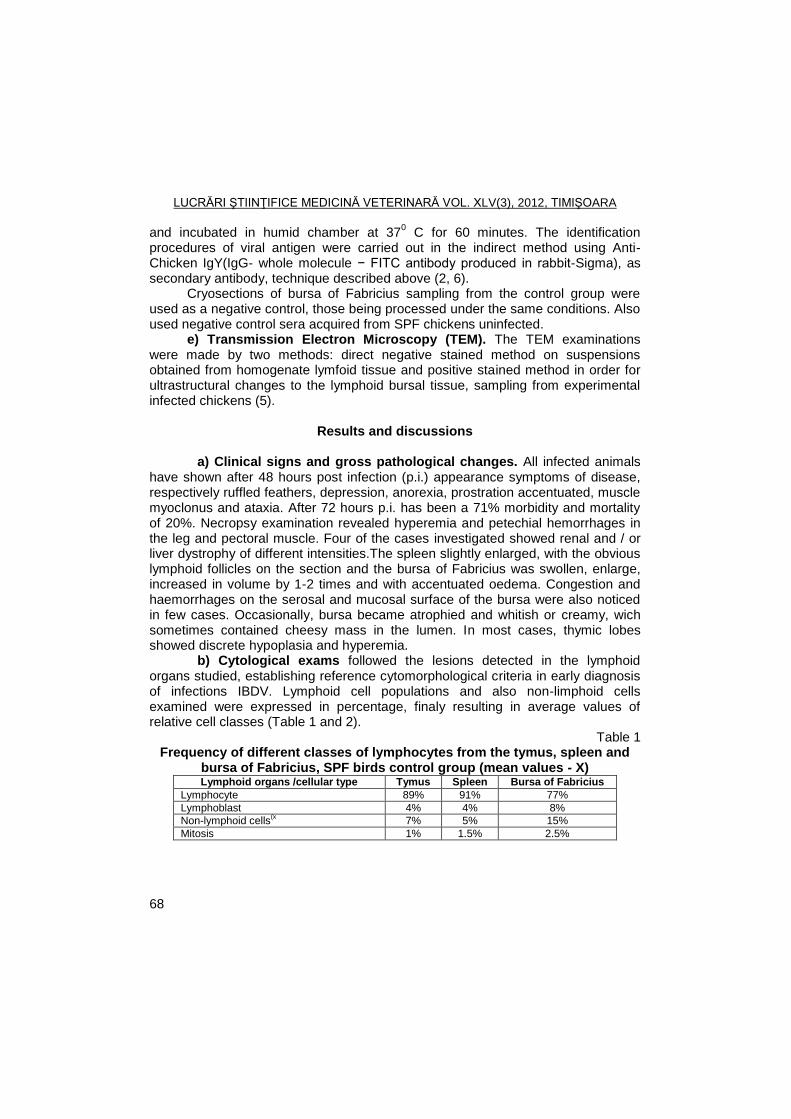
LUCRĂRI ŞTIINŢIFICE MEDICINĂ VETERINARĂ VOL. XLV(3), 2012, TIMIŞOARA
68
and incubated in humid chamber at 370 C for 60 minutes. The identification
procedures of viral antigen were carried out in the indirect method using Anti-Chicken IgY(IgG- whole molecule − FITC antibody produced in rabbit-Sigma), as secondary antibody, technique described above (2, 6).
Cryosections of bursa of Fabricius sampling from the control group were used as a negative control, those being processed under the same conditions. Also used negative control sera acquired from SPF chickens uninfected.
e) Transmission Electron Microscopy (TEM). The TEM examinations were made by two methods: direct negative stained method on suspensions obtained from homogenate lymfoid tissue and positive stained method in order for ultrastructural changes to the lymphoid bursal tissue, sampling from experimental infected chickens (5).
Results and discussions
a) Clinical signs and gross pathological changes. All infected animals have shown after 48 hours post infection (p.i.) appearance symptoms of disease, respectively ruffled feathers, depression, anorexia, prostration accentuated, muscle myoclonus and ataxia. After 72 hours p.i. has been a 71% morbidity and mortality of 20%. Necropsy examination revealed hyperemia and petechial hemorrhages in the leg and pectoral muscle. Four of the cases investigated showed renal and / or liver dystrophy of different intensities.The spleen slightly enlarged, with the obvious lymphoid follicles on the section and the bursa of Fabricius was swollen, enlarge, increased in volume by 1-2 times and with accentuated oedema. Congestion and haemorrhages on the serosal and mucosal surface of the bursa were also noticed in few cases. Occasionally, bursa became atrophied and whitish or creamy, wich sometimes contained cheesy mass in the lumen. In most cases, thymic lobes showed discrete hypoplasia and hyperemia.
b) Cytological exams followed the lesions detected in the lymphoid organs studied, establishing reference cytomorphological criteria in early diagnosis of infections IBDV. Lymphoid cell populations and also non-limphoid cells examined were expressed in percentage, finaly resulting in average values of relative cell classes (Table 1 and 2).
Table 1 Frequency of different classes of lymphocytes from the tymus, spleen and
bursa of Fabricius, SPF birds control group (mean values - X) Lymphoid organs /cellular type Tymus Spleen Bursa of Fabricius
Lymphocyte 89% 91% 77%
Lymphoblast 4% 4% 8%
Non-lymphoid cells(x
7% 5% 15%
Mitosis 1% 1.5% 2.5%
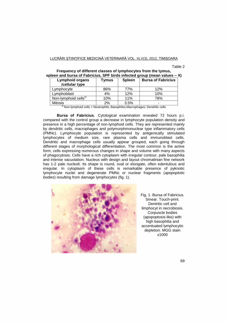
LUCRĂRI ŞTIINŢIFICE MEDICINĂ VETERINARĂ VOL. XLV(3), 2012, TIMIŞOARA
69
Table 2 Frequency of different classes of lymphocytes from the tymus,
spleen and bursa of Fabricius, SPF birds infected group (mean values – X)
Lymphoid organs /cellular type
Tymus Spleen Bursa of Fabricius
Lymphocyte 86% 77% 12%
Lymphoblast 4% 12% 10%
Non-lymphoid cells(x
10% 11% 78%
Mitosis 2% 3.5% - (x
Non-lymphoid cells = Neutrophils; Basophiles;Macrophages; Dendritic cells
Bursa of Fabricius. Cytological examination revealed 72 hours p.i.
compared with the control group a decrease in lymphocyte population density and presence in a high percentage of non-lymphoid cells. They are represented mainly by dendritic cells, macrophages and polymorphmonuclear type inflammatory cells (PMNc). Lymphocyte population is represented by antigenically stimulated lymphocytes of medium size, rare plasma cells and immunoblast cells. Dendritic and macrophage cells usually appear grouped, each going through different stages of morphological differentiation. The most common is the active form, cells expressing numerous changes in shape and volume with many aspects of phagocytosis. Cells have a rich cytoplasm with irregular contour, pale basophilia and intense vacuolation. Nucleus with design and layout chromatinian fine network has 1-2 pale nucleoli. Its shape is round, oval or elongate, often edentulous and irregular. In cytoplasm of these cells is remarkable presence of pyknotic lymphocyte nuclei and degenerate PMNc or nuclear fragments (apopoptotic bodies) resulting from damage lymphocytes (fig. 1).
Fig. 1. Bursa of Fabricius. Smear. Touch-print.
Dentritic cell and limphocyt in necrobiosis.
Corpuscle bodies (apopoptosis-like) with
high basophilia and accentuated lymphocytic
depletion. MGG stain x1000
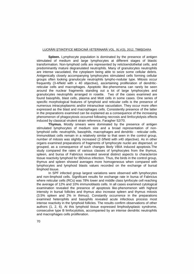
LUCRĂRI ŞTIINŢIFICE MEDICINĂ VETERINARĂ VOL. XLV(3), 2012, TIMIŞOARA
70
Spleen. Lymphocyte population is dominated by the presence of antigen stimulated of medium and large lymphocytes at different stages of blastic transformation. Non-lymphoid cells are represented by reticloendothelial cells, and predominantly mature degranulated neutrophils. Many of granulocytes neutrophils are intense vacuolated, the cytoplasm being able to seize some cellular debris. Antigenically closely accompanying lymphocytes stimulated cells forming cellular groups often looking granulocyte neutrophils lympho-nodular type. Mitosis occur frequently (3-4/field with x 40 objective), ascertaining proliferation of dendritic-reticular cells and macrophages. Apoptotic like-phenomena can rarely be seen around the nuclear fragments standing out a lot of large lymphocytes and granulocytes neutrophils arranged in rosette. Two of the cases examined are found basophils, blast cells, plasma and Mott cells in some cases. One series of specific morphological features of lymphoid and reticular cells is the presence of numerous intracytoplasmic and/or intranuclear vacuolation. They occur more often expressed as the blast and macrophages cells. Consistently presence of the latter in the preparations examined can be explained as a consequence of the increasing phenomenon of phagocytosis occurred following necrosis and limfocytolysis effects induced by classical virulent strain reference, Faragher 52/70.
Thymus. Mostly smears were dominated by the presence of antigen stimulated lymphocytes of medium size and a broad representation of non-lymphoid cells: neutrophils, basophils, macrophages and dendritic - reticular cells. Immunoblast cells remain in a relatively similar to that seen in the control group, number of mitosis was slightly increased (2-3/field with x40 objective). As in other organs examined preparations of fragments of lymphocyte nuclei are dispersed, or grouped, as a consequence of such changes likely VBIA induced apoptosis.The study compared the rates of various classes of lymphocytes from the thymus, spleen, and bursa of Fabricius revealed several distinct aspects to characterize tissue reactivity lymphoid for IBDvirus infection. Thus, the birds in the control group, thymus and spleen showed averages more homogeneous when compared with lymphocytes and lymphoid blasts values recorded on the exchange of bursal limphoid tisuue.
In SPF infected group largest variations were observed with lymphocytes and non-limphoid cells. Significant results for exchange rate in bursa of Fabricius where reticular cells (RCs) was 78% lower and middle class lymfocyte cell reaching the average of 12% and 10% immunoblasts cells. In all cases examined cytological examination revealed the presence of apoptosis like-phenomenon with highest intensity in bursal follicles and thymus also increase spleen and thymus mitosis (3.5% spleen and 2% in thimus). Constantly occurrence in the preparations examined heterophils and basophils revealed acute infectious process most intense reactivity in the lymphoid follicles. The results confirm observations of other authors (1, 2, 6). At this lymphoid tissue expressed limphodysplasic syndrome, consecutive type B limfocytolisis, accompanied by an intense dendritic neutrophils and macrophages cells proliferation.
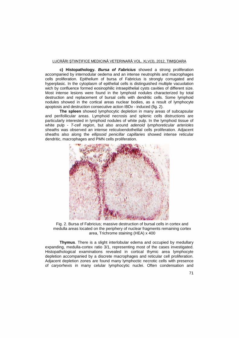
LUCRĂRI ŞTIINŢIFICE MEDICINĂ VETERINARĂ VOL. XLV(3), 2012, TIMIŞOARA
71
c) Histopathology. Bursa of Fabricius showed a strong proliferation accompanied by internodular oedema and an intense neutrophils and macrophages cells proliferation. Epithelium of bursa of Fabricius is strongly corrugated and hyperplasic. In the cytoplasm of epithelial cells is distinguished multiple vacuolation wich by confluence formed eosinophilic intraepithelial cysts cavities of different size. Most intense lesions were found in the lymphoid nodules characterized by total destruction and replacement of bursal cells with dendritic cells. Some lymphoid nodules showed in the cortical areas nuclear bodies, as a result of lymphocyte apoptosis and destruction consecutive action IBDv - induced (fig. 2).
The spleen showed lymphocytic depletion in many areas of subcapsular and perifollicular areas. Lymphoid necrosis and splenic cells distructions are particularly interested in lymphoid nodules of white pulp. In the lymphoid tissue of white pulp - T-cell region, but also around adenoid lymphoreticular arterioles sheaths was observed an intense reticuloendothelilal cells proliferation. Adjacent sheaths also along the ellipsoid penicillar capillaries showed intense reticular dendritic, macrophages and PMN cells proliferation.
Fig. 2. Bursa of Fabricius; massive destruction of bursal cells in cortex and medulla areas located on the periphery of nuclear fragments remaining cortex
area, Trichrome staining (HEA) x 400
Thymus. There is a slight interlobular edema and occupied by medullary expanding, medulla-cortex ratio 3/1, representing most of the cases investigated. Histopathological examinations revealed in cortical thymic area lymphocyte depletion accompanied by a discrete macrophages and reticular cell proliferation. Adjacent depletion zones are found many lymphoctic necrotic cells with presence of caryorhexis in many celular lymphocytic nuclei. Often condensation and
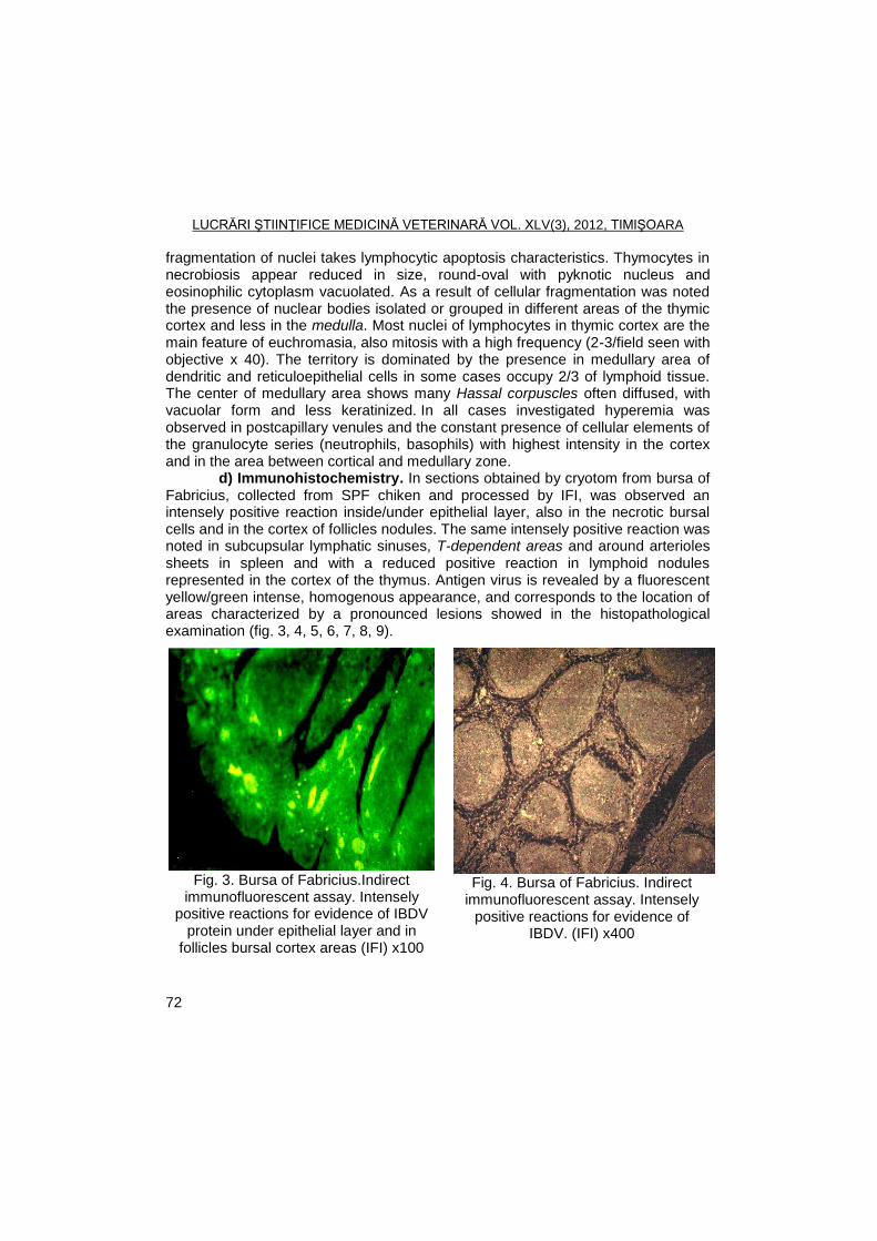
LUCRĂRI ŞTIINŢIFICE MEDICINĂ VETERINARĂ VOL. XLV(3), 2012, TIMIŞOARA
72
fragmentation of nuclei takes lymphocytic apoptosis characteristics. Thymocytes in necrobiosis appear reduced in size, round-oval with pyknotic nucleus and eosinophilic cytoplasm vacuolated. As a result of cellular fragmentation was noted the presence of nuclear bodies isolated or grouped in different areas of the thymic cortex and less in the medulla. Most nuclei of lymphocytes in thymic cortex are the main feature of euchromasia, also mitosis with a high frequency (2-3/field seen with objective x 40). The territory is dominated by the presence in medullary area of dendritic and reticuloepithelial cells in some cases occupy 2/3 of lymphoid tissue. The center of medullary area shows many Hassal corpuscles often diffused, with vacuolar form and less keratinized. In all cases investigated hyperemia was observed in postcapillary venules and the constant presence of cellular elements of the granulocyte series (neutrophils, basophils) with highest intensity in the cortex and in the area between cortical and medullary zone.
d) Immunohistochemistry. In sections obtained by cryotom from bursa of Fabricius, collected from SPF chiken and processed by IFI, was observed an intensely positive reaction inside/under epithelial layer, also in the necrotic bursal cells and in the cortex of follicles nodules. The same intensely positive reaction was noted in subcupsular lymphatic sinuses, T-dependent areas and around arterioles sheets in spleen and with a reduced positive reaction in lymphoid nodules represented in the cortex of the thymus. Antigen virus is revealed by a fluorescent yellow/green intense, homogenous appearance, and corresponds to the location of areas characterized by a pronounced lesions showed in the histopathological examination (fig. 3, 4, 5, 6, 7, 8, 9).
Fig. 3. Bursa of Fabricius.Indirect
immunofluorescent assay. Intensely positive reactions for evidence of IBDV
protein under epithelial layer and in follicles bursal cortex areas (IFI) x100
Fig. 4. Bursa of Fabricius. Indirect
immunofluorescent assay. Intensely positive reactions for evidence of
IBDV. (IFI) x400
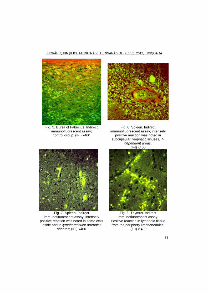
LUCRĂRI ŞTIINŢIFICE MEDICINĂ VETERINARĂ VOL. XLV(3), 2012, TIMIŞOARA
73
Fig. 5. Bursa of Fabricius. Indirect immunofluorescent assay; control group; (IFI) x400
Fig. 6. Spleen. Indirect immunofluorescent assay; intensely
positive reaction was noted in subcupsular lymphatic sinuses, T-
dependent areas; (IFI) x400
Fig. 7. Spleen. Indirect
immunofluorescent assay; intensely positive reaction was noted in some cells inside and in lymphoreticular arterioles
sheaths; (IFI) x400
Fig. 8. Thymus. Indirect immunofluorescent assay.
Positive reaction in lymphoid tissue from the periphery limphonodules;
(IFI) x 400
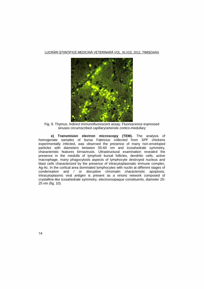
LUCRĂRI ŞTIINŢIFICE MEDICINĂ VETERINARĂ VOL. XLV(3), 2012, TIMIŞOARA
74
Fig. 9. Thymus. Indirect immunofluorescent assay. Fluorescence expressed
sinuses circumscribed capillary/arteriole cortico-medullary
e) Transmision electron microscopy (TEM). The analysis of homogenate samples of bursa Fabricius collected from SPF chickens experimentally infected, was observed the presence of many non-enveloped particles with diameters between 55-60 nm and icosahedrale symmetry, characteristic features birnavirusis. Ultrastructural examination revealed the presence in the medulla of lymphoid bursal follicles, dendritic cells, active macrophage, many phagocytosis aspects of lymphocyte destroyed nucleus and blast cells characterized by the presence of intracytoplasmatic immune complex, Ag-Ac. In the cortical area dominated lymphocytes with nuclei at different stages of condensation and / or disruptive chromatin characteristic apoptosis. Intracytoplasmic viral antigen is present as a virions network composed of crystalline-like icosahedrale symmetry, electronoopaque constituents, diameter 20-25 nm (fig. 10).
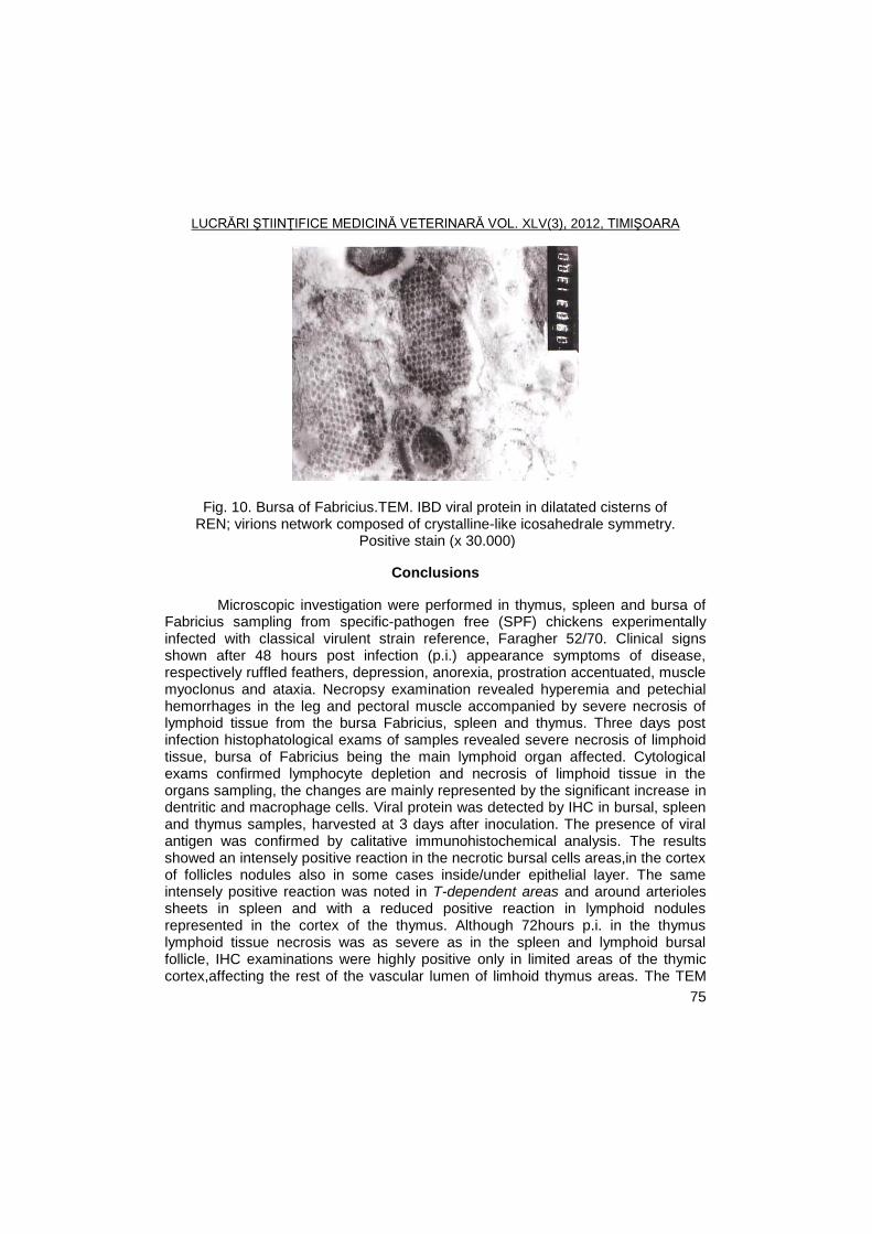
LUCRĂRI ŞTIINŢIFICE MEDICINĂ VETERINARĂ VOL. XLV(3), 2012, TIMIŞOARA
75
Fig. 10. Bursa of Fabricius.TEM. IBD viral protein in dilatated cisterns of REN; virions network composed of crystalline-like icosahedrale symmetry.
Positive stain (x 30.000)
Conclusions
Microscopic investigation were performed in thymus, spleen and bursa of Fabricius sampling from specific-pathogen free (SPF) chickens experimentally infected with classical virulent strain reference, Faragher 52/70. Clinical signs shown after 48 hours post infection (p.i.) appearance symptoms of disease, respectively ruffled feathers, depression, anorexia, prostration accentuated, muscle myoclonus and ataxia. Necropsy examination revealed hyperemia and petechial hemorrhages in the leg and pectoral muscle accompanied by severe necrosis of lymphoid tissue from the bursa Fabricius, spleen and thymus. Three days post infection histophatological exams of samples revealed severe necrosis of limphoid tissue, bursa of Fabricius being the main lymphoid organ affected. Cytological exams confirmed lymphocyte depletion and necrosis of limphoid tissue in the organs sampling, the changes are mainly represented by the significant increase in dentritic and macrophage cells. Viral protein was detected by IHC in bursal, spleen and thymus samples, harvested at 3 days after inoculation. The presence of viral antigen was confirmed by calitative immunohistochemical analysis. The results showed an intensely positive reaction in the necrotic bursal cells areas,in the cortex of follicles nodules also in some cases inside/under epithelial layer. The same intensely positive reaction was noted in T-dependent areas and around arterioles sheets in spleen and with a reduced positive reaction in lymphoid nodules represented in the cortex of the thymus. Although 72hours p.i. in the thymus lymphoid tissue necrosis was as severe as in the spleen and lymphoid bursal follicle, IHC examinations were highly positive only in limited areas of the thymic cortex,affecting the rest of the vascular lumen of limhoid thymus areas. The TEM
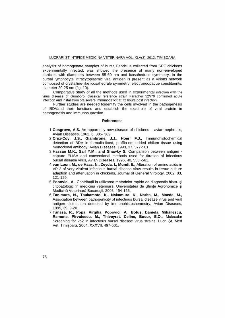
LUCRĂRI ŞTIINŢIFICE MEDICINĂ VETERINARĂ VOL. XLV(3), 2012, TIMIŞOARA
76
analysis of homogenate samples of bursa Fabricius collected from SPF chickens experimentally infected, was showed the presence of many non-enveloped particles with diameters between 55-60 nm and icosahedrale symmetry. In the bursal lymphocyte intracytoplasmic viral antigen is present as a virions network composed of crystalline-like icosahedrale symmetry, electronoopaque constituents, diameter 20-25 nm (fig. 10).
Comparative study of all the methods used in experimental infection with the
virus disease of Gumboro, classical reference strain Faragher 52\/70 confirmed acute infection and installation ofa severe immunodeficit at 72 hours post infection.
Further studies are needed toidentify the cells involved in the pathogenesis of IBDVand their functions and establish the exactrole of viral protein in pathogenesis and immunosupresion.
References
1. Cosgrove, A.S. An apparently new disease of chickens – avian nephrosis, Avian Diseases, 1962, 6, 385- 389.
2. Cruz-Coy, J.S., Giambrone, J.J., Hoerr F.J., Immunohistochemical detection of BDV in formalin-fixed, praffin-embedded chiken tissue using monoclonal antibody, Avian Diseases, 1993, 37, 577-581.
3. Hassan M.K., Saif Y.M., and Shawky S. Comparison between antigen - capture ELISA and conventional methods used for titration of infectious bursal disease virus, Avian Diseases, 1996, 40, 553 -561.
4. van Loon, M., de Haas, N., Zeyda, I., Mundt E., Alteration of amino acids in VP 2 of very virulent infectious bursal diseasa virus results in tissue culture adaption and attenuation in chickens, Journal of General Virology, 2002, 83, 121-129.
5. Popovici, A., Contribuţii la utilizarea metodelor rapide de diagnostic histo- şi citopatologic în medicina veterinară. Universitatea de Ştiinţe Agronomice şi Medicină Veterinară Bucureşti, 2003, 154-165.
6. Tanimura, N., Tsukamoto, K., Nakamura, K., Narita, M., Maeda, M., Association between pathogenicity of infectious bursal disease virus and viral antigen distribution detected by immunohistochemestry, Avian Diseases, 1995, 39, 9-20.
7. Tănasă, R., Popa, Virgilia, Popovici, A., Botuş, Daniela, Mihăilescu, Ramona, Pîrvulescu, M., Thiveyrat, Celine, Bucur, E.O., Molecular Screening for vp2 in infectious bursal disease virus strains, Lucr. Şt. Med Vet. Timişoara, 2004, XXXVII, 497-501.

LUCRĂRI ŞTIINŢIFICE MEDICINĂ VETERINARĂ VOL. XLV(3), 2012, TIMIŞOARA
77
OCCURRENCE OF RENIBACTERIUM SALMONINARUM IN RAINBOW TROUT FARM IN SERBIA
V. RADOSAVLJEVIŠ, SVETLANA JEREMIC, JADRANKA ZUTIC, VESNA
MILICEVIC
Institute of Veterinary Medicine of Serbia, V. Toze Street No. 14, Belgrade, Serbia
E-mail: [email protected]
Summary
Bacterial kidney disease (BKD) is a typically chronic systemic infection of
salmonids, commonly causing high mortality. The infection is characterized by granulomatous lesions in the kidney and other organs of diseased fish. The disease is widespread and occurs in Europe, North America, Asia and South America, and represents a serious threat to salmonid aquaculture all over the world. A disease outbreak occurred in one trout farm during the May of 2011, among imported one-year-old rainbow trout (Oncorhynchus mykiss). Diseased fish showed no apparent lesions except for dark skin pigmentation, exopthalmos and a swelling in the abdominal cavity. Internally, affected specimens showed paleness of liver, kidney swelling, and presence of white granulomatous areas of infection within the kidney and liver tissue. During the outbreak 35% of the rainbow trout yearlings died. FAT and PCR revealed presence of Renibacterium salmoninarum, and the bacterium has been isolated from kidneys of diseased trouts.
Key words: Bacterial kidney disease, Renibacterium salmoninarum, rainbow trout
Bacterial kidney disease (BKD, Renibacteriosis) is chronic disease caused
by a Gram-positive bacillus, Renibacterium salmoninarum, present worldwide in populations of cultured and salmonids in wild (3, 6). The disease is widespread and is reported from North and South America, in several countries in Europe and in Japan (2, 3, 4, 5, 7). Because BKD is a slowly developing infection the disease seldom occurs in fish until they are 6 to 12 months old or about 3 inches in length. (8). Outbreaks of BKD occur in cultured trout populations more often in the spring than at any other time of the year (april to june). Rising temperatures greatly influence the onset and mortality rate of BKD. The external signs of BKD are quite variable and they occur primarily during the terminal stages of the disease (3). Kidney is the organ in which macroscopic lesions most often are observed in connection with R. salmoninarum infection. R. salmoninarum produces creamy-yellow, convex, smooth colonies on KDM-2 (10).
Material and methods
A sample of 20 affected yearlings, with mildly distended abdomen,
haemorrhaging in anterior eye chamber, dark-colored skin and ataxia, were taken for the laboratory examination. A sample of kidneys was spread directly on KDM-2 and tryptic soy agar (TSA) and examined by Gram staining and
LUCRĂRI ŞTIINŢIFICE MEDICINĂ VETERINARĂ VOL. XLV(3), 2012, TIMIŞOARA
78
immunofluorescence. For DFAT staining, fixed smears of tissue or bacterial cultures were
covered with affinity-purified, goat immunoglobulin to R. salmoninarum, conjugated with fluorescein isothiocyanate (Kirkegaard and Perry Laboratories, Inc., Gaithersburg, MD, USA), incubated in a moist chamber at 37 ºC for 60 min. and rinsed with phosphate-buffered saline (PBS). The slides were covered with glycerol and examined under an epifluorescence microscope at a magnification x1000.
DNA was extracted using the QIAamp DNA Mini Kit (Qiagen, Venlo, Netherlands) according to the manufacturer’s instructions. For the detection of R. salmoninarum a nested PCR assay was used, as described by Chase & Pascho (1, 9). Two pairs of oligonucleotide primers are used in the nested PCR protocol. The primers used in the first round were: forward (5'-AGC-TTC-GCA-AGG-TGA-AGG-G-3') and reverse (5'-GCA-ACA-GGT-TTA-TTT-GCC-GGG-3). The primers used in the second round of amplification reaction were: forward (5'-ATT-CTT-CCA-CTT-CAA-CAG-TAC-AAG-G-3') and reverse (5'-CAT-TAT-CGT-TAC-ACC-CGA-AAC-C-3'). The amplification included denaturation at 94
◦C for 30 s, annealing at 60
◦C for
30 s, and extending at 72oC for 1 min. The first and second round of the PCR
consisted of 30 cycles. The PCR products were examined by electrophoresis in 2% agarose gels using 1 × Tris–borate–EDTA as running buffer at 60V for 33 minutes. The ethidium bromide-stained bands were visualized with UV light.
Results and discussions
In spring of 2011, in one trout farm increased mortality among imported rainbow trout yearlings was registered. Affected fish size ranged between 30 g and 50 g. When the disease outbreak occurred, water had the following characteristics: dissolved oxygen 10.80 mg L
−1, temperature 12.0°C, pH 8.02, total hardness
10.0mg L−1
as CaCO3, alkalinity 31.1 mg L−1
as CaCO3. Unionized ammonia and nitrite in the water were below the detectible levels. Clinical signs of disease included lethargy, distension of abdomen, exophthalmia and dark pigmentation of the skin. Gross external signs included exophthalmos, pale gills, distended abdomen, petechiae around the base of the fins and at the vent. Internally, affected fish displayed a remarkable pathology. The kidneys were swollen and liquified with a pale grayish color, with greyish-white nodules of various sizes on the surface of the organ. In advanced cases, extensive kidney damage was obvious since the whole organ was enlarged, greyish in colour and necrotic. Similar nodules were observed in the liver of 10% of examined trouts. The spleens were increased in size and the livers were bright in color. Internal lesions included splenomegaly, a light-coloured liver and haemorrhagic inflamation of intestines.
R. salmoninarum was isolated from a kidney of clinically diseased fish. After 11 days of incubation, creamy, yellow, convex, smooth colonies of varying sizes were detected on KDM-2 agar plates. Gram-positive rods (0.5-1 µm) forming clusters, pairs and short rows were observed in the Gram staining. All the DFAT
LUCRĂRI ŞTIINŢIFICE MEDICINĂ VETERINARĂ VOL. XLV(3), 2012, TIMIŞOARA
79
smears were positive for R. salmoninarum. The identification of isolate as Renibacterium salmoninarum was based on negative growth on TSA, the observation of Gram-positive rods with typical R. salmoninarum size, shape and arrangement by Gram staining, a negative reaction in the oxidase test, a positive catalase test, and a positive FAT stain. Nested RT-PCR amplification confirmed presence of R. salmoninarum in kidneys of diseased trouts.
Conclusions
Occurence of BKD in Serbia represents significant threat to trout production. In order to prevent further spreading of disease, it is needed to implement all necessary measures, including control of import of fish and eggs, regular clinical and laboratory examination, and sensitive diagnostic methods.
Acknowledgements
This work was supported by the Ministry of Science and Technological Development of the Republic of Serbia, grant TR 31075.
References
1. Chase D.M., Pascho R.J., Development of a nested polymerase chain reaction for amplification of a sequence of the p57 gene of Renibacterium salmoninarum that provides a highly sensitive method for detection of the bbacterium in salmonid kidney, Diseases of Aquatic Organisms, 1998, 34, 223–229.
2. Fryer, J.L., Lannan, C.N., The history and current status of Renibacterium salmoninarum, the causative agent of bacterial kidney disease in Pacific salmon, Fisheries Research, 1993, 17, 15-33.
3. Fryer, J.L., Sanders, J.E., Bacterial kidney disease of salmonid fish, Annual Review of Microbiology, 1981, 35, 273-298.
4. Hoffmann, R., Popp, W., van de Graaff, S., Atypical BKD predominantly causing ocular and skin lesions, Bulletin of the European association of fish pathologists, 1984, 4, 7-9.
5. Jeremiš, Svetlana, Valter, D., Bakterijski nefritis kalifornijskih pastrmki u jednom ribnjaku u SR Srbiji. Veterinarski Glasnik, 1981, 35, 10, 997-1084.
6. Klontz, G.W., Bacterial kidney disease in salmonids: an overview. In: D.P. Anderson, M. Dorson, P.H. Dubourget (eds). Antigen of fish pathogens, Collection Foundation Marcel Marioux, 177-199, 1983.
7. Kinkelin, P., Corynebacteriose des salmonides: premiere observation en France, Bulletin francais de pisciculture, 1974, 254, 1-8.
LUCRĂRI ŞTIINŢIFICE MEDICINĂ VETERINARĂ VOL. XLV(3), 2012, TIMIŞOARA
80
8. Mitchum, D.L., Sherman, L.E., Baxter, G.T., Bacterial kidney disease in feral populations of brook trout (Salvelinus fontinalis), brown trout (Salmo trutta), and rainbow trout Salmo gairdneri), Journal of Fish Research Board Can., 1979, 36, 1370-1376.
9. Pascho, R.J., Chase, D., McKibben C.L., Comparison of the membrane-filtration fluorescent antibody test, the enzyme-linked immunosorbent assay, and the polymerase chain reaction to detect Renibacterium salmoninarum in salmonid ovarian fluid, Journal of Veterinary Diagnostic Investigations, 1998, 10, 60–66.
10. Sanders, J.E., Fryer, J.L., Renibacterium salmoninarum gen. nov., sp. nov., the causative agent of bacterial kidney disease in salmonid fishes, International Journal of Systematic Bacteriology, 1980, 30, 496-502.
LUCRĂRI ŞTIINŢIFICE MEDICINĂ VETERINARĂ VOL. XLV(3), 2012, TIMIŞOARA
81
PRELIMINARY INVESTIGATION ABOUT NECROTIC ENTERITIS IN WEANED PIGS
ANCA SOFIANA SURPAT
1, V. HERMAN
1, O. MEDERLE
2
1Banat's University of Agricultural Sciences and Veterinary Medicine Timisoara,
Faculty of Veterinary Medicine, 300645, Aradului Street No. 119, Timisoara, Romania
2University of Medicine and Pharmacy „Victor Babes‖ Timisoara
E-mail: [email protected]
Summary
Macroscopic and microscopic examination of the gastrointestinal mass coming from weaned pigs, revealed a series of pathological changes that illustrate the presence of necrotic inflammatory process. Of the three staining methods used for obtaining slides, it seems that even H&E is a routine method in medical diagnosis, only Diff-Quick and Giemsa afford visualisation of the intracellular pathogens. For identify the species which caused the lesions, further investigations are necessary.
Keywords: diarrhea, necrotic enteritis, weaned pigs, intracellular pathogens.
Necrotic enteritis, like other inflammatory and degenerative processes of
the gastrointestinal tracts, is among the most important economic problems affecting pig production.
In addition to nutritional causes that may increase the incidence of necrotic enteritis (6), in the etiology of this disease are involved a series of infectious factors, mentioning the implication of some bacterial strains like E.coli (2), Clostridium perfringens type A and C (4), Salmonella choleraesuis, Brachyspira hyodisenteriae, Brachyspira pilosicoli, Lawsonia intracellularis (1, 5). Recently, in etiology of necrotic enteritis was involved porcine circovirus 2, even the role of this virus remains unclear as only a few reports described these lesions in post-weaning multisystemic wasting syndrome in affected pigs (7).
The study of the pathogens associated with enteric disorders and the possible interaction between them may provide important tools for the diagnosis and control of these diseases.
Materials and methods
During February to March 2012, were examined gastrointestinal mass with
necrotic lesions along the small intestine and the first third of colon, coming from weaned piglets aged between 45 and 100 days. Duodenum, jejunum and colon samples were submitted for bacteriological examination using broth, buffer water and nutrient agar. Primary cultures were incubated for 24 hours in aerobic condition. Samples from the affected zones were taken and were examined under
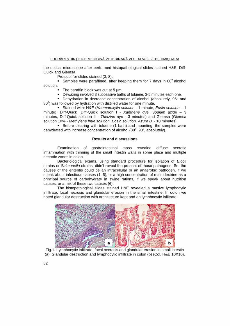
LUCRĂRI ŞTIINŢIFICE MEDICINĂ VETERINARĂ VOL. XLV(3), 2012, TIMIŞOARA
82
the optical microscope after performed histopathological slides stained H&E, Diff-Quick and Giemsa.
Protocol for slides stained (3, 8): Samples were paraffined, after keeping them for 7 days in 80
o alcohol
solution. The paraffin block was cut at 5 µm. Dewaxing involved 3 successive baths of toluene, 3-5 minutes each one. Dehydration in decrease concentration of alcohol (absolutely, 96
o and
80o) was followed by hydration with distilled water for one minute.
Stained with: H&E (Haematoxylin solution - 1 minute, Eosin solution – 1 minute), Diff-Quick (Diff-Quick solution I - Xanthene dye, Sodium azide – 3 minutes, Diff-Quick solution II - Thiazine dye - 3 minutes) and Giemsa (Giemsa solution 10% - Methylene blue solution, Eosin solution, Azure B. - 10 minutes).
Before clearing with toluene (1 bath) and mounting, the samples were dehydrated with increase concentration of alcohol (80
o, 90
o, absolutely).
Results and discussions
Examination of gastrointestinal mass revealed diffuse necrotic
inflammation with thinning of the small intestin walls in some place and multiple necrotic zones in colon.
Bacteriological exams, using standard procedure for isolation of E.coli strains or Salmonella strains, didn’t reveal the present of these pathogens. So, the causes of the enteritis could be an intracellular or an anaerobic pathogen, if we speak about infectious causes (1, 5), or a high concentration of maltodextrine as a principal source of carbohydrate in swine rations, if we speak about nutrition causes, or a mix of these two causes (6).
The histopatological slides stained H&E revealed a masive lymphocytic infiltrate, focal necrosis and glandular erosion in the small intestine. In colon we noted glandular destruction with architecture kept and an lymphocytic infiltrate.
Fig.1. Lymphocytic infiltrate, focal necrosis and glandular erosion in small intestin (a); Glandular destruction and lymphocytic infiltrate in colon (b) (Col. H&E 10X10).
a b
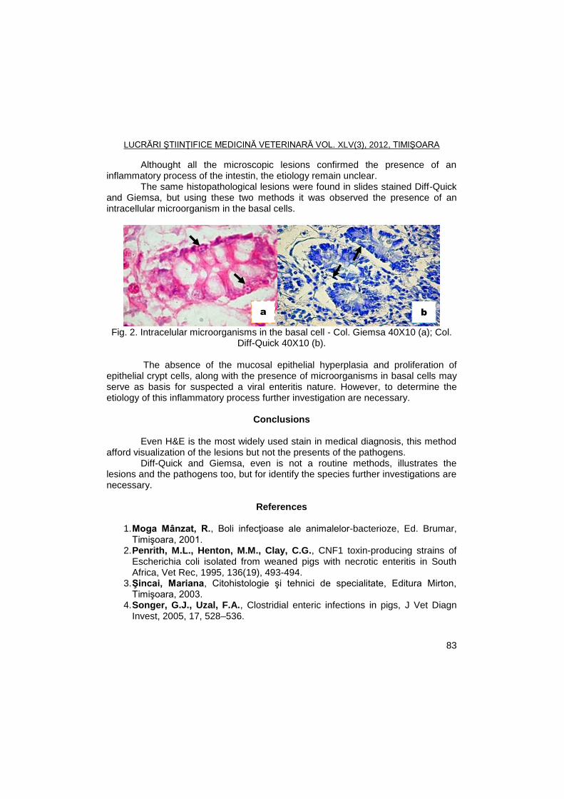
LUCRĂRI ŞTIINŢIFICE MEDICINĂ VETERINARĂ VOL. XLV(3), 2012, TIMIŞOARA
83
Althought all the microscopic lesions confirmed the presence of an inflammatory process of the intestin, the etiology remain unclear.
The same histopathological lesions were found in slides stained Diff-Quick and Giemsa, but using these two methods it was observed the presence of an intracellular microorganism in the basal cells.
Fig. 2. Intracelular microorganisms in the basal cell - Col. Giemsa 40X10 (a); Col.
Diff-Quick 40X10 (b).
The absence of the mucosal epithelial hyperplasia and proliferation of epithelial crypt cells, along with the presence of microorganisms in basal cells may serve as basis for suspected a viral enteritis nature. However, to determine the etiology of this inflammatory process further investigation are necessary.
Conclusions
Even H&E is the most widely used stain in medical diagnosis, this method
afford visualization of the lesions but not the presents of the pathogens. Diff-Quick and Giemsa, even is not a routine methods, illustrates the
lesions and the pathogens too, but for identify the species further investigations are necessary.
References
1. Moga Mânzat, R., Boli infecţioase ale animalelor-bacterioze, Ed. Brumar,
Timişoara, 2001. 2. Penrith, M.L., Henton, M.M., Clay, C.G., CNF1 toxin-producing strains of
Escherichia coli isolated from weaned pigs with necrotic enteritis in South Africa, Vet Rec, 1995, 136(19), 493-494.
3. Şincai, Mariana, Citohistologie şi tehnici de specialitate, Editura Mirton, Timişoara, 2003.
4. Songer, G.J., Uzal, F.A., Clostridial enteric infections in pigs, J Vet Diagn Invest, 2005, 17, 528–536.
a b
LUCRĂRI ŞTIINŢIFICE MEDICINĂ VETERINARĂ VOL. XLV(3), 2012, TIMIŞOARA
84
5. Straw, Barbara E., Zimmerman, J.J., D'Allaire, Sylvie, Taylor, D.J.,
Diseases of Swine, 9th Edition, Ed. Blackwell, 2006. 6. Thymann, T., Moller, H.K., Stoll, B., Stoy, A.C.F., Buddington, R.K.,
Bering, S.B., Jensen, B.B., Olutoye, O.O., Siggers, R.H., Molbak, L., Sangild, P.T., Burrin, D.G., Carbohydrate maldigestion induces necrotizing enterocolitis in preterm pigs, Am J Physiol Gastrointest Liver Physiol, 2009, 297, 1115-1125.
7. Zlotowski, Priscila, Mendes Ribeiro Corrêa, A., Santos Neves de Barcellos, D.E., Ribeiro de Itapema Cardoso, M., Vasconcelos, A.C., Amaral de Castro, L., Driemeier D., Necrotic enterocolitis in pigs naturally infected by porcine circovirus type 2, Cienc Rural, 2009, 39(6), 1801-1807
8. ***http://www.hoslink.com/histo/5.htm#5.2_campy
LUCRĂRI ŞTIINŢIFICE MEDICINĂ VETERINARĂ VOL. XLV(3), 2012, TIMIŞOARA
85
DETERMINING THE SALMONELLA SPP. CARRIER STATE IN THE CASE OF FATTENIG PIGS
I. ŢIBRU, GYÖNGYI DOBAI, ZORIŢA MARIA COCORA
Banat's University of Agricultural Sciences and Veterinary Medicine Timisoara, Faculty
of Veterinary Medicine, 300645, Aradului Street No. 119, Timisoara, Romania E-mail: [email protected]
Summary
Salmonelosis is one of the most reported zoonoses, with major problems in the world. In view of a right handling of this zoonoses attributed to the consumption of pork and pork products, control measures must be taken simultaneously, on all production levels from the farm to the consumer.
By the means of surveillance of the carrier state, by the means of microbiological examination of the faeces, and by simultaneously sanitizing the pork carcasses, I have found that, depending on the state of the queen number, there can be pigs that produce germs and pigs that do not. In order to mark the salmonellas I have used the ISO 6579/2002 method, which gives the possibility of expressing the result after at least five days, in parallel with a fast method, µ-Trac, which gave the possibility of expressing the result in 24 hours. To ensure the sanitizing of the carcasses, I have used an 0, 5% OX Virin solution which was sprayed over the carcasses before they were dried.
Key words: Salmonella spp., zoonoses, carrier, carcasses, identifying.
Animal Salmonelosis represents for most countries with intensive animal breeding, one of the most important sanitary - veterinary problems because of both economic losses and their implications in people's health by causing food poisoning as a result of contaminated products (4).
Salmonelosis is considered to be one of the most important food zoonoses causing health problems and high costs (1). Globally, millions of cases of human Salmonelosis are reported annually (3). In the European Union (EU) Salmonella was the second microorganism that caused health problems reported as zoonoses in 2008 (5). More than 130 000 cases of confirmed human Salmonelosis were reported, reaching 26 cases out of 100 000 inhabitants in 2010 (9). A big part of the infections with Salmonella spp. are not registered, so it is difficult to get a real picture of the cases of human Salmonelosis (1).
In the year 2008, a total of 131,468 cases of human Salmonelosis were reported in 27 countries. The total number of cases of human Salmonelosis reported in the EU dropped constantly from 195 947 cases in 2004, to 133258 cases in 2008 (5). The isolation of salmonellas in animals, food and the environment for the fore mentioned intervals, reach an impressive level, representing 77.3% of the 46.134 isolated and categorized stems (4). The most important food sources were, in countdown, eggs and egg products (23.1%), pork and pork products (10.2%), and
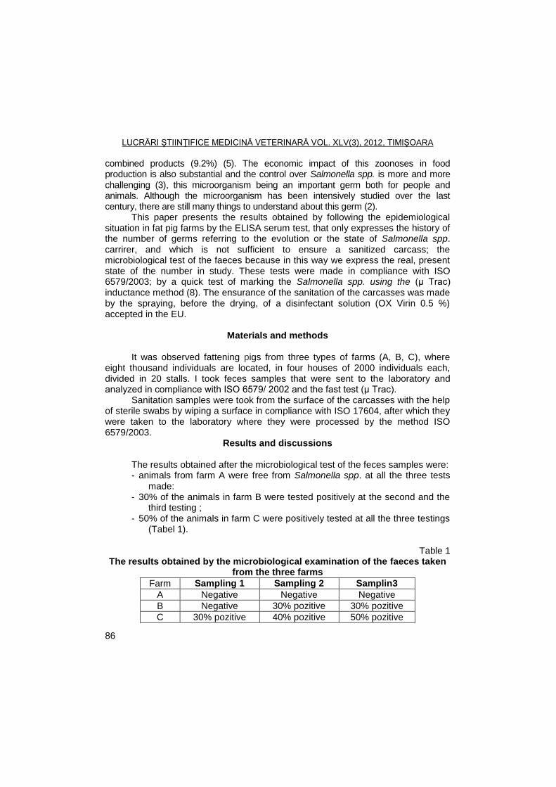
LUCRĂRI ŞTIINŢIFICE MEDICINĂ VETERINARĂ VOL. XLV(3), 2012, TIMIŞOARA
86
combined products (9.2%) (5). The economic impact of this zoonoses in food production is also substantial and the control over Salmonella spp. is more and more challenging (3), this microorganism being an important germ both for people and animals. Although the microorganism has been intensively studied over the last century, there are still many things to understand about this germ (2).
This paper presents the results obtained by following the epidemiological situation in fat pig farms by the ELISA serum test, that only expresses the history of the number of germs referring to the evolution or the state of Salmonella spp. carrirer, and which is not sufficient to ensure a sanitized carcass; the microbiological test of the faeces because in this way we express the real, present state of the number in study. These tests were made in compliance with ISO 6579/2003; by a quick test of marking the Salmonella spp. using the (µ Trac) inductance method (8). The ensurance of the sanitation of the carcasses was made by the spraying, before the drying, of a disinfectant solution (OX Virin 0.5 %) accepted in the EU.
Materials and methods
It was observed fattening pigs from three types of farms (A, B, C), where
eight thousand individuals are located, in four houses of 2000 individuals each, divided in 20 stalls. I took feces samples that were sent to the laboratory and analyzed in compliance with ISO 6579/ 2002 and the fast test (µ Trac).
Sanitation samples were took from the surface of the carcasses with the help of sterile swabs by wiping a surface in compliance with ISO 17604, after which they were taken to the laboratory where they were processed by the method ISO 6579/2003.
Results and discussions
The results obtained after the microbiological test of the feces samples were: - animals from farm A were free from Salmonella spp. at all the three tests
made: - 30% of the animals in farm B were tested positively at the second and the
third testing ; - 50% of the animals in farm C were positively tested at all the three testings
(Tabel 1).
Table 1 The results obtained by the microbiological examination of the faeces taken
from the three farms
Farm Sampling 1 Sampling 2 Samplin3
A Negative Negative Negative
B Negative 30% pozitive 30% pozitive
C 30% pozitive 40% pozitive 50% pozitive
LUCRĂRI ŞTIINŢIFICE MEDICINĂ VETERINARĂ VOL. XLV(3), 2012, TIMIŞOARA
87
Based on obtained data, we can say that pigs from farm A, came from a free for Salmonella spp. farm animals, which allowed them to maintain the status of free animals of salmonella.
Analyzing data on farm B, we observed that the animals came from a farm free for Salmonella spp., but during the process, due to uncontrolled conditions, their status was changed, in such a way that part of the animals (30%) became positive.
In farm C, as a result of analyzing the results obtained, it could notice that the animals were positive from the beginning, that all of them came from a positive farm, and because of being collected animals, the number of positive animals increased, finally reaching a percentage of 50 of the animals positive.
The results obtained by identifying the salmonellas by the μ-Trac method. By analyzing the samples through the two parallel methods, the standard method and the μ-Trac method, was noticed that the results were alike, which gives the possibility of using this method to identify the indicator microorganisms in food industry. As the time for obtaining the results for the samples is in accordance with the technological flux, there is still to discuss on the economic aspect of using this method.
The results obtained by identifying the salmonellas on the carcass surface in the slaughter-house. As during the inspection from the local authority DSVSA Timis, carcasses presenting Salmonella spp. on their surface were found, and taking into account the results from the microbiological examination concerning the state of Salmonella spp. carrier, was decided to sprayed the carcasses with an 0.5% OX Virin solution before entering the refrigeration room. The product contains 25% peroxide, 5% peracetic acid, and a mixing component belonging to the producer. After analyzing the results that were obtained, no matter where the pigs came from, there was no other positive sample, so was concluded that using the 0.5% OX Virin solution was ensure the protection of the consumer from the contamination of the surface of the carcass with Salmonella spp.
Conclusions
The microbiological method allows finding out the exact carrier state in the
case of fattening pigs before they are taken to the slaughter-house. The alternative method gives the possibility to find out on time about the
carrier state, being an obvious choice and having the same results as the standard method.
Using the OX Virin solution in 0.5% concentration allows obtaining carcasses free from germs.
LUCRĂRI ŞTIINŢIFICE MEDICINĂ VETERINARĂ VOL. XLV(3), 2012, TIMIŞOARA
88
References
1. De Jong Skierus, B., Human Salmonellosis - Impact of Travel and Trade from a Swedish Perspective, Diss. Karolinska Institutet, Stockholm, 2006.
2. Lin-Hui, S., Cheng-Hsun, C., Salmonella: Clinical Importance and Evolution of Nomenclature, PhD Thesis, Taiwan, 2007.
3. Österberg, Julia, Salmonella in Pigs Infection Dynamics of Different Serotypes, Doctoral Thesis Faculty of Veterinary Medicine and Animal Sciences, Suedia, 2010.
4. Verdeș, N., Boli produse de bacteria din genul Salmonella, In: Mânzat, R., M., (coord) Boli infecțioase ale animalelor-bacterioze, Ed. Brumar, Timișoara, 2001, p.26-34.
5. EFSA, Community summary report on trends and sources of zoonoses and zoonotic agents and food-borne outbreaks in the European Union in 2008. The EFSA Journal, 2010 8(1), 1496.
6. ***ISO 17604/2003 Microbiology of food and animal feeding stuffs - Carcass stampling for microbiological analysis..
7. ***ISO 6579/2003 Microbiologia produselor alimentare şi furajere. Metoda orizontală pentru detectarea bacteriilor din genul Salmonella, June 2003.
8. ***ISO 6570/A1/2007 Microbiologia produselor alimentare şi furajere. Metoda orizontală pentru detectarea bacteriilor din genul Salmonella - Amendament 1: Anexa D: Detecţia Salmonella Spp.din mostrele fecale de animale şi din probe de mediu din etapa de producţie primară, Octombrie.
9. ***SMI Statistik för salmonellainfektion Available from http://www.smittskyddsinstitutet.se/statistik/salmonellainfektion/?t=c#statistics-nav (January 20, 2012).
LUCRĂRI ŞTIINŢIFICE MEDICINĂ VETERINARĂ VOL. XLV(3), 2012, TIMIŞOARA
89
PREVALENCE OF F4 (K88) FIMBRIAL ADHESINS IN E. COLI STRAINS ISOLATED FROM PIGLETS IN THE SUCKLING PERIOD
JADRANKA ŢUTIŠ, O. RADANOVIŠ, V. RADOSAVLJEVIŠ, B. SAVIŠ, V. IVETIŠ, M. ŢUTIŠ
Scientific Veterinary Institute, Belgrade, V. Toze Street No. 14, Belgrade, Serbia
E-mail: [email protected]
Summary
Diarrhea during the neonatal period, caused by ETEC strains, is one of the most
present and economically most significant diseases in intensive pig production. Fimbrial adhesins and enterotoxins are considered as the main factors of the virulence of ETEC strains.
ETEC adhere to the small intestine mucosa in neonatal pigs by means of one or more of the fimbrial adhesins F4/K88), F5(K99), F6(987P), F41.They may also produce an adhesin involved in diffuse adherence (AIDA).They colonize the small intestine and produce one or more of the enterotoxins ST, LT or EAST1. The consequence of the effects of enterotoxins is the accumulation of fluids and electrolytes in the intestine lumen, which is manifested with the appearance of diarrhea. In the more grave cases, animal death occurs as a consequence of dehydration and the appearance of acidosis. In our investigations the presence of fimbrial antigens was established in 66 (76%) of the total of 87 examined strains. The results of investigations indicate a high incidence (68 %) of F4 adhesin. The presence of F6 adhesin was established in 14 (21%) strains and in 7 (11%) strains adhesins F4+F6 were present concurrently. Hemolytic activity was proven in 39 (45%) strains. The results of our investigations indicate that among different pathogens, ETEC with F4 adhesin
plays the main role in the etiology of diarrhea in neonatal piglets on pig farms in Serbia in which no immunoprophylaxis is applied.
Key words: pig, ETEC, fimbrial adhesine
Diseases of the digestive system stand out because of their high incidence and general significance. These diseases appear as a result of different causes, of which the most frequently mentioned are rota and corona viruses, TGE virus, sapovirus, coccidia, E. coli, Clostridium spp. and others (7, 17, 4). Among different pathogens, enterotoxic E.coli (ETEC) has for a number of years been the most significant cause of diseases of the digestive tract in sucklings piglets (6, 3). Diarrhea during the neonatal period, caused by ETEC strains, is one of the most present and economically most significant diseases in intensive pig production (16, 14).
Neonatal diarrhea associated with E.coli is observed most commonly in pigs aged from 0-4 days. Causative strains produce one or more enterotoxins and are referred to as ETEC.ETEC adhere to the small intestine mucosa in neonatal pigs by means of one or more of the fimbrial adhesins F4/K88), F5(K99), F6(987P), F41. They may also produce an adhesin involved in diffuse adherence (AIDA).They
LUCRĂRI ŞTIINŢIFICE MEDICINĂ VETERINARĂ VOL. XLV(3), 2012, TIMIŞOARA
90
colonize the small intestine and produce one or more of the enterotoxins ST, LT or EAST1 (5). Newborn piglets ingest ETEC found in their enviroment especially from the mammary glands of the mother and the farrowing crate. Factors that promote development of diarrhea include poor hygiene, inadequate disinfection, a continuous-farrowing system, an ambient temperature of less than 25
oC, or
excessive air currents. Diarrhea during the neonatal period implies the disease in susceptible
piglets infected with enterotoxic strains of E. coli. After entering the host organism, enterotoxic strains of E. coli adhere to targetted cells, multiply and produce enterotoxins. The consequence of the effects of enterotoxins is the accumulation of fluids and electrolytes in the intestine lumen, which is manifested with the appearance of diarrhea (2). In the more grave cases, animal death occurs as a consequence of dehydration and the appearence of acidosis.
Fimbrial adhesins and enterotoxins are considered as the main factors of the virulence of ETEC strains (3). More than 30 types of fimbrial antigens have been discovered so far, and F4 adhesin is considered as the one with the highest incidence (15, 10).
Acceptance through adhesin F4 is specific mostly for piglets; however, certain animals do not possess a receptor in intestine epithelial cells for this adhesin, which is why they are naturally resistent to infection with ETEC strains that contain F4 adhesins. The F4 adhesin initially thought to be K antigen (K88), was subseqently found to be fine fibrilar structure. F4 (K88) fimbriae mediate bacterial adherence to the intestinal epithelium throughout most of the small intestine of pigs of all ages.Hence, colonization of the intestinal mucosa by F4 positive ETEC occurs in both neonatal and postweaning pigs and may be obsreved in finisher pigs.There are three variants of F4, namely ab,ac and ad (1).The most commonly encountered variant is ac. Up to 50% of pigs lack the receptors for the F4 adhesin.
Since E. coli is an integral part of the intestine microbiome, its isolation during the diagnostic procedure does not confirm an etiological connection with the incidence of enteritis in piglets. It is necessary at the same time to establish factors of virulence of the isolated strains, such as production of enterotoxins, adhesin, and other. Due to its practical implementation, the method of determining the antigen structure of the isolated strains is often applied in the laboratory. The reason for this is that there is a high degree of correlation between certain antigens, possibilities for enterotoxin production, and the presence of fimbrial adhesins (2, 13). Enterotoxic strains of E. coli produce two main classes of enterotoxins, thermo-stable ST and thermo-labile LT (5), and shiga toxin-producing strains STEC have also been discovered (8).
The most frequently observed ETEC strains in cases of piglet diarrhea during the neonatal period belonged to classic serogroups O8, O147, O149 and O157, with dominant F4 fimbrial adhesins (15).
LUCRĂRI ŞTIINŢIFICE MEDICINĂ VETERINARĂ VOL. XLV(3), 2012, TIMIŞOARA
91
The objective of the investigations was to establish the presence and distribution of certain fimbrial adhesins, and hemolytic activity of strains of E. coli isolated from piglets. Samples were collected from 2 different farms located in regions of eastern Serbia in the years 2010-2011.
Materials and methods
The samples for the investigations were rectal swabs of piglets with
clinically manifest diarrhea. Samples of the small intestine on which pathoanatomical changes were observed were taken from dead piglets. The piglets were maintained on a concrete floor with or without straw and a floor heating system, and they originated from mothers non-vaccinated against E. coli infections. In most cases, samples were taken with the first appearance of diarrhea. All the samples originated from suckling piglets aged 0-30 days.
On the same day that they were taken, the samples were inoculated on agar with 5% sheep’s blood, nutritive and MacConkey agar (Himedia) and incubated in aerobic conditions at a temperature of 37
oC for 24 hours. Colonies suspected of
being E. coli were subcultivation, and upon obtaining a pure culture, speedy identification was performed of the biochemical characteristics by inoculated on TSI agar and carrying out the IMVIC test. The identification was confirmed using BBL Crystal, E/N, ID kit (Becton Dickinson)
For the identification of fimbrial antigen, commercial antiserums F4(K88), F5(K99) and 987P (Toxigenic E. coli pili antisera, Denka Seiken, Japan) were used. The cultivation of the strains for the identification of fimbrial adhesins, as well as the performance of the agglutination test and the interpretation of the results were carried out according to the instructions of the antiserum manufacturer. Hemolysin production was monitored on blood agar, and only those strains with a clearly expressed hemolysis zone were considered hemolytic strains.
Results and discussions
The presence of fimbrial antigens was established in 66 (76%) of the total
of 87 examined strains.The dominance of F4 fimbrial adhesin was established, as it was discovered in 45 (68 %) strains. The presence of F6 adhesin was established in 14 strains (21 %) and in 7(11%) strains F4+F6 adhesins were present concurently (Figure 1).
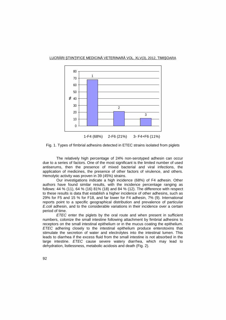
LUCRĂRI ŞTIINŢIFICE MEDICINĂ VETERINARĂ VOL. XLV(3), 2012, TIMIŞOARA
92
1-F4 (68%) 2-F6 (21%) 3- F4+F6 (11%)
Fig. 1. Types of fimbrial adhesins detected in ETEC strains isolated from piglets
The relatively high percentage of 24% non-serotyped adhesin can occur due to a series of factors. One of the most significant is the limited number of used antiserums, then the presence of mixed bacterial and viral infections, the application of medicines, the presence of other factors of virulence, and others. Hemolytic activity was proven in 39 (45%) strains.
Our investigations indicate a high incidence (68%) of F4 adhesin. Other authors have found similar results, with the incidence percentage ranging as follows: 44 % (11), 64 % (16) 81% (18) and 84 % (12). The difference with respect to these results is data that establish a higher incidence of other adhesins, such as 29% for F5 and 15 % for F18, and far lower for F4 adhesin, 7% (9). International reports point to a specific geographical distribution and prevalence of particular E.coli adhesin, and to the considerable variations in their incidence over a certain period of time.
ETEC enter the piglets by the oral route and when present in sufficient numbers, colonize the small intestine following attachment by fimbrial adhesins to receptors on the small intestinal epithelium or in the mucus coating the epithelium. ETEC adhering closely to the intestinal epithelium produce enterotoxins that stimulate the secretion of water and electrolytes into the intestinal lumen. This leads to diarrhea if the excess fluid from the small intestine is not absorbed in the large intestine. ETEC cause severe watery diarrhea, which may lead to dehydration, listlessness, metabolic acidosis and death (Fig. 2).
1
2
3
0
10
20
30
40
50
60
70
80%
LUCRĂRI ŞTIINŢIFICE MEDICINĂ VETERINARĂ VOL. XLV(3), 2012, TIMIŞOARA
93
Fig, 2. Pathogenesis of ETEC
(http://www.ecl-lab.ca/en/ecoli/pathogenesis.asp#etec)
It is an undeniable fact that today, in intensive pig production, neonatal diarrhea is one of the most present and economically most significant diseases that leads to major losses in the early postnatal period. In the epidemiology of this disease, the hygiene of the environment has the greatest role. Under non-hygienic conditions, as soon as they are born piglets come into contact with dirt and pathogenic strains of different species of microorganisms, including ETEC strains.
Stress factors of the environment that contribute to the occurrence of diseases include bad ambient conditions, especially sudden changes in temperature and humidity, the presence of different gases, dust with numerous microorganisms, overcrowding, as well as intercurrent infections with various other pathogens. Furthermore, in the event that piglets are for any reason unable to take colostrum they are extremely susceptible to infections.
Conclusions
The results of our investigations indicate that among different pathogens, E.coli, with F4 adhesin plays the main role in the etiology of diarrhea in neonatal piglets on pig farms in Serbia in which no immunoprophylaxis is applied.
We consider that it is necessary not only to continue, but even to expand this type of investigations, with the objective to identify other enteric pathogens.
LUCRĂRI ŞTIINŢIFICE MEDICINĂ VETERINARĂ VOL. XLV(3), 2012, TIMIŞOARA
94
In addition to the health aspect and the feasibility of production, there is the knowledge that, after the presence of shiga toxin-producing E. coli strains (STEC) has been established in piglets, there is the potential threat that piglets could become a reservoir and source of infection in humans.
Acknowledgments
This research was supported by a grant from the Ministry of Education and
Science of the Republic of Serbia, Project No. TR.31079.
References
1. Alexa, P., Štouraţova, K., Hamrik, J., Rychlik, I., Gene typing of the colonisation factors K88(F4) in enterotoxigenic Escherichia coli strains isolated from diarrhoeic piglets, Vet Med Czech, 2001, 46, 46-9.
2. Blanco, M., Lazo, L., Blanco, J.E., Dahbi, G., Mora, A., Lopez, C., Serotypes, virulence genes and PFGE patterns of enteropathogenic E. coli isolated from Cuban pigs with diarrhea, Int Microbiol, 2006, 9, 1, 53-60.
3. Chen, X., Zhao, L., Gao, S., Miao, X., Jiao, X., Liu, X.,Virulence genes of pathogenic Escherichia coli from ill pigs in China and their relationship with O-serogroups, Wei Sheng Wu Xue Bao, 2008, 48, 7, 857-62.
4. Darong, C., Yuan, Z.S., Lang, C.X., Pan, G.X., Wei, D.W., Chang, S.H., Rapid diagnosis of ETEC and HPIharboring E. coli infection in newborn piglets with diarrhea, Afr J Microbiol Res, 2010,7, 575-80.
5. Fairbrother, J.M., Gyles,C.l., Escherichia coli infections In:Diseases of swine., Ed:Straw BE, Zimmerman JJ, DAllaire S, Taylor DJ, Blackwell.2006.
6. Francis, DH., Enterotoxigenic Escherichia coli infection in pigs and its diagnosis, J Swine Health Prod,2002, 10, 4, 171-5.
7. Katsuda, K., Kohmoto, M., Kawashima, K., Tsunemitsu, H., Frequency of enteropathogen detection in suckling and weaned pigs with diarrhea in Japan, J Vet Diagn Invest, 2006, 18, 350-4.
8. Kim, Y.J., Kim, J.H., Hur, J., Lee, J.H., Isolation of Escherichia coli from piglets in South Korea with diarrhea and characteristics of the virulence genes, Can J Vet Res,2010, 74, 1, 59-64.
9. Lee, S.I., Kang, S.G., Kang, M.L., Yoo, HS., Development of multiplex polymerase chain reaction assays for detecting enterotoxigenic Escherichia coli and their application to field isolates from piglets with diarrhea, J Vet Diagn Invest,2008, 20, 4, 492-6.
10. Moon, H.W., Cornick, N.A., Hoffman, L.J., Bosworth, B.T., Prevalence of virulence factors among hemolytic Escherichia coli, 2010, http://www.extension.iastate.edu
11. Vidotto, M.C., deLima, N.C.S., Fritzen, J.T.T., deFreitas, J.C., Venancio, E.J., Ono, M.A., Frequency of virulence genes in Escherichia coli
LUCRĂRI ŞTIINŢIFICE MEDICINĂ VETERINARĂ VOL. XLV(3), 2012, TIMIŞOARA
95
strains isolated from piglets with diarrhea in the North Parana State Brazil, Braz J Microbiol, 2009, 40, 1, 199-204.
12. Vu-Khac, H., Holoda, E., Majerţiak, M., Gaspar, G., Pilipţinec, E.,Genotyping of fimbrial adhesions in Escherichia coli strains isolated from Slovak piglets suffering from diarrhea, Folia Microbiologica, 2004, 49, 1, 59-63.
13. Vu-Khac, H., Holoda, E., Pilipţinec, E., Blanco, M., Blanco, J.E., Dahbi, G.,Serotypes, virulencegenes, intimin types and PFGE profiles of Escherichia coli isolated from piglets with diarrhea in Slovakia, Vet J, 2007, 174, 1, 176-87.
14. White, M., Neonatal colibacillosis, NADIS, Pig Health, 2009, 1-3. 15. Yan, X., Huang, X., Ren, J., Zou, Z., Yang, S., Ouyang, J., Distribution of
Escherichia coli F4 adhesin phenotypes in pigs of 15 chinese and western breeds and a white DurocxErhulian intercross, J Med Microbiol, 2009, 58, 1112-7.
16. Zhang, W., Zhao, M., Ruesch, L., Omot, A., Francis, D., Prevalence of virulence genes in Escherichia coli strains recently isolated from young pigs with diarrhea in the US, Vet Microbiol, 2007,123, 145- 52.
17. Zhang, W., Shen, Q., Hua, X., Cui, L., Liu, J., Yang, S.,The first Chinese porcine Sapovirus strain that contributed to an outbreak of gastroenteritis in piglets, Virology, 2008, 82, 16, 8239-40.
18. Zutic, Jadranka., Asanin, Jelena., Misic, D., Jakic-Dimic, D., Milic, N., Asanin, R., Stojanovic, D., Zutic M., Isolation of ETEC strains from piglets with diarrhea in the neonatal period and their typization based on somatic and fimbrial antigen, Acta Veterinaria, 2010,.60, 5-6, 497-506.
LUCRĂRI ŞTIINŢIFICE MEDICINĂ VETERINARĂ VOL. XLV(3), 2012, TIMIŞOARA
96
INTESTINAL PARASITES INFECTIONS IN DOGS POPULATION FROM TIMIS AND ARAD COUNTIES
SIDONIA ANDREI, M.S. ILIE, GH. DĂRĂBUŞ
Banat's University of Agricultural Sciences and Veterinary Medicine Timisoara, Faculty
of Veterinary Medicine, 300645, Aradului Street No. 119, Timisoara, Romania E-mail: [email protected]
Summary
This study has been conducted to determine the frequency of intestinal parasites infections in dogs population from Timis and Arad counties. For the purpose, 320 fecal samples were examined using simple flotation, flotation and sedimentation method and direct faecal smear techniques. 20 dogs have been necropsied and examined in detail for the presence of adult parasites. The study animals were dogs from shelter, stray dogs, kennels and dogs with owner. The overall prevalence of intestinal parasites in dogs was 73.44%. In this study have been recorded infestations with only one species 46.88% and multiple species 26.56%. Ancylostoma caninum (41.25%) and Toxocara canis (21.25%) were most commonly detected, followed by Trichocephalus vulpis (20.62), Uncynaria stenocephala 12.81%, Dipilidium caninum 3.43%, Isospora species 1.87%, Stongyloides stercoralis 1.56% and Toxascaris leonina 0.62%. The only species that has not been found in mixed infestation was Strongyloides stercoralis. The most parasitized, were the dogs from shelter 90.74% and stray dogs 88.64% followed by dogs with owner 59.35% and dogs from kennel 46.15%. There were statistically significant differences in the prevalence of parasite infection between ages of dogs (p= 0.04), also between categories of dogs from shelter and owner dogs where (p= 0.001), but no difference between sexes p ˃0.999999. In conclusion, the prevalence of intestinal parasites in dogs is high, and the presences of these are a real risk for public health.
Key words: dog, intestinal parasites, prevalence, zoonosis, Timis and Arad
Intestinal parasites are present in dog population with infections reported from all parts of the world. Dogs are definitive host for several endoparasites that cause important diseases in man and animals. Dog feces harbouring infective parasitic forms are potential sources of environmental contamination representing a high risk of infection for the people (11).
Besides dogs with owners, stray dogs are major factors in the spread of parasitic diseases therefore have been recognized as a significant public health problem worldwide especially in rural area. In our country there is no available recent data about the incidence of different parasitic infections in dogs.
The aim of the present work is to determine the diversity and prevalence of the canine intestinal parasites from Timis and Arad counties. We estimate the prevalence of intestinal parasites and the epidemiological significance of the identified parasite species.
LUCRĂRI ŞTIINŢIFICE MEDICINĂ VETERINARĂ VOL. XLV(3), 2012, TIMIŞOARA
97
Materials and methods
Study area and period The study was performed on animals from Timis and Arad counties during
the period between October 2010 and March 2012. Study animals The study animals were dogs from four sources (shelter, dog with owners,
stray dogs and kennels). From 320 dogs examined, 20 death animals have been necropsied and examined in detail. The sex and approximate age using dentition formulae have been recorded. The fecale samples were collected from other 300 dogs. Some of these dogs have never been dewormed. The numbers of male and female dogs were 234 and 86, respectively.
Coproscopic examination The faecal samples were collected directly from rectum of the dogs, and
were also collected from floor cages or ground and then transferred into sterile containers. Samples were brought to the Parasitological laboratory, Faculty of Veterinary Medicine, Timisoara for further examination. Samples were stored in a refrigerator at 4°C until processing. Coprological examination for the detection of parasite eggs was performed using simple floatation, flotation and sedimentation method and direct faecal smear techniques (9, 8). Each faecal sample was examined microscopically for intestinal parasite eggs. All parasite eggs have been identified based on their morphological characteristic. Identification was made by measuring each egg and by observing their characters (21).
Postmortem examination Gastrointestinal tract was dissected out. The esophagus, stomach, small
intestine, ceacum and colon were separated. Each of the portions was opened and examined for the presence of parasites. Contents of small intestine were collected by flushing with warm water. The mucosal surface scraped was made using a gut runner. Sedimentation method was performed to determine the presence of parasites. Identification and counting of parasites were made by Soulsby 1982 (20).
Statistical analysis Collected data were entered, stored and analyzed using statistic excel
spreadsheet. Descriptive statistics were generated and presented as tables. For the
epidemiological studies the prevalence was calculated by dividing the number of animals harbouring any parasites by the total animals examined.
Statistical analysis was carried out by using Fisher's Exact test and chi-square tests, and the differences were considered significant when p ≤ 0.05.
Results and discussions
The overall prevalence of intestinal parasites in dog population from Timis and Arad counties was 73.44% (235/320) as indicated in Table 1.
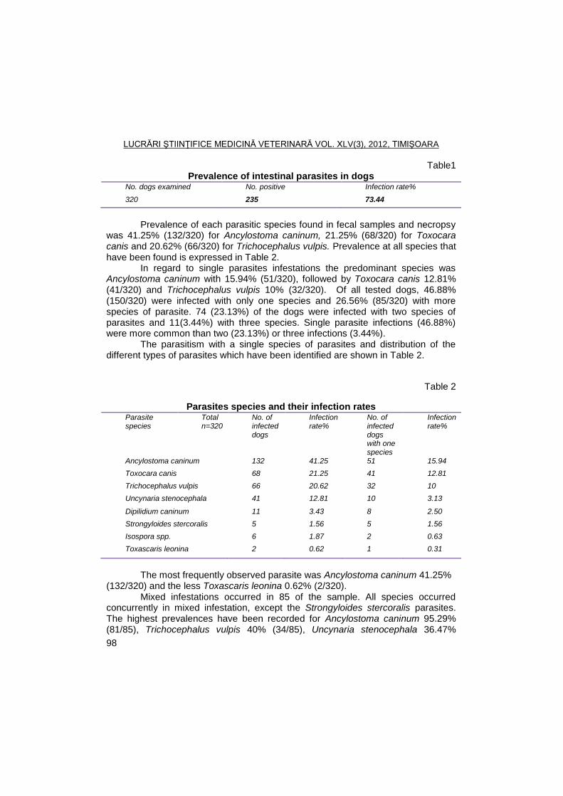
LUCRĂRI ŞTIINŢIFICE MEDICINĂ VETERINARĂ VOL. XLV(3), 2012, TIMIŞOARA
98
Table1 Prevalence of intestinal parasites in dogs
No. dogs examined No. positive Infection rate%
320 235 73.44
Prevalence of each parasitic species found in fecal samples and necropsy
was 41.25% (132/320) for Ancylostoma caninum, 21.25% (68/320) for Toxocara canis and 20.62% (66/320) for Trichocephalus vulpis. Prevalence at all species that have been found is expressed in Table 2.
In regard to single parasites infestations the predominant species was Ancylostoma caninum with 15.94% (51/320), followed by Toxocara canis 12.81% (41/320) and Trichocephalus vulpis 10% (32/320). Of all tested dogs, 46.88% (150/320) were infected with only one species and 26.56% (85/320) with more species of parasite. 74 (23.13%) of the dogs were infected with two species of parasites and 11(3.44%) with three species. Single parasite infections (46.88%) were more common than two (23.13%) or three infections (3.44%).
The parasitism with a single species of parasites and distribution of the different types of parasites which have been identified are shown in Table 2.
Table 2
Parasites species and their infection rates Parasite species
Total n=320
No. of infected dogs
Infection rate%
No. of infected dogs with one species
Infection rate%
Ancylostoma caninum 132 41.25 51 15.94
Toxocara canis 68 21.25 41 12.81
Trichocephalus vulpis 66 20.62 32 10
Uncynaria stenocephala 41 12.81 10 3.13
Dipilidium caninum 11 3.43 8 2.50
Strongyloides stercoralis 5 1.56 5 1.56
Isospora spp. 6 1.87 2 0.63
Toxascaris leonina 2 0.62 1 0.31
The most frequently observed parasite was Ancylostoma caninum 41.25%
(132/320) and the less Toxascaris leonina 0.62% (2/320). Mixed infestations occurred in 85 of the sample. All species occurred
concurrently in mixed infestation, except the Strongyloides stercoralis parasites. The highest prevalences have been recorded for Ancylostoma caninum 95.29% (81/85), Trichocephalus vulpis 40% (34/85), Uncynaria stenocephala 36.47%
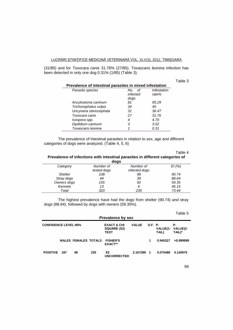
LUCRĂRI ŞTIINŢIFICE MEDICINĂ VETERINARĂ VOL. XLV(3), 2012, TIMIŞOARA
99
(31/85) and for Toxocara canis 31.76% (27/85). Toxascaris leonina infection has
been detected in only one dog 0.31% (1/85) (Table 3).
Table 3 Prevalence of intestinal parasites in mixed infestation
Parasite species No. of infected dogs
Infestation rate%
Ancylostoma caninum 81 95.29
Trichocephalus vulpis 34 40
Uncynaria stenocephala 31 36.47
Toxocara canis 27 31.76
Isospora spp. 4 4.70
Dipilidium caninum 3 3.52
Toxascaris leonina 1 0.31
The prevalence of intestinal parasites in relation to sex, age and different
categories of dogs were analyzed. (Table 4, 5, 6)
Table 4 Prevalence of infections with intestinal parasites in different categories of
dogs
The highest prevalence have had the dogs from shelter (90.74) and stray
dogs (88.64), followed by dogs with owners (59.35%).
Table 5 Prevalence by sex
CONFIDENCE LEVEL:95% EXACT & CHI SQUARE (Χ2) TEST
VALUE D.F. P-VALUE(1-TAIL)
P-VALUE(2-TAIL)*
MALES FEMALES TOTALS FISHER'S EXACT**
1 0.945327 ˃0.999999
POSITIVE 187 48 235 Χ2 UNCORRECTED
2.167289 1 0.070488 0.140975
Category Number of tested dogs
Number of infected dogs
EI (%)
Shelter 108 98 90.74 Stray dogs 44 39 88.64
Owners dogs 155 92 59.35 Kennels 13 6 46.15
Total 320 235 73.44
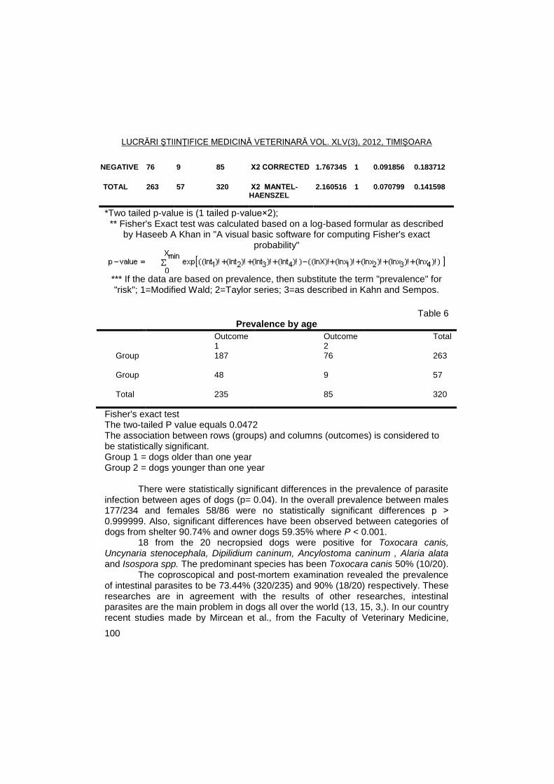
LUCRĂRI ŞTIINŢIFICE MEDICINĂ VETERINARĂ VOL. XLV(3), 2012, TIMIŞOARA
100
NEGATIVE 76 9 85 Χ2 CORRECTED 1.767345 1 0.091856 0.183712
TOTAL 263 57 320 Χ2 MANTEL-HAENSZEL
2.160516 1 0.070799 0.141598
*Two tailed p-value is (1 tailed p-value×2); ** Fisher's Exact test was calculated based on a log-based formular as described
by Haseeb A Khan in "A visual basic software for computing Fisher's exact probability"
*** If the data are based on prevalence, then substitute the term "prevalence" for "risk"; 1=Modified Wald; 2=Taylor series; 3=as described in Kahn and Sempos.
Table 6
Prevalence by age
Outcome 1
Outcome 2
Total
Group 1
187 76 263
Group 2
48 9 57
Total 235 85 320
Fisher's exact test The two-tailed P value equals 0.0472 The association between rows (groups) and columns (outcomes) is considered to be statistically significant. Group 1 = dogs older than one year Group 2 = dogs younger than one year
There were statistically significant differences in the prevalence of parasite
infection between ages of dogs (p= 0.04). In the overall prevalence between males 177/234 and females 58/86 were no statistically significant differences p ˃ 0.999999. Also, significant differences have been observed between categories of dogs from shelter 90.74% and owner dogs 59.35% where P < 0.001.
18 from the 20 necropsied dogs were positive for Toxocara canis, Uncynaria stenocephala, Dipilidium caninum, Ancylostoma caninum , Alaria alata and Isospora spp. The predominant species has been Toxocara canis 50% (10/20).
The coproscopical and post-mortem examination revealed the prevalence of intestinal parasites to be 73.44% (320/235) and 90% (18/20) respectively. These researches are in agreement with the results of other researches, intestinal parasites are the main problem in dogs all over the world (13, 15, 3,). In our country recent studies made by Mircean et al., from the Faculty of Veterinary Medicine,
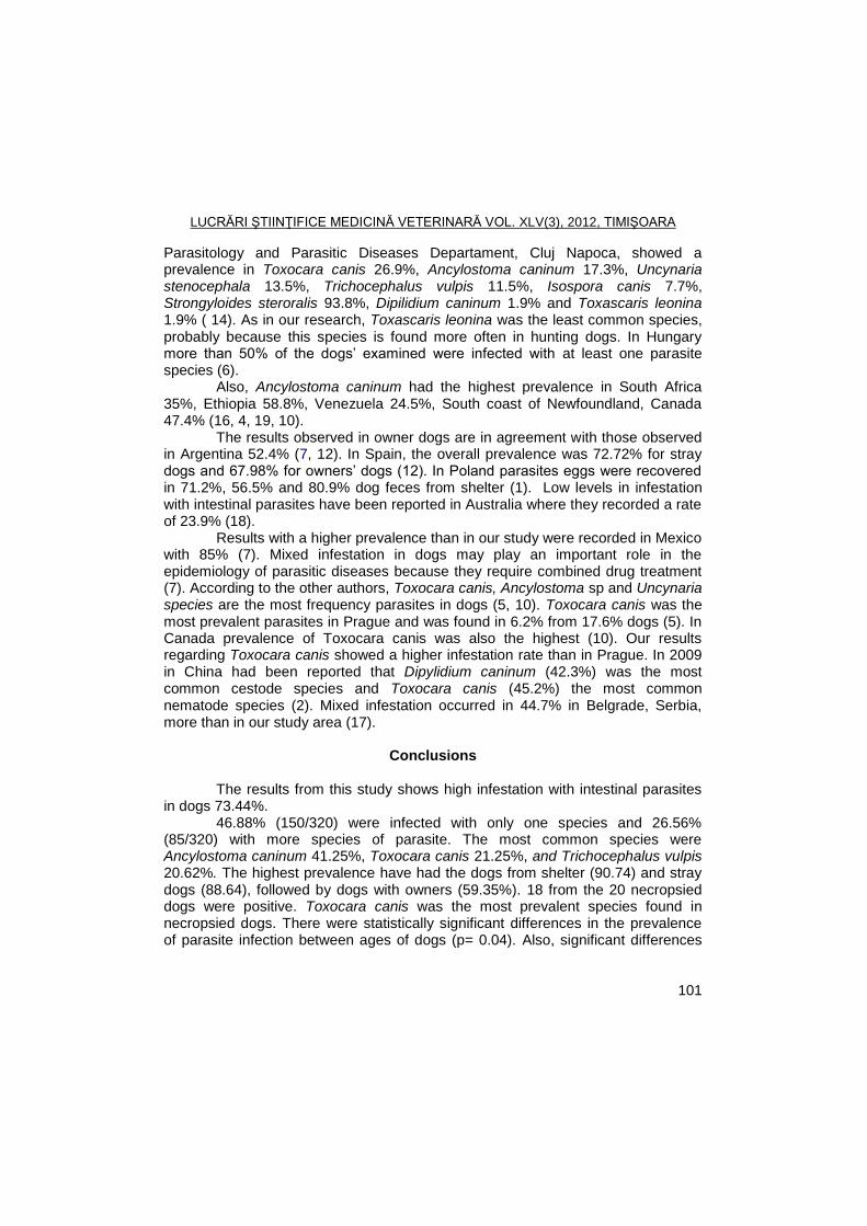
LUCRĂRI ŞTIINŢIFICE MEDICINĂ VETERINARĂ VOL. XLV(3), 2012, TIMIŞOARA
101
Parasitology and Parasitic Diseases Departament, Cluj Napoca, showed a prevalence in Toxocara canis 26.9%, Ancylostoma caninum 17.3%, Uncynaria stenocephala 13.5%, Trichocephalus vulpis 11.5%, Isospora canis 7.7%, Strongyloides steroralis 93.8%, Dipilidium caninum 1.9% and Toxascaris leonina 1.9% ( 14). As in our research, Toxascaris leonina was the least common species, probably because this species is found more often in hunting dogs. In Hungary more than 50% of the dogs’ examined were infected with at least one parasite species (6).
Also, Ancylostoma caninum had the highest prevalence in South Africa 35%, Ethiopia 58.8%, Venezuela 24.5%, South coast of Newfoundland, Canada 47.4% (16, 4, 19, 10).
The results observed in owner dogs are in agreement with those observed in Argentina 52.4% (7, 12). In Spain, the overall prevalence was 72.72% for stray dogs and 67.98% for owners’ dogs (12). In Poland parasites eggs were recovered in 71.2%, 56.5% and 80.9% dog feces from shelter (1). Low levels in infestation with intestinal parasites have been reported in Australia where they recorded a rate of 23.9% (18).
Results with a higher prevalence than in our study were recorded in Mexico with 85% (7). Mixed infestation in dogs may play an important role in the epidemiology of parasitic diseases because they require combined drug treatment (7). According to the other authors, Toxocara canis, Ancylostoma sp and Uncynaria species are the most frequency parasites in dogs (5, 10). Toxocara canis was the most prevalent parasites in Prague and was found in 6.2% from 17.6% dogs (5). In Canada prevalence of Toxocara canis was also the highest (10). Our results regarding Toxocara canis showed a higher infestation rate than in Prague. In 2009 in China had been reported that Dipylidium caninum (42.3%) was the most common cestode species and Toxocara canis (45.2%) the most common nematode species (2). Mixed infestation occurred in 44.7% in Belgrade, Serbia, more than in our study area (17).
Conclusions
The results from this study shows high infestation with intestinal parasites
in dogs 73.44%. 46.88% (150/320) were infected with only one species and 26.56%
(85/320) with more species of parasite. The most common species were Ancylostoma caninum 41.25%, Toxocara canis 21.25%, and Trichocephalus vulpis 20.62%. The highest prevalence have had the dogs from shelter (90.74) and stray dogs (88.64), followed by dogs with owners (59.35%). 18 from the 20 necropsied dogs were positive. Toxocara canis was the most prevalent species found in necropsied dogs. There were statistically significant differences in the prevalence of parasite infection between ages of dogs (p= 0.04). Also, significant differences
LUCRĂRI ŞTIINŢIFICE MEDICINĂ VETERINARĂ VOL. XLV(3), 2012, TIMIŞOARA
102
have been observed between categories of dogs from shelter 90.74% and owner dogs 59.35% where P < 0.001.
Between males 177/234 and females 58/86 were no statistically significant differences p ˃ 0.999999. All the species of parasites recorded in the present work have potential zoonotic importance. Strategic deworming and public education in preventing transmission of diseases from dog and control of zoonotic diseases have major importance. It is important to know the significance of zoonotic diseases caused by intestinal parasites to take measures for further control, regarding the spread and environmental contamination.
References
1. Borecka, A., Prevalence of intestinal nematodes of dogs in the Warsaw
area, Poland, Helminthologia, 2005, 42, 1:35-39. 2. Dai, R.S., Li, Z.Y., Li, F., Liu, D.X., Liu, W., Liu, G.H., He, S.W., Tan, M.Y.,
Lin, R.Q., Liu, Y., Zhu, X.Q., Severe infection of adult dogs with helminths in Hunan Province, China poses significant public health concerns, Veterinary Parasitology, 2009, 160, 348–350.
3. Davoust, B., Normand, T., Bourry, O., Dang, H., Leroy, E., Bourdoiseau, G., Epidemiological survey on gastro-intestinal and blood-borne helminths of dogs in North-East Gabon, Onderstepoort Jornal Veterinary Research, 2008, 75(4): 359-364.
4. Degefu, H., Tefera Abyot, Yohannes, M., Zoonotic helminth parasites in faecal samples of household dogs in Jimma Town, Ethiopia, Journal of Public Health and Epidemiology, 2011, 3(4),138-143.
5. Dubna, S., Langrova, I., Nápravník, J., Jankovská, I., Vadlejch, J., Pekár, S., Fechtner, J., The prevalence of intestinal parasites in dogs from Prague, rural areas, and shelters of the Czech Republic, Veterinary Parasitology, 2007,145(1-2):120-8.
6. Fok, E., Szatmári, V., Busák, K., Rozgonyi, F., Prevalence of intestinal parasites in dogs in some urban and rural areas of Hungary, Veterinary Quarterly, 2001, 23 (2), 96-8.
7. Fontanarrosa, M.F., Vezzani, D., Basabe, J., Eiras, D.F., An epidemiological study of gastrointestinal parasites of dogs from Southern Greater Buenos Aires (Argentina): age, gender, breed, mixed infections, and seasonal and spatial patterns, Veterinary Parasitolology, 2006, 136, 283–295.
8. Hendrix, C.M., Diagnostic Veterinary Technicians, 3rd Edition, Mosby, Inc., USA, 2006.
9. Hendrix, C.M., Sirois. M., Laboratory Procedures for Veterinary Technicians, Mosby, Inc., USA, 2007.
LUCRĂRI ŞTIINŢIFICE MEDICINĂ VETERINARĂ VOL. XLV(3), 2012, TIMIŞOARA
103
10. Hugh Whitney, Bridger Kimberly E., Gastrointestinal parasites in dogs from the Island of St. Pierre off the south coast of Newfoundland, Veterinary Parasitology, 2009, 162, 1–2, 26, 167–170.
11. Lavallén, C.M., Dopchiz, M.C., Lobianco, E., Hollmann, P., Denegri, G., Intestinal parasites of zoonotic importance in dogs from the District of General Pueyrredón (Buenos Aires, Argentina). Rev. vet, 2011, 22: 1, 19–24.
12. Martinez-Moreno, F.J., Hernandez, S., Lopez-Cobos, E., Becerra, C., Acosta, I., Martinez-Moreno, A., Estimation of canine intestinal parasites in Cordoba (Spain) and their risk to public health, Veterinary Parasitology, 2007, 143: 7-13.
13. Minnaar, W.N., Krecek, R.C., Fourie, L.J., Helminths in dogs from a peri-urban resource-limited community in Free State Province, South Africa. Parasitology, 2002, 107(4): 343-349.
14. Mircean, Viorica, Adriana, Gyӧrke, Vasile C., Prevalence and risk factors of Giardia duodenalis in dogs from Romania, Veterinary Parasitology, 2012,184: 325-329.
15. Muhairwa, A.P., Nonga, H.E., Kusiluka, L.J., A retrospective study of intestinal helminthosis as a cause of clinical disease in dogs, Tanzania Veterinary Journal, 2008 25(1): 24-30.
16. MukaratirwaI, S., Singh, V.P., Prevalence of gastrointestinal parasites of stray dogs impounded by the Society for the Prevention of Cruelty to Animals (SPCA), Durban and Coast, South Africa, Journal of the South African Veterinary Association, Pretoria 2010, 81, 2.
17. Nikoliš Aleksandra , Dimitrijeviš Sanda, Katiš-Radivojeviš Sofija , Klun Ivana, Bobiš Branko, Djurkoviš-Djakoviš Olgica, High prevalence of intestinal zoonotic parasites in dogs from Belgrade, Serbia — Short communication, Acta Veterinaria Hungarica, 2008, 56(3),335-340.
18. Palmer, C.S., Andrew Thomson, R.C., Traub, Rebeca, J., Rees, R., Roberton, I.D., National sudy of the gastrointestinal parasites of dogs and cats in Australia, Veterinary Parasitology, 2008,151: 181-190.
19. Ram´ırez-Barrios, R.A., Barboza-Mena, G., Muñoz, J., Angulo-Cubillán, F., Hernández Elena, Fany González Escalona, F., Prevalence of intestinal parasites in dogs under veterinary care in Maracaibo, Venezuela, Veterinary Parasitology,2004, 121,11–20.
20. Soulsby, E.L.J., Helminths, Arthropodes and Protozoa of Domestic Animals. 7th Edn., The English Language Book Society and Bailliere Tindall, London, 1982.
21. Yakob, H.T., Ayele,T., Fikru, R., Basu A.K., Gastrointestinal nematodes in dogs from Debre Zeit: Ethiopia. Veterinary Parasitology, 2007, 148: 144-148.
LUCRĂRI ŞTIINŢIFICE MEDICINĂ VETERINARĂ VOL. XLV(3), 2012, TIMIŞOARA
104
TOXOPLASMA GONDII SEROPREVALENCE IN CATS AND SHEEP FROM CARAS-SEVERIN COUNTY, ROMANIA
IONELA HOTEA, M.S. ILIE, MIRELA IMRE, DENISA SORESCU, D. INDRE,
ILEANA BRUDIU, OLIMPIA COLIBAR, GH. DĂRĂBUŞ
Banat’s University of Agricultural Sciences and Veterinary Medicine, Faculty of Veterinary Medicine, 300645, Aradului Street, no. 119, Timisoara, Romania
E-mail: [email protected]
Summary
To determine the seroprevalence of Toxoplasma gondii infection in cats and sheep in the Caras-Severin county ELISA method was used. From cats were collected 62 samples of which 48 (77.42%) were positive for T. gondii infection and of the 450 studied sheep, 276 (61.33%) had anti-Toxoplasma antibodies.
Coproscopic, in a fecal sample from cats were identified Toxoplasma type oocysts and in other samples were observed different parasite species: eggs of Ancylostoma/Uncinaria spp., Isospora spp. type oocysts, eggs of Toxocara cati and Taenia spp.
Key words: Toxoplasma gondii, cats, sheep, Caras-Severin, Romania
Toxoplasmosis is one of the most common parasites in humans and
animals. Toxoplasmosis causes economic losses in livestock, with abortion, embryonic mortality and non-viable products (2, 11). Intermediate hosts are numerous species of birds and mammals, including humans. The serological surveillance was found that T. gondii infection is widespread in farm animals, sheep being the most susceptible specie (2). At felide, definitive hosts, T. gondii develop in the small intestine, while in the intermediate hosts, the parasite forms pseudocysts and cysts in the brain and then the muscles and other organs (1, 2). The epidemiology is influenced by many factors, such as deficitary growth management and maintenance of farm animals, cats density, climatic conditions affecting the oocysts sporulation (temperature, humidity, wind), geographical area, and various culinary and cultural habits of consumers (1, 3). At European level, interest of specialists to toxoplasmosis is outstanding (4, 6, 8). In Romania, research on this zoonosis are developing (5, 7, 11), which motivates the purpose of this study.
Materials and methods
To assess the prevalence of Toxoplasma gondii infection, in Caras-
Severin, during 2008 - 2010, 62 cats and 450 sheep were examined. Cats were divided into two age categories: cats aged three months and
one year, representing the category of youth and adult cats over the age of one
LUCRĂRI ŞTIINŢIFICE MEDICINĂ VETERINARĂ VOL. XLV(3), 2012, TIMIŞOARA
105
year and two months. 36 cats were European breed, and 26 were of other breeds, seven of the Persian breed, 12 Burmese, six Siamese and one of Russian Blue breed. Cats were from different backgrounds: urban or rural, maintained in apartment or house, with or without outdoors access.
Studied sheep were aged between 3 and 5 years, female and belonged to non-business units. Samples were collected from nine localities of the County, from each locality every 50 samples. The farms contained between 350 and 2300 sheep.
For the study, sheep blood was harvested from the jugular vein, in containers without anticoagulant and than the serum was separated and stored in a freezer (-20 ° C).
From all cats were collected, individual, fecal and blood samples. For serological processing, blood was collected from the cephalic vein forearm, and samples were collected in individual and numbered containers. Microscopic examination of feces from definitive host (cat) was achieved by flotation method with sugar solution which had the same technique so as Willis method, except that instead of saturated NaCl solution, we used a mixture of 300 ml water, 500 g sugar and 6.5 g of phenol crystals (1). Serum samples from both species were processed in the laboratory of Parasitology and Parasitic Diseases of the Faculty of Veterinary Medicine, Timisoara by indirect ELISA using VET ID Screen Multi-species kits (ID-VET Innovative Diagnostics , France). Interpretation of results was done according to the values of S / P: ≤ 40% - negative, 40% ≥ - ≤ 50% - uncertain, 50% ≥ - ≤ 200% - positive, ≥ 200% - strongly positive.
The results were statistically processed with Minitab and EpiInfo 2000 programs.
Results and discussions
Of the 62 examined cats, 48 were found positive for Toxoplasma gondii
infection (77.42%), and 14 were negative. Table 1 presents the percentages of positivity obtained and the influence of different factors on disease prevalence. It is noted that toxoplasmosis is common in adult cats in rural areas with access to outdoors and who eat raw meat, the males and cats of European race.
After examining the feces in one fecal sample were identified oocysts of Toxoplasma gondii/Hammondia hammondi, but no tests were performed to identify the molecular species (fig. 1). By ELISA, in this sample were identified specific anti-Toxoplasma antibodies. In the other samples were not identified as oocysts.
Some feces had other parasitic species. Of 62 examined samples were identified 23 positive samples. Of these, three samples showed eggs of Ancylostoma/Uncinaria spp., seven samples - Isospora spp. type oocysts, eight samples were positive for Toxocara cati, and five of the samples was revealed Taenia spp. 17, of 24 coproscopic positive cats revealed anti-Toxoplasma IgG antibodies.
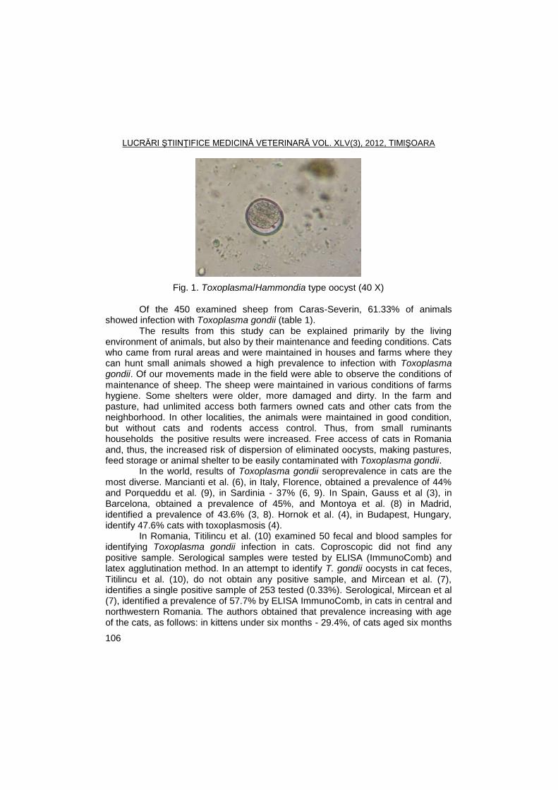
LUCRĂRI ŞTIINŢIFICE MEDICINĂ VETERINARĂ VOL. XLV(3), 2012, TIMIŞOARA
106
Fig. 1. Toxoplasma/Hammondia type oocyst (40 X) Of the 450 examined sheep from Caras-Severin, 61.33% of animals
showed infection with Toxoplasma gondii (table 1). The results from this study can be explained primarily by the living
environment of animals, but also by their maintenance and feeding conditions. Cats who came from rural areas and were maintained in houses and farms where they can hunt small animals showed a high prevalence to infection with Toxoplasma gondii. Of our movements made in the field were able to observe the conditions of maintenance of sheep. The sheep were maintained in various conditions of farms hygiene. Some shelters were older, more damaged and dirty. In the farm and pasture, had unlimited access both farmers owned cats and other cats from the neighborhood. In other localities, the animals were maintained in good condition, but without cats and rodents access control. Thus, from small ruminants households the positive results were increased. Free access of cats in Romania and, thus, the increased risk of dispersion of eliminated oocysts, making pastures, feed storage or animal shelter to be easily contaminated with Toxoplasma gondii.
In the world, results of Toxoplasma gondii seroprevalence in cats are the most diverse. Mancianti et al. (6), in Italy, Florence, obtained a prevalence of 44% and Porqueddu et al. (9), in Sardinia - 37% (6, 9). In Spain, Gauss et al (3), in Barcelona, obtained a prevalence of 45%, and Montoya et al. (8) in Madrid, identified a prevalence of 43.6% (3, 8). Hornok et al. (4), in Budapest, Hungary, identify 47.6% cats with toxoplasmosis (4).
In Romania, Titilincu et al. (10) examined 50 fecal and blood samples for identifying Toxoplasma gondii infection in cats. Coproscopic did not find any positive sample. Serological samples were tested by ELISA (ImmunoComb) and latex agglutination method. In an attempt to identify T. gondii oocysts in cat feces, Titilincu et al. (10), do not obtain any positive sample, and Mircean et al. (7), identifies a single positive sample of 253 tested (0.33%). Serological, Mircean et al (7), identified a prevalence of 57.7% by ELISA ImmunoComb, in cats in central and northwestern Romania. The authors obtained that prevalence increasing with age of the cats, as follows: in kittens under six months - 29.4%, of cats aged six months
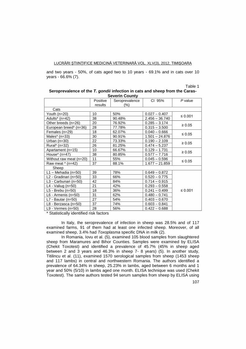
LUCRĂRI ŞTIINŢIFICE MEDICINĂ VETERINARĂ VOL. XLV(3), 2012, TIMIŞOARA
107
and two years - 50%, of cats aged two to 10 years - 69.1% and in cats over 10 years - 66.6% (7).
Table 1
Seroprevalence of the T. gondii infection in cats and sheep from the Caras-Severin County
Positive results
Seroprevalence (%)
CI 95% P value
Cats
Youth (n=20) 10 50% 0.027 – 0.407 ≤ 0.001
Adults* (n=42) 38 90.48% 2.456 – 36.740
Other breeds (n=26) 20 76.92% 0.285 – 3.174 ≥ 0.05
European breed* (n=36) 28 77.78% 0.315 – 3.500
Females (n=29) 18 62.07% 0.040 – 0.666 ≤ 0.05
Males* (n=33) 30 90.91% 1.501 – 24.876
Urban (n=30) 22 73.33% 0.190 – 2.109 ≥ 0.05
Rural* (n=32) 26 81.25% 0.474 – 5.237
Apartament (n=15) 10 66.67% 0.129 – 1.731 ≥ 0.05
House* (n=47) 38 80.85% 0.577 – 7.716
Without raw meat (n=20) 11 55% 0.045 – 0.596 ≤ 0.05
Raw meat * (n=42) 37 88.1% 1.677 – 21.859
Sheep
L1 – Mehadia (n=50) 39 78% 0.649 – 0.872
≤ 0.001
L2 - Gradinari (n=50) 33 66% 0.520 – 0.775
L3 - Carbunari (n=50) 42 84% 0.714 – 0.915
L4 - Valiug (n=50) 21 42% 0.293 – 0.558
L5 - Brebu (n=50) 18 36% 0.241 – 0.499
L6 - Armenis (n=50) 31 62% 0.480 – 0.741
L7 - Bautar (n=50) 27 54% 0.403 – 0.670
L8 - Berzasca (n=50) 37 74% 0.603 – 0.841
L9 - Vermes (n=50) 28 56% 0.422 – 0.688
* Statistically identified risk factors
In Italy, the seroprevalence of infection in sheep was 28.5% and of 117 examined farms, 91 of them had at least one infected sheep. Moreover, of all examined sheep, 3.4% had Toxoplasma specific DNA in milk (2).
In Romania, Iovu et al. (5), examined 105 blood samples from slaughtered sheep from Maramures and Bihor Counties. Samples were examined by ELISA (Chekit Toxotest) and identified a prevalence of 45.7% (45% in sheep aged between 2 and 3 years and 46.3% in sheep 7- 8 years) (5). In another study, Titilincu et al. (11), examined 1570 serological samples from sheep (1453 sheep and 117 lambs) in central and northwestern Romania. The authors identified a prevalence of 64.34% in sheep, 25.23% in lambs, aged between 6 months and 1 year and 50% (5/10) in lambs aged one month. ELISA technique was used (Chekit Toxotest). The same authors tested 94 serum samples from sheep by ELISA using
LUCRĂRI ŞTIINŢIFICE MEDICINĂ VETERINARĂ VOL. XLV(3), 2012, TIMIŞOARA
108
a kit prepared in the laboratory with Toxoplasma gondii RH strain. Samples were retested with the IFAT technique. The prevalence obtained was 55.3% by ELISA, 50% by IFAT technique and 64% by commercial ELISA - Chekit Toxotest. Correlation between the three tests was good (k = 0.81) (12).
Thus, increased prevalence obtained in this study, both in cats and sheep, pull the alarm about the possibility of transmission of Toxoplasma gondii infection in the population of Romania, especially in risk groups (pregnant women, immunosuppressed individuals), but also other species of carnivores or omnivores.
Conclusions
In Caras-Severin County, the Toxoplasma gondii seroprevalence in cats,
was 77.42% and 61.33% in sheep, with variations between 36 and 84%. Toxoplasmosis seropositivity in cats can be influenced by age, sex, breed,
place of origin and maintenance, the outdoors acces of cats and feeding cats with raw meat.
In one sample were identified Toxoplasma gondii type oocysts and specific anti-Toxoplasma IgG antibodies which directs us to the diagnosis of toxoplasmosis.
On microscopic examination of feces samples were identified parasitic associated infestations with: Toxocara cati, Isospora spp., Ancylostoma/ Uncinaria spp. and Taenia spp.
Free and uncontrolled acces of cats in shelters and feed storage was the main risk factor for Toxoplasma gondii infection, identified in sheep farms in the Caras-Severin County.
Acknowledgements
This work was supported by CNCSIS, Bd, grant No. 87/2008 obtained by
Ionela Hotea and by CNMP, grant PC No. 51-013/2007.
References
1. Anderson, B.C., Patterns of shedding of cryptosporidial oocysts in Idaho calves, J. Am. Vet. Med. Assoc., 1981, 181, 484-485.
2. Fusco, Giovanna, Rinaldib, Laura, Guarinoa, A., Yolande, Therese, Rose, Prorogaa, Antonella, Pescea, De Marco, Giuseppinaa, Cringolib, G., Toxoplasma gondii in sheep from the Campania region (Italy), Vet. Parasitol., 2007, 149, 271-274.
3. Gauss, C.B., Almería, S., Ortuño, A., Garcia, F., Dubey, J.P., Seroprevalence of Toxoplasma gondii antibodies in domestic cats from Barcelona, Spain, Vet. Bull., 2004, 74, 353.
LUCRĂRI ŞTIINŢIFICE MEDICINĂ VETERINARĂ VOL. XLV(3), 2012, TIMIŞOARA
109
4. Hornok, S., Edelhofer, R., Joachim, A., Farkas, R., Berta, K., Répási, A., Lakatos, B., Seroprevalence of Toxoplasma gondii and Neospora caninum infection of cats in Hungary, Acta Vet. Hung., 2008, 56, 81-88.
5. Iovu, Anamaria, Titilincu, Adriana, Mircean, Viorica, Blaga, R., Bejan, A., Cozma, V., Serosurvey of Toxoplasma gondii in sheep for human consumption in two slaughter-house, Bulletin UASMV, Veterinary Medicine, 2008, 65, 40-43.
6. Mancianti, F., Nardoni, S., Ariti, G., Parlanti, D., Giuliani, G., Papini, R.A., Cross-sectional survey of Toxoplasma gondii infection in colony cats from urban Florence (Italy), J. Feline Med. Surg., 2010, 12, 351-354.
7. Mircean, Viorica, Titilincu, Adriana, Cozma, V., Prevalence of Toxoplasma gondii infection in domestic cats from Romania, Toxoplasma Centennial Congress, Abstract book, 2008, 103.
8. Montoya, A., Miró, G., Mateo, M., Ramírez, C., Fuentes, I., Detection of Toxoplasma gondii in cats by comparing bioassay in mice and polymerase chain reaction (PCR), Vet. Parasitol., 2010, 160, 159-162.
9. Porqueddu, M., Scala, A., Tilocca, V., Principal endoparasitoses of domestic cats in Sardinia, Vet. Bull., 2004, 74, 1454.
10. Titilincu, Adriana, Mircean, Viorica, Blaga, R., Chiţimia, Lidia, Cernea, M., Mirescu, F., Cozma, V., Research regarding the prevalence of Toxoplasma gondii infection in cats, Rev. Rom. Med. Vet., 2008, 18, 108-122.
11. Titilincu, Adriana, Mircean, Viorica, Iovu, Anamaria, Blaga, R., Onac, Diana, Cozma, V., Seroprevalence of Toxoplasma gondii infection in small ruminants from Romania, World Association for the Advancement of Veterinary Parasitology, Canada, 2009, Abstracts PO1.26, 126.
12. Titilincu, Adriana, Mircean, Viorica, Iovu, Anamaria, Cozma, V., Development of an indirect ELISA test using tachyzoite crude antigen for sero-diagnosis of sheep Toxoplasma gondii infection, Bulletin UASMV, Veterinary Medicine, 2009, 66, 137-141.
LUCRĂRI ŞTIINŢIFICE MEDICINĂ VETERINARĂ VOL. XLV(3), 2012, TIMIŞOARA
110
THE THERAPEUTIC EFFICACY OF GARDAL 10% (ALBENDAZOLE SULFOXIDE) AGAINST DIGESTIVE AND
PULMONARY PARASITIC FAUNA IN NATURALLY INFESTED GOATS
OLIMPIA C. IACOB
University of Agriculture Science and Veterinary Medicine „Ion Ionescu de la Brad‖,
700490, Mihail Sadoveanu Alley No. 6-8, Iasi, Romania E-mail: [email protected]
Summary
Investigations were conducted in 2011 on a number of 1500 goats, Carpathian breed and half breed with Saanen goats from Costești-Botoșani area, the main goal being to test the efficacy of Gardal 10% (albendazolesulfoxide) on digestive and pulmonary parasitic fauna. Samples of feces were collected from 5% of the herd, considering the age, sex and physiological state, pre- and post therapy. The samples were analyzed by ovoscopic and larvoscopic qualitative methods (Willis, Vajda) and quantitative methods (McMaster, Euzeby), determining the pre- and post- therapeutic dynamics of intensity (OPG) and extensivity (E%) of digestive and pulmonary invasive elements. The results were statistically interpreted by Student test.
The obtained coproscopic results revealed the different infestation of the herd concerning the age, with species of Eimeria (OPG x:50 in adults, 115 in fawns, 245 in young goats with E of 30-60%), species of Moniezia (OPG x:395 in fawns, 490 in young goats, 270 in adults, with E of 20-50%), species of Nematodirus (OPG x:1545 in fawns, 1375 in young goats, 545 in adults, with E of 100%), other species of Trichostrongylidae (OPG x:1240 in fawns, 1395 in young goats, 2230 in adults, with E of 90-100%), species from Protostrongylusgenus (Lpg X:3,6 in young goats, 9,2 in adults with E of 40-80%), Dictyocaulusfilaria have not been identified in the studied herd (Lpg:0).
Gardal 10% therapy in a dosis of 7,5mg/kg body weight recorded a different efficacy on cestodes and digestive and pulmonary nematodes. Thus, in adults, the therapeutic efficacy have been of 100% on nematodes from the folowing genus: Protostrongylus, Nematodirus and Moniezia; in young goats the efficacy have been of 100% against Protostrongylus and 90% against Nematodirus, 80% against cestodes and 40%
against other Trichostrongylidae; in fawns the efficacy have been of 80% against Nematodirus, 60% against cestodes. The comparative pre- and post therapy results (Student test) showed significantly differences regarding the efficacy ogGardal 10% on Nematodirus genus in adults and in young goats; significantly differences in fawns;
significantly differences regarding the efficacy against other Trichostrongylidae in adults and young goats. Also, significantly differences have been recorded for Protostrongylus.
Key words: goats, digestive and, pulmonary infestation, albendazolesulfoxide,
efficacy
Small ruminantsparasitosis benefits of plenty therapeutic formulas with great efficacy just for a limited period of time. The excessive use of a single drug
LUCRĂRI ŞTIINŢIFICE MEDICINĂ VETERINARĂ VOL. XLV(3), 2012, TIMIŞOARA
111
within animal population leads to the appearance of resistance to drugs and diminish their efficacy (4). The therapeutic formulas used until now (albendazole, fenbendazole, ivermectine, etc.) against parasitic fauna do not lead to maximum efficacy against all parasitic species from the targeted organic segments (3, 5, 6).
Regarding the therapy of small ruminantsparasitosis, in our country, sheeps and goats are treated as a single population with the same features, an unjust fact. The goats have a different metabolism, more dynamic and with numerous species particularities, requiring bigger therapeutic doses. The reactivity of goats against parasitic aggression is different requiring a specific approach of pathology in general and of parasitosis in particular. The aim of this study have been to test for the first in a herd a new therapeutic formula – albendazolesulfoxide (Gardal 10%) against digestive and pulmonary parasitic infestation in goats and to appreciate the therapeutic efficacy.
Material and methods
The investigations were conducted in 2011 at the end of grazing season on
a herd of Carpathian goats and half breed Saanen goats. The samples of feces were collected by rectal examination from 5% of the herd, considering the age, physiological state and sex and were ovoscopic and larvoscopic analyzed by qualitative (Willis and Vajda) and quantitative (McMaster and Euzeby) methods. There have been studied the dynamics of intensity (OPG, Lpg) and extensivity (E%) of invasive elements and have been drawn parasitograms. The therapy have been done with albendazolesulfoxide as suspension in the medical product Gardal 10%, used for the first time in the studied herd of goats, in a doses of 7,5 mg/kg body weight, orally administered, with an autodosing gun. The appreciation of efficacy have been done by repeating the coproscopic exams after 7 days since the start of the treatment. The obtained results were classified in tables and graphically represented. There have been calculated level statistical parameters (average and average error and dispersion parameters (minimum and maximum value, variation amplitude, dispersion, standard deviation and variation coefficient (v%)). The results were statistically interpreted by Student test.
Results and discussions
The coproscopic results regarding the digestive and pulmonary infestation
in fawns in October and statistical indices are shown in table 1; the invasive elements dynamics is observed in figure 1.
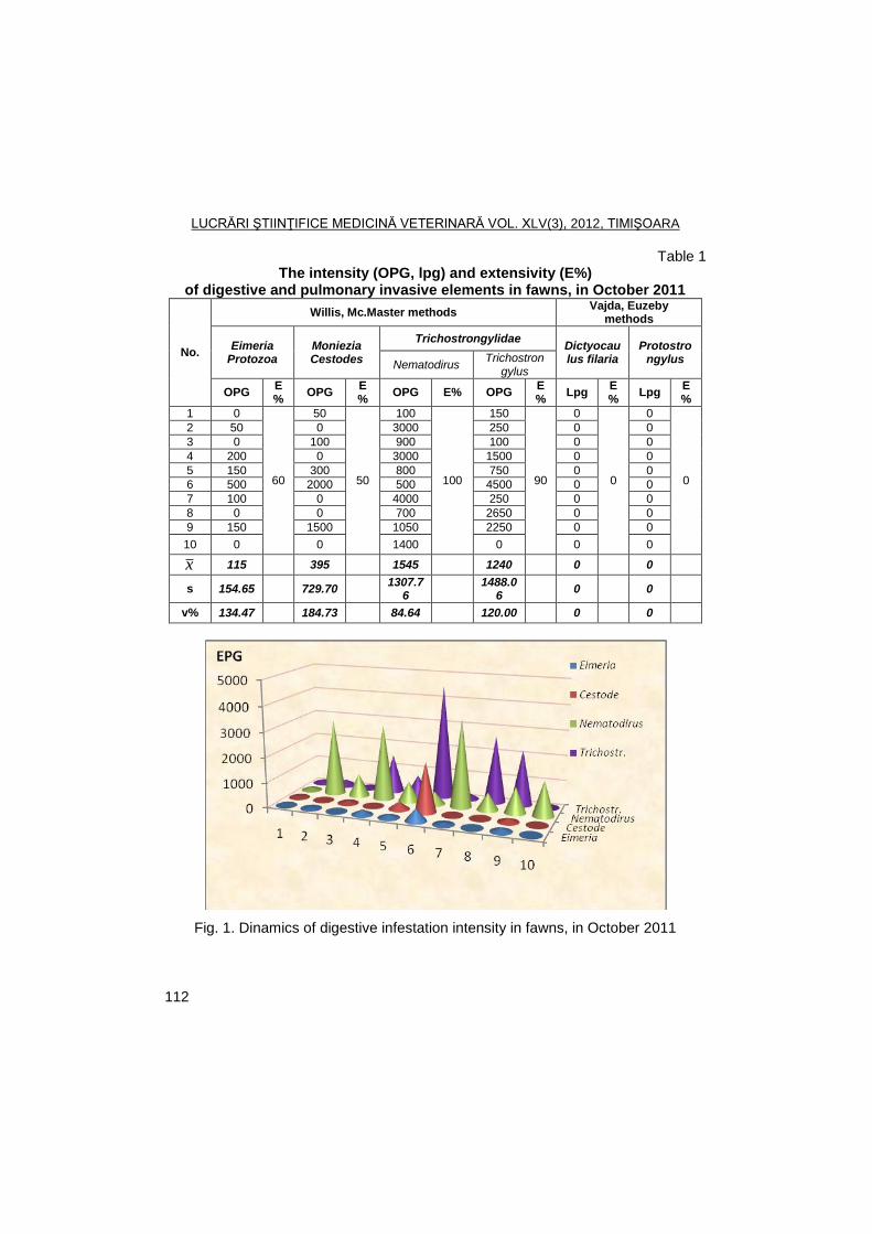
LUCRĂRI ŞTIINŢIFICE MEDICINĂ VETERINARĂ VOL. XLV(3), 2012, TIMIŞOARA
112
Table 1 The intensity (OPG, lpg) and extensivity (E%)
of digestive and pulmonary invasive elements in fawns, in October 2011
No.
Willis, Mc.Master methods Vajda, Euzeby
methods
Eimeria Protozoa
Moniezia Cestodes
Trichostrongylidae Dictyocau lus filaria
Protostrongylus
Nematodirus Trichostron
gylus
OPG E%
OPG E%
OPG E% OPG E%
Lpg E%
Lpg E%
1 0
60
50
50
100
100
150
90
0
0
0
0
2 50 0 3000 250 0 0
3 0 100 900 100 0 0
4 200 0 3000 1500 0 0
5 150 300 800 750 0 0
6 500 2000 500 4500 0 0
7 100 0 4000 250 0 0
8 0 0 700 2650 0 0
9 150 1500 1050 2250 0 0
10 0 0 1400 0 0 0
x 115 395 1545 1240 0 0
s 154.65 729.70 1307.7
6
1488.06
0 0
v% 134.47 184.73 84.64 120.00 0 0
Fig. 1. Dinamics of digestive infestation intensity in fawns, in October 2011
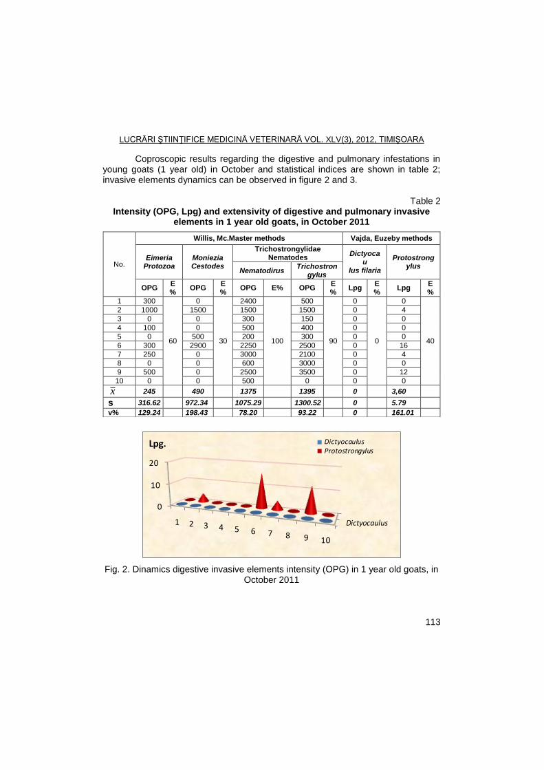
LUCRĂRI ŞTIINŢIFICE MEDICINĂ VETERINARĂ VOL. XLV(3), 2012, TIMIŞOARA
113
Coproscopic results regarding the digestive and pulmonary infestations in young goats (1 year old) in October and statistical indices are shown in table 2; invasive elements dynamics can be observed in figure 2 and 3.
Table 2
Intensity (OPG, Lpg) and extensivity of digestive and pulmonary invasive elements in 1 year old goats, in October 2011
Dictyocaulus
0
10
20
1 2 3 4 5 6 7 8 9 10
DictyocaulusProtostrongylus
Lpg.
Fig. 2. Dinamics digestive invasive elements intensity (OPG) in 1 year old goats, in October 2011
No.
Willis, Mc.Master methods Vajda, Euzeby methods
Eimeria Protozoa
Moniezia Cestodes
Trichostrongylidae Nematodes
Dictyocau
lus filaria
Protostrongylus
Nematodirus Trichostron
gylus
OPG E%
OPG E%
OPG E% OPG E%
Lpg E%
Lpg E%
1 300
60
0
30
2400
100
500
90
0
0
0
40
2 1000 1500 1500 1500 0 4
3 0 0 300 150 0 0
4 100 0 500 400 0 0
5 0 500 200 300 0 0
6 300 2900 2250 2500 0 16
7 250 0 3000 2100 0 4
8 0 0 600 3000 0 0
9 500 0 2500 3500 0 12
10 0 0 500 0 0 0
x 245 490 1375 1395 0 3,60
s 316.62 972.34 1075.29 1300.52 0 5.79
v% 129.24 198.43 78.20 93.22 0 161.01
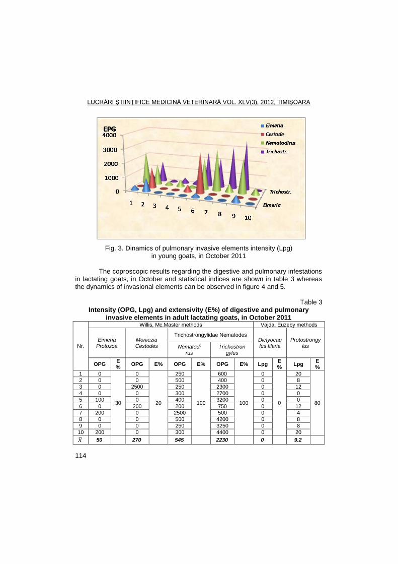
LUCRĂRI ŞTIINŢIFICE MEDICINĂ VETERINARĂ VOL. XLV(3), 2012, TIMIŞOARA
114
Fig. 3. Dinamics of pulmonary invasive elements intensity (Lpg) in young goats, in October 2011
The coproscopic results regarding the digestive and pulmonary infestations
in lactating goats, in October and statistical indices are shown in table 3 whereas the dynamics of invasional elements can be observed in figure 4 and 5.
Table 3
Intensity (OPG, Lpg) and extensivity (E%) of digestive and pulmonary invasive elements in adult lactating goats, in October 2011
Nr.
Willis, Mc.Master methods Vajda, Euzeby methods
Eimeria Protozoa
Moniezia Cestodes
Trichostrongylidae Nematodes Dictyocau lus filaria
Protostrongylus Nematodi
rus Trichostron
gylus
OPG E%
OPG E% OPG E% OPG E% Lpg E%
Lpg E%
1 0
30
0
20
250
100
600
100
0
0
20
80
2 0 0 500 400 0 8
3 0 2500 250 2300 0 12
4 0 0 300 2700 0 0
5 100 0 400 3200 0 0
6 0 200 200 750 0 12
7 200 0 2500 500 0 4
8 0 0 500 4200 0 8
9 0 0 250 3250 0 8
10 200 0 300 4400 0 20
x 50 270 545 2230 0 9.2
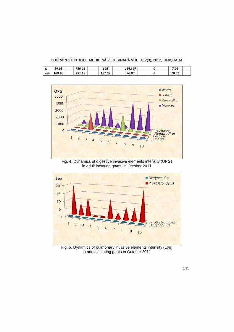
LUCRĂRI ŞTIINŢIFICE MEDICINĂ VETERINARĂ VOL. XLV(3), 2012, TIMIŞOARA
115
s 84.98 786.05 695 1562.97 0 7.06
v% 169.96 291.13 127.52 70.08 0 76.82
Fig. 4. Dynamics of digestive invasive elements intensity (OPG)
in adult lactating goats, in October 2011
Fig. 5. Dynamics of pulmonary invasive elements intensity (Lpg)
in adult lactating goats in October 2011
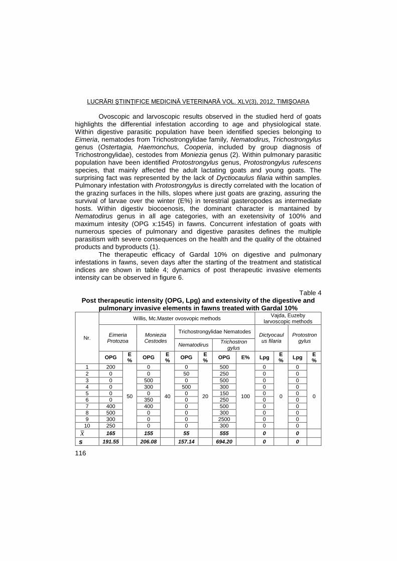
LUCRĂRI ŞTIINŢIFICE MEDICINĂ VETERINARĂ VOL. XLV(3), 2012, TIMIŞOARA
116
Ovoscopic and larvoscopic results observed in the studied herd of goats highlights the differential infestation according to age and physiological state. Within digestive parasitic population have been identified species belonging to Eimeria, nematodes from Trichostrongylidae family, Nematodirus, Trichostrongylus genus (Ostertagia, Haemonchus, Cooperia, included by group diagnosis of Trichostrongylidae), cestodes from Moniezia genus (2). Within pulmonary parasitic population have been identified Protostrongylus genus, Protostrongylus rufescens species, that mainly affected the adult lactating goats and young goats. The surprising fact was represented by the lack of Dyctiocaulus filaria within samples. Pulmonary infestation with Protostrongylus is directly correlated with the location of the grazing surfaces in the hills, slopes where just goats are grazing, assuring the survival of larvae over the winter (E%) in terestrial gasteropodes as intermediate hosts. Within digestiv biocoenosis, the dominant character is mantained by Nematodirus genus in all age categories, with an exetensivity of 100% and maximum intesity (OPG x:1545) in fawns. Concurrent infestation of goats with numerous species of pulmonary and digestive parasites defines the multiple parasitism with severe consequences on the health and the quality of the obtained products and byproducts (1).
The therapeutic efficacy of Gardal 10% on digestive and pulmonary infestations in fawns, seven days after the starting of the treatment and statistical indices are shown in table 4; dynamics of post therapeutic invasive elements intensity can be observed in figure 6.
Table 4
Post therapeutic intensity (OPG, Lpg) and extensivity of the digestive and pulmonary invasive elements in fawns treated with Gardal 10%
Nr.
Willis, Mc.Master ovosvopic methods Vajda, Euzeby
larvoscopic methods
Eimeria Protozoa
Moniezia Cestodes
Trichostrongylidae Nematodes Dictyocaulus filaria
Protostron gylus
Nematodirus Trichostron
gylus
OPG E%
OPG E%
OPG E%
OPG E% Lpg E%
Lpg E%
1 200
50
0
40
0
20
500
100
0
0
0
0
2 0 0 50 250 0 0
3 0 500 0 500 0 0
4 0 300 500 300 0 0
5 0 0 0 150 0 0
6 0 350 0 250 0 0
7 400 400 0 500 0 0
8 500 0 0 300 0 0
9 300 0 0 2500 0 0
10 250 0 0 300 0 0
x 165 155 55 555 0 0
s 191.55 206.08 157.14 694.20 0 0
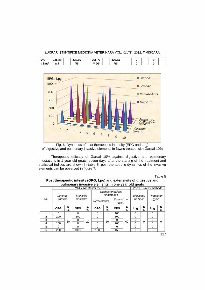
LUCRĂRI ŞTIINŢIFICE MEDICINĂ VETERINARĂ VOL. XLV(3), 2012, TIMIŞOARA
117
v% 116.09 132.96 285.71 125.08 0 0
t Stud NS NS ** DS NS 0 0
Fig. 6. Dynamics of post therapeutic intensity (EPG and Lpg)
of digestive and pulmonary invasive elements in fawns treated with Gardal 10%
Therapeutic efficacy of Gardal 10% against digestive and pulmonary infestations in 1 year old goats, seven days after the starting of the treatment and statistical indices are shown in table 5; post therapeutic dynamics of the invasive elements can be observed in figure 7.
Table 5
Post therapeutic intesity (OPG, Lpg) and extensivity of digestive and pulmonary invasive elements in one year old goats
Nr.
Willis, Mc.Master methods Vajda, Euzeby methods
Eimeria Protozoa
Moniezia Cestodes
Trichostrongylidae Nematodes Dictyocau
lus filaria Protostron
gylus Nematodirus
Trichostron gylus
OPG E%
OPG E%
OPG E%
OPG E%
Lpg E%
Lpg E%
1 0
50
0
20
0
10
100
60
0
0
0
0
2 200 500 0 500 0 0
3 0 0 0 0 0 0
4 100 0 0 200 0 0
5 0 0 0 0 0 0
6 250 1000 100 150 0 0
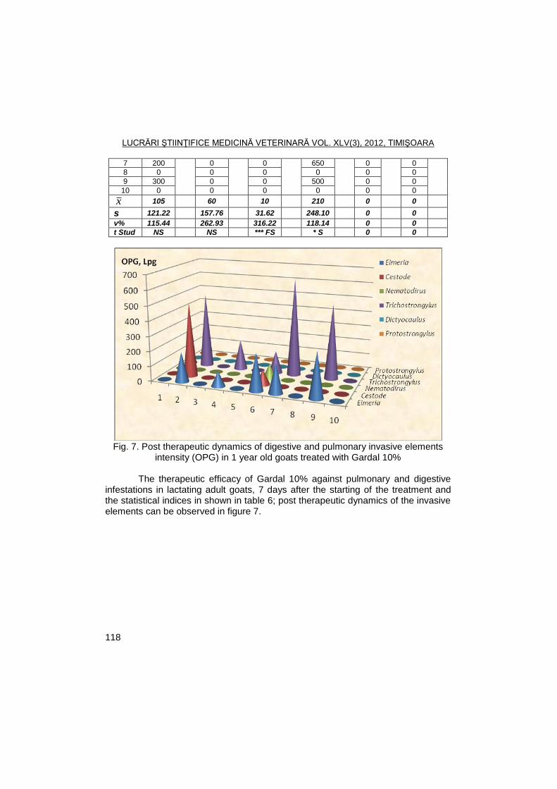
LUCRĂRI ŞTIINŢIFICE MEDICINĂ VETERINARĂ VOL. XLV(3), 2012, TIMIŞOARA
118
7 200 0 0 650 0 0
8 0 0 0 0 0 0
9 300 0 0 500 0 0
10 0 0 0 0 0 0
x 105 60 10 210 0 0
s 121.22 157.76 31.62 248.10 0 0
v% 115.44 262.93 316.22 118.14 0 0
t Stud NS NS *** FS * S 0 0
Fig. 7. Post therapeutic dynamics of digestive and pulmonary invasive elements
intensity (OPG) in 1 year old goats treated with Gardal 10% The therapeutic efficacy of Gardal 10% against pulmonary and digestive
infestations in lactating adult goats, 7 days after the starting of the treatment and the statistical indices in shown in table 6; post therapeutic dynamics of the invasive elements can be observed in figure 7.
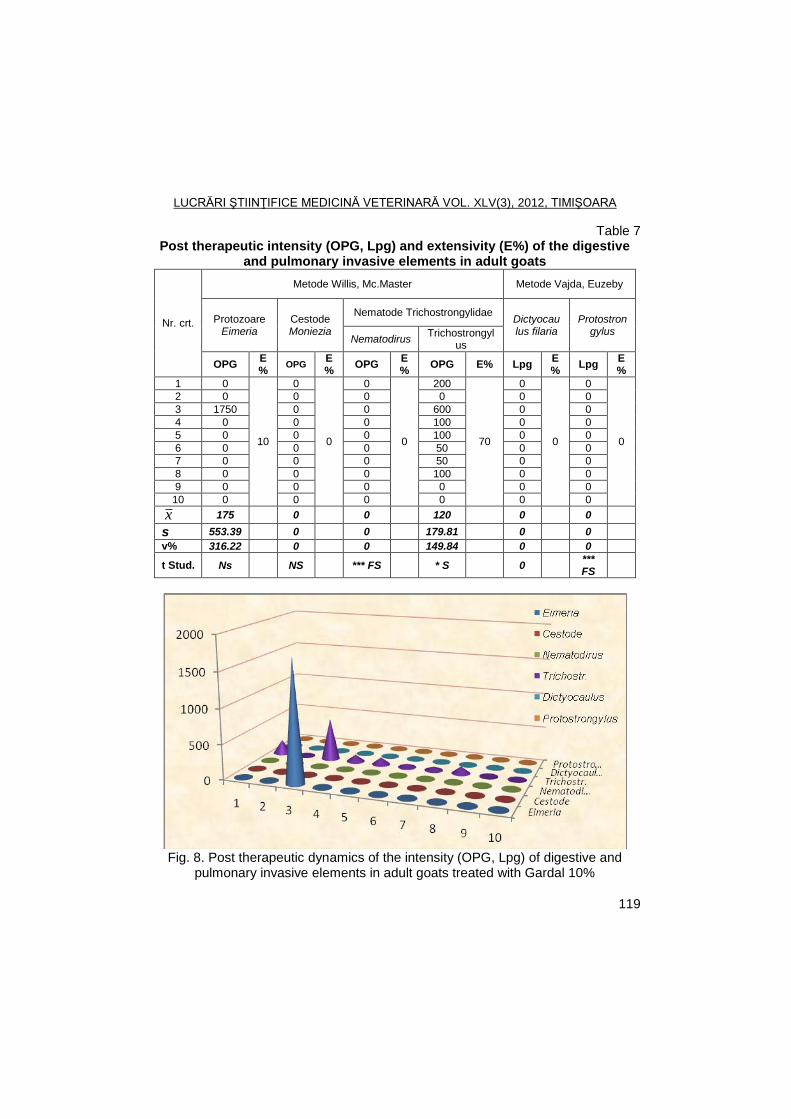
LUCRĂRI ŞTIINŢIFICE MEDICINĂ VETERINARĂ VOL. XLV(3), 2012, TIMIŞOARA
119
Table 7 Post therapeutic intensity (OPG, Lpg) and extensivity (E%) of the digestive
and pulmonary invasive elements in adult goats
Nr. crt.
Metode Willis, Mc.Master Metode Vajda, Euzeby
Protozoare Eimeria
Cestode Moniezia
Nematode Trichostrongylidae Dictyocau lus filaria
Protostrongylus
Nematodirus Trichostrongyl
us
OPG E%
OPG E%
OPG E%
OPG E% Lpg E%
Lpg E%
1 0
10
0
0
0
0
200
70
0
0
0
0
2 0 0 0 0 0 0
3 1750 0 0 600 0 0
4 0 0 0 100 0 0
5 0 0 0 100 0 0
6 0 0 0 50 0 0
7 0 0 0 50 0 0
8 0 0 0 100 0 0
9 0 0 0 0 0 0
10 0 0 0 0 0 0
x 175 0 0 120 0 0
s 553.39 0 0 179.81 0 0
v% 316.22 0 0 149.84 0 0
t Stud. Ns NS *** FS * S 0 *** FS
Fig. 8. Post therapeutic dynamics of the intensity (OPG, Lpg) of digestive and
pulmonary invasive elements in adult goats treated with Gardal 10%
LUCRĂRI ŞTIINŢIFICE MEDICINĂ VETERINARĂ VOL. XLV(3), 2012, TIMIŞOARA
120
Post therapeutic ovoscopic and larvoscopic results that were analyzed seven days after the starting of the treatment with Gardal 10% (Albendazole sulfoxide) within the herd revealed the different efficacy of this product against nematodes and cestodes according to the age of the treated subjects. Thus, in adult goats, the therapeutic efficacy has been of 100% against nematodes from following genus: Protostrongylus, Nematodirus and Moniezia; in young goats, the efficacy has been of 100% against Protostrongylus, 90% against Nematodirus, 80% against cestodes and 40% against other species of Trichostrongylidae; in fawns, the efficacy has been of 80% against Nematodirus and 60% against cestodes.
The comaparative pre and post therapeutic statistic results (Student test) recorded significant differences regarding the efficacy of Gardal 10% against Nematodirus in adult and young goats and distinctly significant differences in fawns; significant differences regarding the efficacy of the product against other species of Trichostrongylidae have been recorded in adult and young goats. Also, very significant differences have been observed against Protostrongylus in adult goats.
Conclusions
Testing for the first time of the Gardal 10% (Albendazole sulfoxide) on a herd
of Carpathian and half breed Saanen goats highlighted the different efficacy against numerous species of digestive and pulmonary parasites, with great results against digestive nematodes from Nematodirus genus (very significant differeces) and against pulmonary nematodes from Protostrongylus genus (very significant differences), recommending the product in the prophylaxis and therapy of these parasitosis in goats.
References
1. Jas, R., Datta, S., Ghosh, J.D., Economic impact of gastrointestinal
nematodosis in goat on meat production, Journal of Veterinary Parasitology, 2007, 21 (2), 109-112.
2. Meshram, M.D., Shirale, S.Y., Khillare, K.P., Prevalence of helminthic infection in goats, Indian Veterinary Journal, 2007, 84, (9), 992.
3. Molina, J.M., Ruiz, A., Hernandez, B., Gonzalez, J.F., Eprinomectin pour-on ant the shedding of Haemonchus contortus eggs in experimentaly infected goats, Australian Veterinary Journal, 2008, 86(11), 444-445.
4. Paraud, C. Kulo, A., Pors, I., Resistance of goat nematodes to multiple anthelmintics on a farm in France, Veterinary Record, 2009, 164(18), 563-564.
5. Prakash, Ved, Bano, Siddiqua, Comparative study on treatment of gastro-intestinal nematodes of goats in Kanpur, Asian Journal of Animal Science, 2008, 3(1), 77-78.
6. Sahoo, N., Ray, J.P., Efficacy of oral ivermectin against haemonchosis in goats, Indian Veterinary Journal, 2008, 85(8), 889.
LUCRĂRI ŞTIINŢIFICE MEDICINĂ VETERINARĂ VOL. XLV(3), 2012, TIMIŞOARA
121
PREVALENCE OF HYPODERMA INFESTATION IN DEER IN WESTERN ROMANIA
M.S. ILIE, MIRELA IMRE, IONELA HOTEA, K. IMRE, IONELA DENISA
SORESCU, SIDONIA ANDREI, P. ONITA, I. OPRESCU, S. MORARIU, C. MIHALI, GH. DĂRĂBUȘ
Banat's University of Agricultural Sciences and Veterinary Medicine Timisoara, Faculty
of Veterinary Medicine, 300645, Aradului Street No. 119, Timisoara, Romania E-mail: [email protected]
Summary
In this study was to establish prevalence of Hypoderma infestation in roe deer in hunting found from western Romania and to investigate possible relations between positivity and the age and gender of the tested animals.The presence of subcutaneous Hypoderma spp. larvae was investigated in 99 roe deer finding death in four hunting found from December 2011 to January 2012. The causes that led to death of animals were toxic nature, according with hemorrhagic syndrome (cumarinic agents raticides poisoning) showed at necropsy. The overall prevalence was 58.6% andmean intensity of infestation was 28.3 larvae/host. All larvae collected were identified as H. diana. No statistical significant differences could be detected between age or gender groups and intensity values.The results of this study highlighted the importance and widespread occurrence of these parasites in Romania, previously confirmed by other investigations in the neighbouring countries. However, for a more comprehensive etiological picture further studies, supported by molecular tools, are still required.
Key words: hypodermosis, roe deer, survey
Bovidae and Cervidae of the Holarctic region can be affected by
hypodermosis, a common myiasis produced by flies of the genus Hypoderma (Diptera: Oestridae). Larvae of Hypodermaaffect domesticated and wild ruminants including cattle, buffaloes, sheep, goats, deer, and reindeer. Adult flies are commonly known as heel flies, warble flies, bomb flies, gad flies, deerfly orroebuckfly. The species of Hypoderma in deer (roe deer, red deer, and reindeer) in Europe are Hypodermadiana, Hypodermaactaeon, and Hypodermatarandi (1, 7, 10, 13).
Diseases cause serious economic losses in herds reared for meat and dairy production. Hypodermosis in European Cervidae is of high economic importance in regard of the losses for leather processing (5, 6, 12). Little is known about the infestation of deer, and few studies have discussed the influence of environmental factors on the biology of flies and their resulting distribution. At the same time, the pathogenicity of larvae for host deer remains unknown.
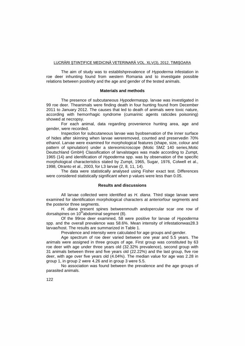
LUCRĂRI ŞTIINŢIFICE MEDICINĂ VETERINARĂ VOL. XLV(3), 2012, TIMIŞOARA
122
The aim of study was to establishprevalence of Hypoderma infestation in roe deer inhunting found from western Romania and to investigate possible relations between positivity and the age and gender of the tested animals.
Materials and methods
The presence of subcutaneous Hypodermaspp. larvae was investigated in
99 roe deer. Theanimals were finding death in four hunting found from December 2011 to January 2012. The causes that led to death of animals were toxic nature, according with hemorrhagic syndrome (cumarinic agents raticides poisoning) showed at necropsy.
For each animal, data regarding provenience hunting area, age and gender, were recorded.
Inspection for subcutaneous larvae was byobservation of the inner surface of hides after skinning when larvae wereremoved, counted and preservedin 70% ethanol. Larvae were examined for morphological features (shape, size, colour and pattern of spinulation) under a stereomicroscope (Motic SMZ 140 series,Motic Deutschland GmbH) Classification of larvalstages was made according to Zumpt, 1965 (14) and identification of Hypoderma spp. was by observation of the specific morphological characteristics stated by Zumpt, 1965, Sugar, 1976, Colwell et al., 1998, Otranto et al., 2003, for L3 larvae (2, 8, 11, 14).
The data were statistically analysed using Fisher exact test. Differences were considered statistically significant when p values were less than 0.05.
Results and discussions
All larvae collected were identified as H. diana. Third stage larvae were
examined for identification morphological characters at anteriorfour segments and the posterior three segments.
H. diana present spines betweenmouth andopercular scar one row of dorsalspines on 10
thabdominal segment (8).
Of the 99roe deer examined, 58 were positive for larvae of Hypoderma spp. and the overall prevalence was 58.6%. Mean intensity of infestationwas28.3 larvae/host. The results are summarized in Table 1.
Prevalence and intensity were calculated for age groups and gender. Age spectrum of roe deer varied between one year and 5.5 years. The
animals were assigned in three groups of age. First group was constituted by 63 roe deer with age under three years old (32.32% prevalence), second group with 31 animals between three and five years old (22.22%) and the last group, five roe deer, with age over five years old (4.04%). The median value for age was 2.28 in group 1, in group 2 were 4.26 and in group 3 were 5.5.
No association was found between the prevalence and the age groups of parasited animals.
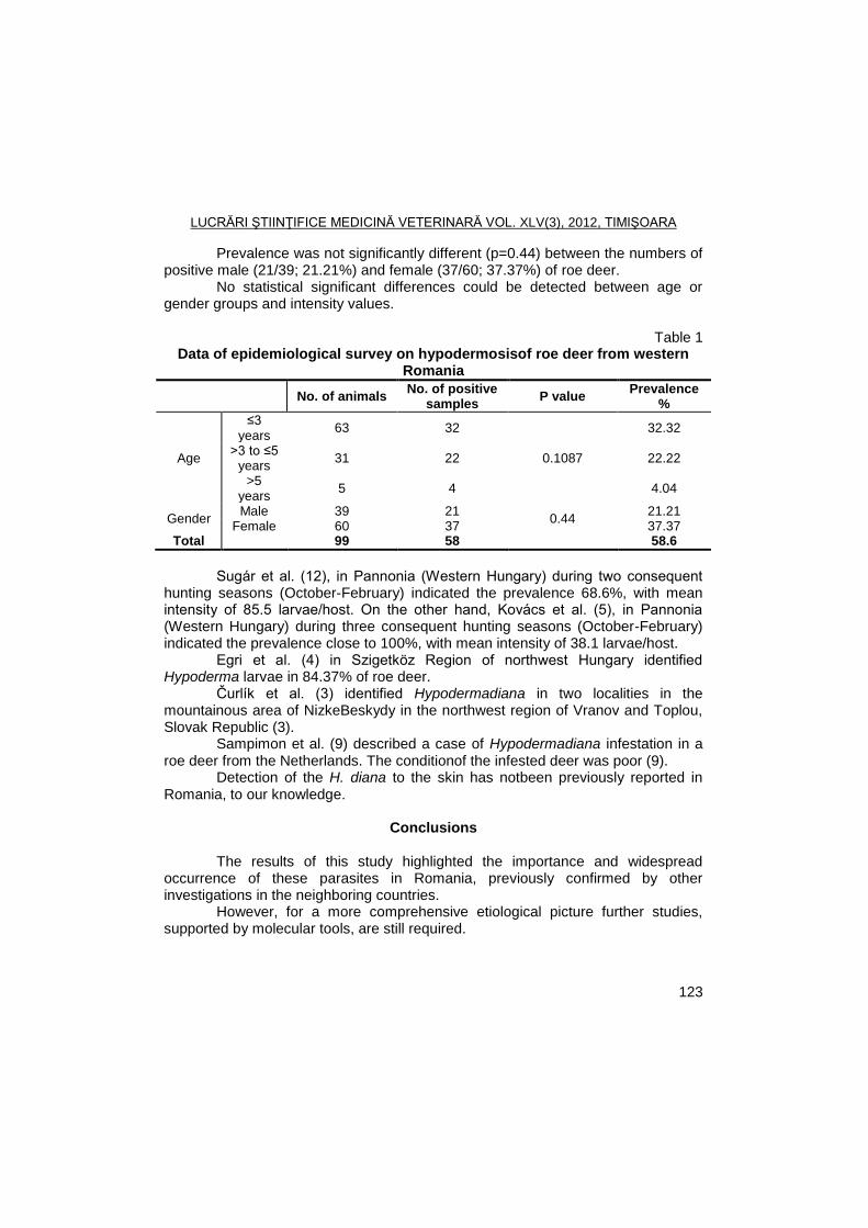
LUCRĂRI ŞTIINŢIFICE MEDICINĂ VETERINARĂ VOL. XLV(3), 2012, TIMIŞOARA
123
Prevalence was not significantly different (p=0.44) between the numbers of positive male (21/39; 21.21%) and female (37/60; 37.37%) of roe deer.
No statistical significant differences could be detected between age or gender groups and intensity values.
Table 1
Data of epidemiological survey on hypodermosisof roe deer from western Romania
No. of animals No. of positive
samples P value
Prevalence %
Age
≤3 years
63 32
0.1087
32.32
>3 to ≤5 years
31 22 22.22
>5 years
5 4 4.04
Gender Male 39 21
0.44 21.21
Female 60 37 37.37 Total 99 58 58.6
Sugár et al. (12), in Pannonia (Western Hungary) during two consequent
hunting seasons (October-February) indicated the prevalence 68.6%, with mean intensity of 85.5 larvae/host. On the other hand, Kovács et al. (5), in Pannonia (Western Hungary) during three consequent hunting seasons (October-February) indicated the prevalence close to 100%, with mean intensity of 38.1 larvae/host.
Egri et al. (4) in Szigetköz Region of northwest Hungary identified Hypoderma larvae in 84.37% of roe deer.
Čurlík et al. (3) identified Hypodermadiana in two localities in the mountainous area of NizkeBeskydy in the northwest region of Vranov and Toplou, Slovak Republic (3).
Sampimon et al. (9) described a case of Hypodermadiana infestation in a roe deer from the Netherlands. The conditionof the infested deer was poor (9).
Detection of the H. diana to the skin has notbeen previously reported in Romania, to our knowledge.
Conclusions
The results of this study highlighted the importance and widespread
occurrence of these parasites in Romania, previously confirmed by other investigations in the neighboring countries.
However, for a more comprehensive etiological picture further studies, supported by molecular tools, are still required.
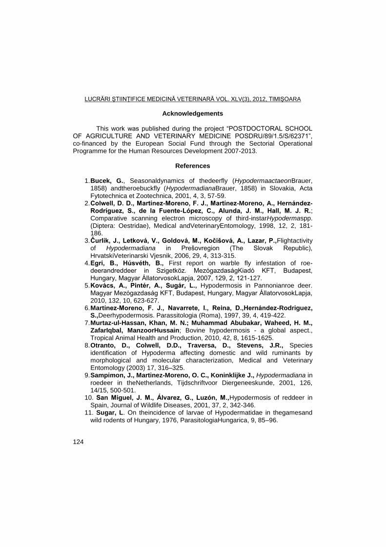
LUCRĂRI ŞTIINŢIFICE MEDICINĂ VETERINARĂ VOL. XLV(3), 2012, TIMIŞOARA
124
Acknowledgements This work was published during the project ―POSTDOCTORAL SCHOOL
OF AGRICULTURE AND VETERINARY MEDICINE POSDRU/89/1.5/S/62371‖, co-financed by the European Social Fund through the Sectorial Operational Programme for the Human Resources Development 2007-2013.
References
1. Bucek, G., Seasonaldynamics of thedeerfly (HypodermaactaeonBrauer,
1858) andtheroebuckfly (HypodermadianaBrauer, 1858) in Slovakia, Acta Fytotechnica et Zootechnica, 2001, 4, 3, 57-59.
2. Colwell, D. D., Martínez-Moreno, F. J., Martínez-Moreno, A., Hernández-Rodríguez, S., de la Fuente-López, C., Alunda, J. M., Hall, M. J. R.; Comparative scanning electron microscopy of third-instarHypodermaspp. (Diptera: Oestridae), Medical andVeterinaryEntomology, 1998, 12, 2, 181-186.
3. Ţurlík, J., Letková, V., Goldová, M., Koţišová, A., Lazar, P.,Flightactivity of Hypodermadiana in Prešovregion (The Slovak Republic), HrvatskiVeterinarski Vjesnik, 2006, 29, 4, 313-315.
4. Egri, B., Húsvéth, B., First report on warble fly infestation of roe-deerandreddeer in Szigetköz. MezógazdaságKiadó KFT, Budapest, Hungary, Magyar ÁllatorvosokLapja, 2007, 129, 2, 121-127.
5. Kovács, A., Pintér, A., Sugár, L., Hypodermosis in Pannonianroe deer. Magyar Mezógazdaság KFT, Budapest, Hungary, Magyar ÁllatorvosokLapja, 2010, 132, 10, 623-627.
6. Martínez-Moreno, F. J., Navarrete, I., Reina, D.,Hernández-Rodriguez, S.,Deerhypodermosis. Parassitologia (Roma), 1997, 39, 4, 419-422.
7. Murtaz-ul-Hassan, Khan, M. N.; Muhammad Abubakar, Waheed, H. M., ZafarIqbal, ManzoorHussain; Bovine hypodermosis - a global aspect., Tropical Animal Health and Production, 2010, 42, 8, 1615-1625.
8. Otranto, D., Colwell, D.D., Traversa, D., Stevens, J.R., Species identification of Hypoderma affecting domestic and wild ruminants by morphological and molecular characterization, Medical and Veterinary Entomology (2003) 17, 316–325.
9. Sampimon, J., Martinez-Moreno, O. C., Koninklijke J., Hypodermadiana in roedeer in theNetherlands, Tijdschriftvoor Diergeneeskunde, 2001, 126, 14/15, 500-501.
10. San Miguel, J. M., Álvarez, G., Luzón, M.,Hypodermosis of reddeer in Spain, Journal of Wildlife Diseases, 2001, 37, 2, 342-346.
11. Sugar, L. On theincidence of larvae of Hypodermatidae in thegamesand wild rodents of Hungary, 1976, ParasitologiaHungarica, 9, 85–96.
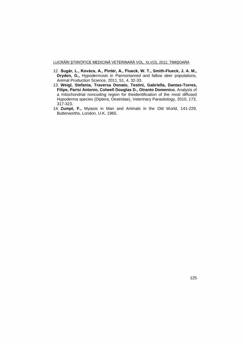
LUCRĂRI ŞTIINŢIFICE MEDICINĂ VETERINARĂ VOL. XLV(3), 2012, TIMIŞOARA
125
12. Sugár, L., Kovács, A., Pintér, A., Flueck, W. T., Smith-Flueck, J. A. M., Dryden, G., Hypodermosis in Pannonianred and fallow deer populations, Animal Production Science, 2011, 51, 4, 32-33.
13. Weigl, Stefania, Traversa Donato, Testini, Gabriella, Dantas-Torres, Filipe, Parisi Antonio, Colwell Douglas D., Otranto Domenico, Analysis of a mitochondrial noncoding region for theidentification of the most diffused Hypoderma species (Diptera, Oestridae), Veterinary Parasitology, 2010, 173, 317-323.
14. Zumpt, F., Myiasis in Man and Animals in the Old World, 141-229, Butterworths, London, U.K, 1965.
LUCRĂRI ŞTIINŢIFICE MEDICINĂ VETERINARĂ VOL. XLV(3), 2012, TIMIŞOARA
126
OCCURENCE OF SARCOCYSTIS SPP. IN ROE DEER (CAPREOLUS CAPREOLUS) FROM WESTERN ROMANIA-
PRELIMINARY RESULTS
K. IMRE1, CLAUDIA SALA
1, ADRIANA MORAR
1, M.S. ILIE
1, MIRELA IMRE
1,
P. ONIȚĂ2, GH. DĂRĂBUȘ
1
1Banat's University of Agricultural Sciences and Veterinary Medicine from
Timişoara, Faculty of Veterinary Medicine, 300645, Calea Aradului No. 119, Timisoara, Romania
2Veterinary and Public Health Authority (DSV) Arad;
E-mail: [email protected]
Summary
Fresh muscle tissues from 55 roe deer (Capreolus capreolus), originating from five hunting funds located in two western counties (Arad and Timis) of Romania, were screened for the presence of Sarcocystis spp. (muscle sarcocystosis) using a modified magnetic stirrer method. Overall, thirty-seven (67.3%) deer’s were found positive for Sarcocystis spp. All the examined hunting funds were found to be Sarcocystis positive and the percentage of infected deer’s varied between 50 and 85.7%. Statistical analysis showed significant differences (p<0.05) of infection between female (23/31; 74.1%) and male (14/24; 58.3%) deer’s. The results of this study showed a widespread occurrence of muscle sarcocystosis in a five hunting funds from western Romania and highlighted the risk that is poses when the infected deer meat is consumed raw or undercooked by humans.
Key words: survey, Sarcocystis spp., roe deer
Species of the genus Sarcocystis are apicomplexan parasites, with
heteroxenous life cycle, infecting a wide variety of mammals, reptiles and birds (2, 10). In definitive hosts, usually represented by predators (carnivores), the infections appeared to be asymptomatic. In intermediate hosts (herbivores and omnivores), which can be acquire the parasite through the ingestion of oocysts shed from definitive hosts, specific (eosinophilic myositis with flaccid paralysis) and nespecific (anorexia, pyrexia, anemia, cahexia, enlarged palpable lymph nodes and excessive salivation) symptoms have been frequently reported (3, 10). In humans (which serve as definitive hosts), infection occurs when raw or undercooked meat is ingested, and the most important symptoms included anorexia, nausea, abdominal pain, diarrhea, vomiting, dyspnoea and tachycardia (2).
Several studies (1, 3, 4, 5, 6, 8) have shown that the infection with Sarcocystis spp. in roe deer host is common. The prevalence data have varied significantly among and within countries depending on the study design and tested population. In Romania, no information is available about the occurence of muscle sarcocystosis in wildlife. However, taking into account the high possibilities of prey-
LUCRĂRI ŞTIINŢIFICE MEDICINĂ VETERINARĂ VOL. XLV(3), 2012, TIMIŞOARA
127
predator relationsheep between wildlife, the transmission of Sarcocystis species in this area can be very frequently.
The aim of this study was to investigate the occurence of Sarcocystis spp. (muscle sarcocystosis) in roe deer (Capreolus capreolus), in five hunting funds located in two western counties (Arad and Timiș) of Romania, and to evaluate the risk that may pose to humans via consumption of contaminated deer meat.
Materials and methods
The study was conducted between January and February 2012, in five hunting funds (Andrei Șaguna, Curtici, Dorobanț, Iratoșu and Variaș) from western Romania. A total of 55 samples represented by the pillar of the diaphragm were collected from death deers after an poisoning episode and were assayed for the presence of matures sarcocysts "Miescher's Tubules".
Each sample was prepared through the modified magnetic stirrer method (2) and examined under light microscopy. Briefly, the muscle samples were cut into small pieces, removing all non-muscle tissues and were blended 20-30 s in a blender. The blended meat (1-2 g) was place in a beaker containing a magnet and tap water was added over into the volume 1:20. After a good homogenization, the mix was placed in 50 ml conical vials for sedimentation. In 15-20 minutes the supernatant was removed by placing the suction pump at about 2 cm from the bottom of the beaker being careful not to remove the sediment, which contains the "Miescher's Tubules" (2). This procedure was repeated several times until the supernatant was fairly transparent. Finally, the supernatant was removed and the sediment was placed in a Petri dish and examined under light microscope (at x10 magnification) for the presence of matures sarcocysts.
The data were statistically analyzed using StatPac Inc® (Statistic
Calculator, 2012). Differences were considered statistically significant when p values were less than 0.05.
Results and discussions
Morphologically, under light microscopy, the cysts appear with characteristic elongated shape (measuring 1-3 mm in length x 0.2-0.4 mm in diameter), brown colored and with rounded ends (Fig. 1).
The results are summarized in Table 1. Thirty-seven (67.3%) out of 55 examined deer’s were found positive for Sarcocystis spp. ("Miescher's Tubules‖). All the examined hunting funds were found to be sarcocysts positive and the percentage of infected deer’s varied between 50 and 85.7 %.
The prevalence of muscle sarcocystosis in three age groups was 69.2% (≤2 years), 62.1% (>2 to ≤4 years) and 76.9% (>4 years), respectively. Statistical analysis showed significant differences (p<0.05) of infection between female (23/31; 74.1%) and male (14/24; 58.3%) deer’s.
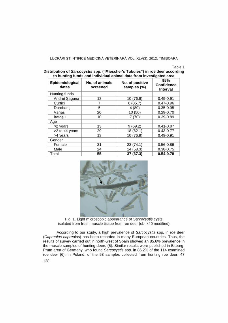
LUCRĂRI ŞTIINŢIFICE MEDICINĂ VETERINARĂ VOL. XLV(3), 2012, TIMIŞOARA
128
Table 1 Distribution of Sarcocystis spp. ("Miescher's Tubules”) in roe deer according
to hunting funds and individual animal data from investigated area
Epidemiological datas
No. of animals screened
No. of positive samples (%)
95% Confidence
Interval
Hunting funds
Andrei Șaguna 13 10 (76.9) 0.49-0.91
Curtici 7 6 (85.7) 0.47-0.96
Dorobanț 5 4 (80) 0.35-0.95
Variaș 20 10 (50) 0.29-0.70
Iratoșu 10 7 (70) 0.39-0.89
Age
≤2 years 13 9 (69.2) 0.41-0.87
>2 to ≤4 years 29 18 (62.1) 0.43-0.77
>4 years 13 10 (76.9) 0.49-0.91
Gender
Female 31 23 (74.1) 0.56-0.86
Male 24 14 (58.3) 0.38-0.75
Total 55 37 (67.3) 0.54-0.78
Fig. 1. Light microscopic appearance of Sarcocystis cysts isolated from fresh muscle tissue from roe deer (ob. x40 modified)
According to our study, a high prevalence of Sarcocystis spp. in roe deer
(Capreolus capreolus) has been recorded in many European countries. Thus, the results of survey carried out in north-west of Spain showed an 85.6% prevalence in the muscle samples of hunting deers (5). Similar results were published in Bitburg-Prum area of Germany, who found Sarcocystis spp. in 86.2% of the 114 examined roe deer (6). In Poland, of the 53 samples collected from hunting roe deer, 47
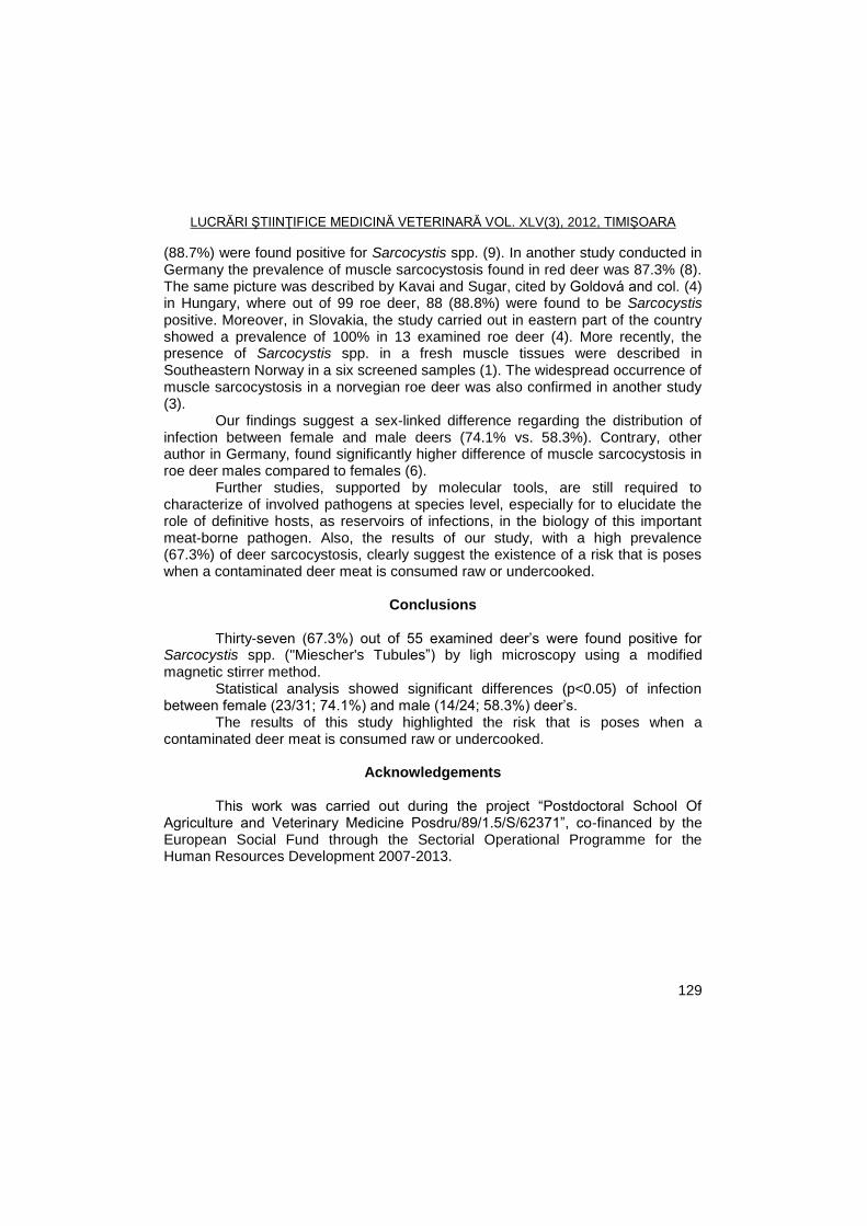
LUCRĂRI ŞTIINŢIFICE MEDICINĂ VETERINARĂ VOL. XLV(3), 2012, TIMIŞOARA
129
(88.7%) were found positive for Sarcocystis spp. (9). In another study conducted in Germany the prevalence of muscle sarcocystosis found in red deer was 87.3% (8). The same picture was described by Kavai and Sugar, cited by Goldová and col. (4) in Hungary, where out of 99 roe deer, 88 (88.8%) were found to be Sarcocystis positive. Moreover, in Slovakia, the study carried out in eastern part of the country showed a prevalence of 100% in 13 examined roe deer (4). More recently, the presence of Sarcocystis spp. in a fresh muscle tissues were described in Southeastern Norway in a six screened samples (1). The widespread occurrence of muscle sarcocystosis in a norvegian roe deer was also confirmed in another study (3).
Our findings suggest a sex-linked difference regarding the distribution of infection between female and male deers (74.1% vs. 58.3%). Contrary, other author in Germany, found significantly higher difference of muscle sarcocystosis in roe deer males compared to females (6).
Further studies, supported by molecular tools, are still required to characterize of involved pathogens at species level, especially for to elucidate the role of definitive hosts, as reservoirs of infections, in the biology of this important meat-borne pathogen. Also, the results of our study, with a high prevalence (67.3%) of deer sarcocystosis, clearly suggest the existence of a risk that is poses when a contaminated deer meat is consumed raw or undercooked.
Conclusions
Thirty-seven (67.3%) out of 55 examined deer’s were found positive for Sarcocystis spp. ("Miescher's Tubules‖) by ligh microscopy using a modified magnetic stirrer method.
Statistical analysis showed significant differences (p<0.05) of infection between female (23/31; 74.1%) and male (14/24; 58.3%) deer’s.
The results of this study highlighted the risk that is poses when a contaminated deer meat is consumed raw or undercooked.
Acknowledgements
This work was carried out during the project ―Postdoctoral School Of Agriculture and Veterinary Medicine Posdru/89/1.5/S/62371‖, co-financed by the European Social Fund through the Sectorial Operational Programme for the Human Resources Development 2007-2013.
LUCRĂRI ŞTIINŢIFICE MEDICINĂ VETERINARĂ VOL. XLV(3), 2012, TIMIŞOARA
130
References
1. Dahlgren, S., Gjerde, B., Sarcocystis in Norwegian roe deer (Capreolus capreolus): molecular and morphological identification of Sarcocystis oviformis n. sp. and Sarcocystis gracilis and their phylogenetic relationship with other Sarcocystis species, Parasitol Res, 2009, 104, 993-1003.
2. Dubey, J.P., Speer, C.A., Fayer, R., Sarcocystosis of animals and man. Ed. Boca Raton, CRC Press, Florida, 1989, pp. 215.
3. Gjede, B., Morphological and molecular characterization and phylogenetic placement of Sarcocystis capreolicanis and Sarcocystis silva n. sp. from roe deer (Capreolus capreolus) in Norway. Parasitol Res, 2012, 110, 1225-1237.
4. Goldová, M., Tóth, Š., Letková, V., Mojţišová, J., Ciberej, J., Konjeviš, D., Koţišová, A., Slavica, A., Sarcocystosis in cloven-hoofed game in Slovak Republic. Nat, Croat, 2008, 17, 303-309.
5. Lopez, C., Panadero, R., Bravo, A., Paz, A., Sanchez-Andrale, R., Diezz-Banos, P., Morrodo, P., Sarcocystis spp. infection in roe deer (Capreolus capreolus) from the north-west of Spain, Z Jagdwiss, 2003, 49, 211-218.
6. Partenheimer–Hannemann, C., Untersuchung zum Workommen von Sarkosporidien bei Reh-und Rotwild im Raum Bitburrg-Prüm area (Rheinland-Pfalz). Inaugural-Dissertation, Hannover, Germany: Tierärztliche Hochschule Hannover, 1991, pp. 114.
7. Pozio, E., La Rosa, G., PCR derived methods for the identificationof Trichinella parasites from animal and human samples, Meth Mol Biol, 2003, 216, 299-309.
8. Spickschen, C., Pohlmeyer, K., Investigation on the occurrence of Sarcosporidia in roe deer, red deer, and mouflon from two different natural habitats in Lower Saxony, Zeitsch, Jagdwiss, 2002, 48, 35-48.
9. Tropilo, J., Katkiewicz, M.T., Wisniewski, J., Sarcocystis spp. infection in free-living animals: wild boar (Sus scrofa) deer (Cervus elaphus) roe deer (Capreolus capreolus), Pol J Vet Sci, 2001, 4, 15-18.
10. Sedlaczek, J., Wesemeier, H., On the diagnostics and nomenclature of Sarcocystis species (Sporozoa) in roe deer (Capreolus capreolus), Appl Parasitol, 1995, 36, 73-82.
LUCRĂRI ŞTIINŢIFICE MEDICINĂ VETERINARĂ VOL. XLV(3), 2012, TIMIŞOARA
131
PREVALENCE OF TICK SPECIES IN DOGS USING CLASSICAL METHODS AND SEM
MIRELA IMRE
1, M.S. ILIE
1, C.V. MIHALI
2, I. OPRESCU
1, S. MORARIU
1, IONELA
HOTEA1, K. IMRE
1, ALINA ILIE
1, M. PALCA
3, GH. DĂRĂBUȘ
1
1Banat's University of Agricultural Sciences and Veterinary Medicine from
Timişoara, Faculty of Veterinary Medicine, 300645, Calea Aradului No. 119, Timisoara, Romania
2Institute of Life Sciences, ―Vasile Goldis‖ Western University of Arad
3Veterinary Practician
E-mail: [email protected]
Summary
Tick specimens removed from 48 dogs living in three counties (Arad, Bihor and Timiş) of western Romania were collected between April 2010 and March 2012. Out of 258 collected ticks, 156 (60.4%) were identified as Rhipicephalus sanguineus, 77 (29.8%) as Ixodes ricinus and 23 (8.91%) as Dermacentor reticulatus, respectively. Only two (0.77%) specimens were found as Haemaphysalis punctata. Regarding the dogs, 30 (62.5%) were parasitized only with I. ricinus, 9 (18.75%) animals were infested with R. sanguineus and 5 (10.41%) with D. reticulatus. H. punctata was found on one (2.08%) dog but on the same animal co-infestation with I. ricinus was recorded. Out of the tick species identified, some are known as reservoirs for specific pathogens, or are being involved in the spreading of different infections in animals and/or humans.
Key words: tick, dog, SEM, tick-borne infections
Hard ticks (suborder Ixodida) are one of the most important group of
vectors for pathogens within the phylum Arthropoda. These hematophagous, temporary ectoparasites, are reservoirs and transmit many pathogens including bacteria, helminths, protozoa and viruses (3, 7, 13).
In the last years canine population increased due to their role as guard dogs and their social role, developing a close relationship with humans in both rural and urban areas. This poses new concerns for human public health being reservoir hosts for zoonotic pathogen agents (11).
Some tick species are known as reservoirs for specific pathogens. Also, some pathogens, ingested by ticks, are transmitted trans-stadially and/or trans-ovarially, increasing the risk for infection (13).
Canine babesiosis is transmitted, especially, by Dermacentor spp. (Babesia canis canis), but also by Rhipicephalus sanguineus (B. canis canis, B. canis vogeli and B. gibsoni) and Haemaphysalis spp. (B. canis rossi, B. gibsoni) (13).
In Europe, canine ehrlichiosis is transmitted by Rh. sanguineus and anaplasmosis by Ixodes spp. and R. sanguineus. Lyme borreliosis is transmited by
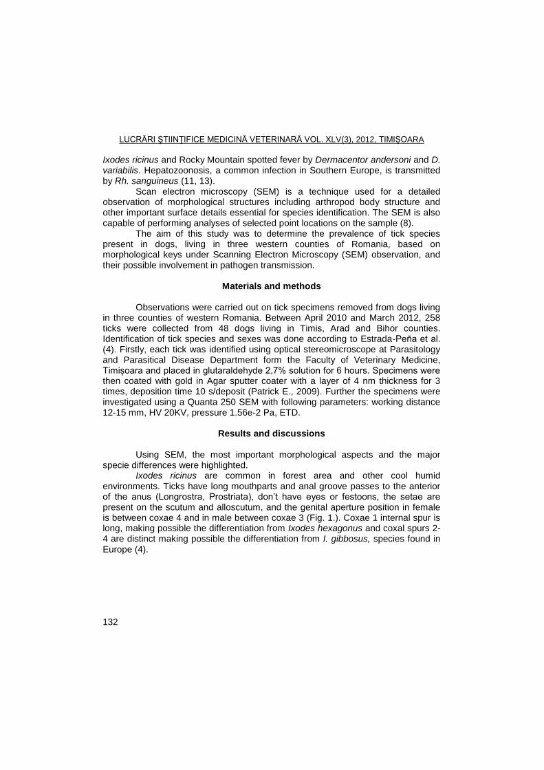
LUCRĂRI ŞTIINŢIFICE MEDICINĂ VETERINARĂ VOL. XLV(3), 2012, TIMIŞOARA
132
Ixodes ricinus and Rocky Mountain spotted fever by Dermacentor andersoni and D. variabilis. Hepatozoonosis, a common infection in Southern Europe, is transmitted by Rh. sanguineus (11, 13).
Scan electron microscopy (SEM) is a technique used for a detailed observation of morphological structures including arthropod body structure and other important surface details essential for species identification. The SEM is also capable of performing analyses of selected point locations on the sample (8).
The aim of this study was to determine the prevalence of tick species present in dogs, living in three western counties of Romania, based on morphological keys under Scanning Electron Microscopy (SEM) observation, and their possible involvement in pathogen transmission.
Materials and methods Observations were carried out on tick specimens removed from dogs living
in three counties of western Romania. Between April 2010 and March 2012, 258 ticks were collected from 48 dogs living in Timis, Arad and Bihor counties. Identification of tick species and sexes was done according to Estrada-Peña et al. (4). Firstly, each tick was identified using optical stereomicroscope at Parasitology and Parasitical Disease Department form the Faculty of Veterinary Medicine, Timișoara and placed in glutaraldehyde 2,7% solution for 6 hours. Specimens were then coated with gold in Agar sputter coater with a layer of 4 nm thickness for 3 times, deposition time 10 s/deposit (Patrick E., 2009). Further the specimens were investigated using a Quanta 250 SEM with following parameters: working distance 12-15 mm, HV 20KV, pressure 1.56e-2 Pa, ETD.
Results and discussions
Using SEM, the most important morphological aspects and the major
specie differences were highlighted. Ixodes ricinus are common in forest area and other cool humid
environments. Ticks have long mouthparts and anal groove passes to the anterior of the anus (Longrostra, Prostriata), don’t have eyes or festoons, the setae are present on the scutum and alloscutum, and the genital aperture position in female is between coxae 4 and in male between coxae 3 (Fig. 1.). Coxae 1 internal spur is long, making possible the differentiation from Ixodes hexagonus and coxal spurs 2-4 are distinct making possible the differentiation from I. gibbosus, species found in Europe (4).
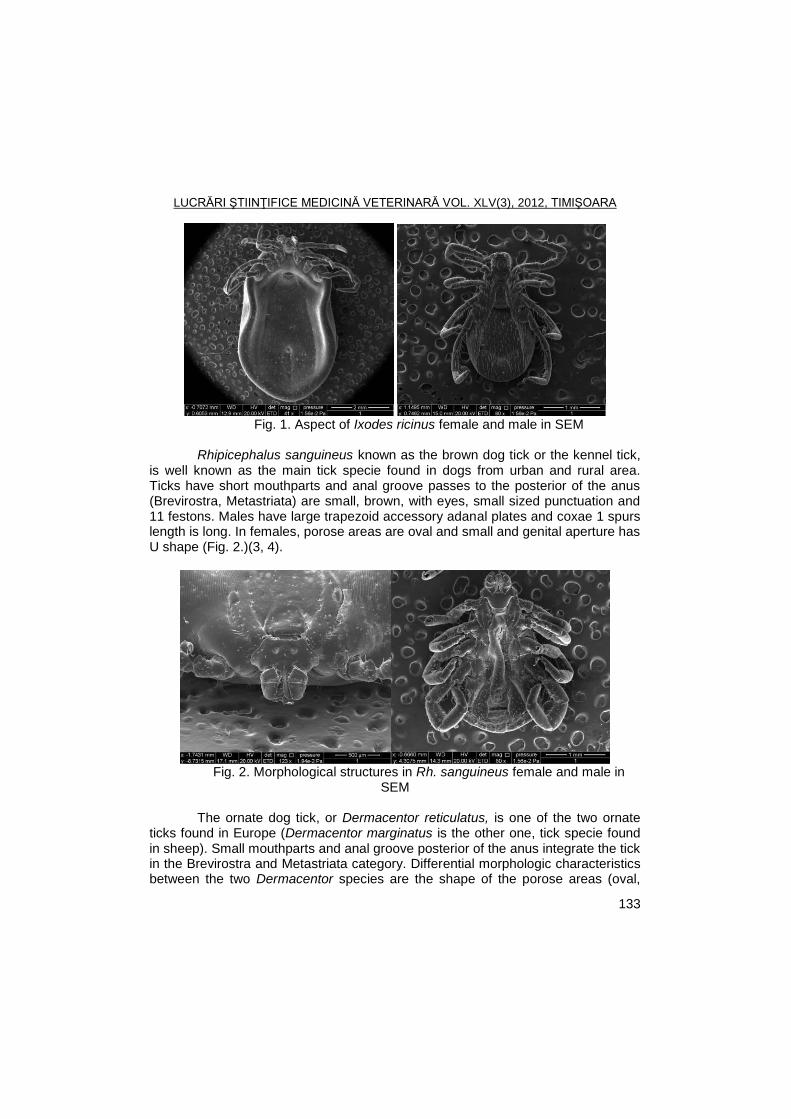
LUCRĂRI ŞTIINŢIFICE MEDICINĂ VETERINARĂ VOL. XLV(3), 2012, TIMIŞOARA
133
Fig. 1. Aspect of Ixodes ricinus female and male in SEM
Rhipicephalus sanguineus known as the brown dog tick or the kennel tick,
is well known as the main tick specie found in dogs from urban and rural area. Ticks have short mouthparts and anal groove passes to the posterior of the anus (Brevirostra, Metastriata) are small, brown, with eyes, small sized punctuation and 11 festons. Males have large trapezoid accessory adanal plates and coxae 1 spurs length is long. In females, porose areas are oval and small and genital aperture has U shape (Fig. 2.)(3, 4).
Fig. 2. Morphological structures in Rh. sanguineus female and male in
SEM The ornate dog tick, or Dermacentor reticulatus, is one of the two ornate
ticks found in Europe (Dermacentor marginatus is the other one, tick specie found in sheep). Small mouthparts and anal groove posterior of the anus integrate the tick in the Brevirostra and Metastriata category. Differential morphologic characteristics between the two Dermacentor species are the shape of the porose areas (oval,
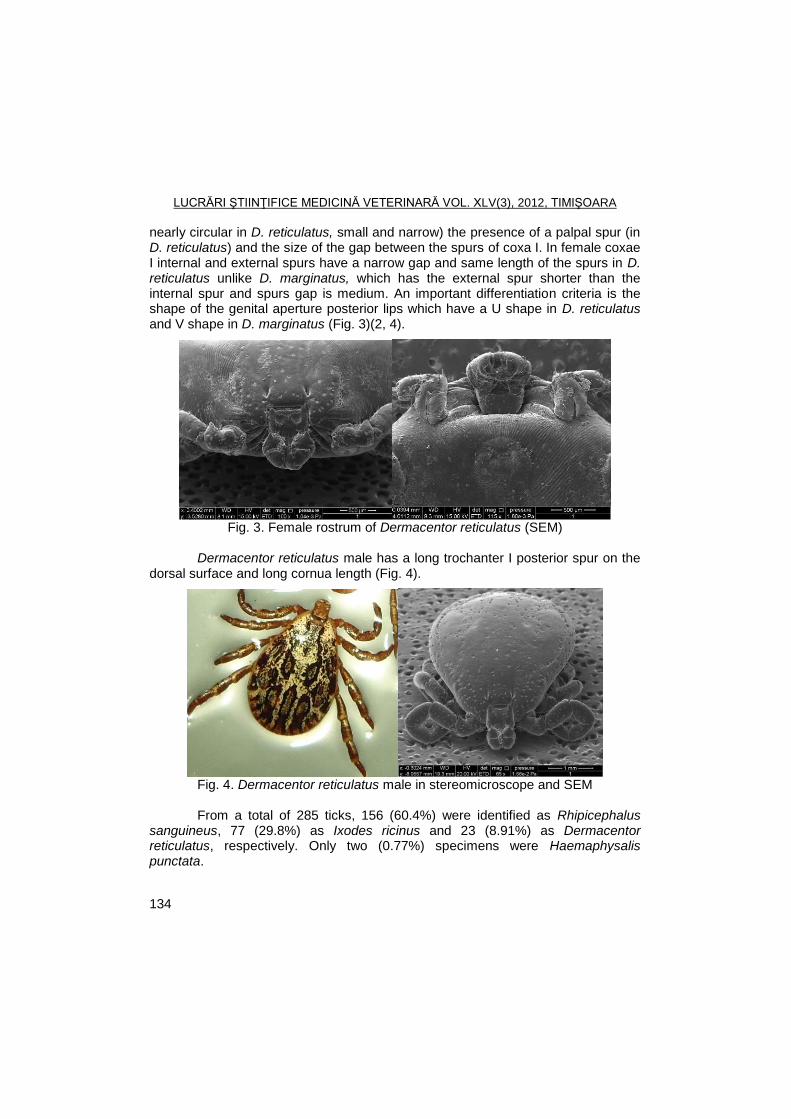
LUCRĂRI ŞTIINŢIFICE MEDICINĂ VETERINARĂ VOL. XLV(3), 2012, TIMIŞOARA
134
nearly circular in D. reticulatus, small and narrow) the presence of a palpal spur (in D. reticulatus) and the size of the gap between the spurs of coxa I. In female coxae I internal and external spurs have a narrow gap and same length of the spurs in D. reticulatus unlike D. marginatus, which has the external spur shorter than the internal spur and spurs gap is medium. An important differentiation criteria is the shape of the genital aperture posterior lips which have a U shape in D. reticulatus and V shape in D. marginatus (Fig. 3)(2, 4).
Fig. 3. Female rostrum of Dermacentor reticulatus (SEM)
Dermacentor reticulatus male has a long trochanter I posterior spur on the
dorsal surface and long cornua length (Fig. 4).
Fig. 4. Dermacentor reticulatus male in stereomicroscope and SEM
From a total of 285 ticks, 156 (60.4%) were identified as Rhipicephalus
sanguineus, 77 (29.8%) as Ixodes ricinus and 23 (8.91%) as Dermacentor reticulatus, respectively. Only two (0.77%) specimens were Haemaphysalis punctata.
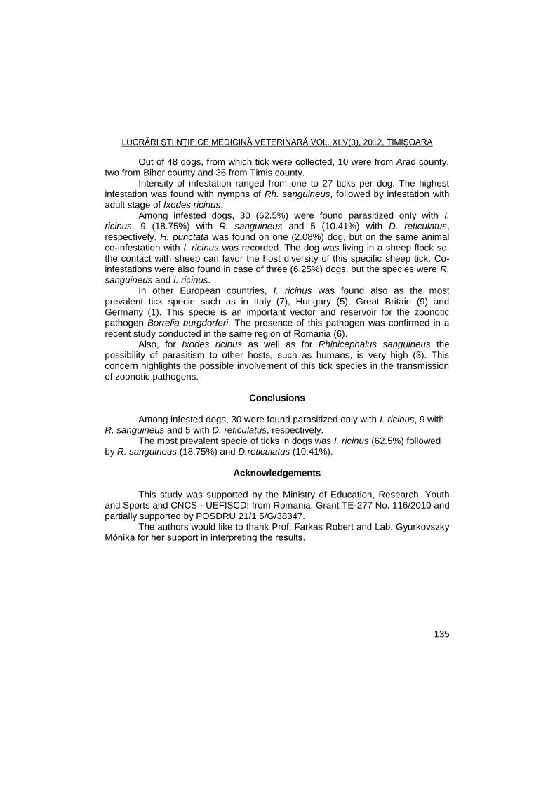
LUCRĂRI ŞTIINŢIFICE MEDICINĂ VETERINARĂ VOL. XLV(3), 2012, TIMIŞOARA
135
Out of 48 dogs, from which tick were collected, 10 were from Arad county, two from Bihor county and 36 from Timis county.
Intensity of infestation ranged from one to 27 ticks per dog. The highest infestation was found with nymphs of Rh. sanguineus, followed by infestation with adult stage of Ixodes ricinus.
Among infested dogs, 30 (62.5%) were found parasitized only with I. ricinus, 9 (18.75%) with R. sanguineus and 5 (10.41%) with D. reticulatus, respectively. H. punctata was found on one (2.08%) dog, but on the same animal co-infestation with I. ricinus was recorded. The dog was living in a sheep flock so, the contact with sheep can favor the host diversity of this specific sheep tick. Co-infestations were also found in case of three (6.25%) dogs, but the species were R. sanguineus and I. ricinus.
In other European countries, I. ricinus was found also as the most prevalent tick specie such as in Italy (7), Hungary (5), Great Britain (9) and Germany (1). This specie is an important vector and reservoir for the zoonotic pathogen Borrelia burgdorferi. The presence of this pathogen was confirmed in a recent study conducted in the same region of Romania (6).
Also, for Ixodes ricinus as well as for Rhipicephalus sanguineus the possibility of parasitism to other hosts, such as humans, is very high (3). This concern highlights the possible involvement of this tick species in the transmission of zoonotic pathogens.
Conclusions
Among infested dogs, 30 were found parasitized only with I. ricinus, 9 with R. sanguineus and 5 with D. reticulatus, respectively.
The most prevalent specie of ticks in dogs was I. ricinus (62.5%) followed by R. sanguineus (18.75%) and D.reticulatus (10.41%).
Acknowledgements
This study was supported by the Ministry of Education, Research, Youth
and Sports and CNCS - UEFISCDI from Romania, Grant TE-277 No. 116/2010 and partially supported by POSDRU 21/1.5/G/38347.
The authors would like to thank Prof. Farkas Robert and Lab. Gyurkovszky Mónika for her support in interpreting the results.
LUCRĂRI ŞTIINŢIFICE MEDICINĂ VETERINARĂ VOL. XLV(3), 2012, TIMIŞOARA
136
References
1. Beichel, E., Petney, T.N., Hassler, D., Brückner, M., Maiwald, M., Tick infestation patterns and prevalence of Borrelia burgdorferi in ticks collected at a veterinary clinic in Germany, Vet Parasitol, 1996, 65, 147-155.
2. Chiţimia, Lidia, Căpuşe Ixodide, Ed. Mirton, Timişoara, 2007. 3. Dantas-Torres, F., Biology and ecology of the brown dog tick, Rhipicephalus
sanguineus, Parasit Vectors, 2010, 3, 26. 4. Estrada-Peña, A., Bouattour, A., Camicas, J.L., Walker, A.R., Ticks of
domestic animals in the Mediterranean region. A guide to identification of species, Univ. Zaragoza, Spain, 2004.
5. Földvári, G., Farkas, R., Ixodid tick species attaching to dogs in Hungary, Vet. Parasitol., 2005, 129, 125–131.
6. Ilie, M. S., Darabus, G., Imre, Mirela, Imre, K., Hotea, Ionela, Ilie, Alina, Morariu, S., Immunofluorescence antibody test surveillance of Canine borreliosis in Western Romania, Current Opinion in Biotechnology, 2011, 22, Suppl 1: 110.
7. Iori, Albertina, Gabrielli, Simona, Calderini, Pietro, Moretti, Annabella, Pietrobelli, Mario, Tampieri, Maria-Paola, Galuppi, Roberta, Cancrini, Gabriella, Tick reservoirs for piroplasms in central and northern Italy, Vet. Parasitol., 2010, 170, 291–296.
8. Ludwig, R., Scanning Electron Microscopy: Physics of Image Formation and Microanalysis, 1998, pp.449.
9. Ogden, N.H., Cripps, P., Davison, C.C., Owen, G., Parry, J.M., Timms, B.J., Forbes, A.B., The ixodid tick species attaching to domestic dogs and cats in Great Britain and Ireland, Med. Vet. Entomol, 2000, 14, 332–338.
10. Otranto, D., Dantas-Torres, F., Breitschwerdt, E.B., Managing canine vector-borne diseases of zoonotic concern: part one, Trends Parsitol., 2010, 25, 157-163.
11. Otranto, D., Dantas-Torres, F., Canine and feline vector –borne diseases in Italy: current situation and perspectives, Parasit. Vectors, 2010, 3, 2.
12. Patrick E., Handbook of Sample Preparation for Scanning Electron Microscopy and X-Ray Microanalysis, 2009, pp. 323.
13. Shaw, Susan, Day, M.J., Birtles, R.J., Breitschwerdt, E.B., Tick-borne infectious diseases of dogs, Trends in Parasitology, 2001, 17, 74-80.
LUCRĂRI ŞTIINŢIFICE MEDICINĂ VETERINARĂ VOL. XLV(3), 2012, TIMIŞOARA
137
VERMINOUS PNEUMONIA AND TRACHEOBRONCHITIS IN FOXES AND THEIR ZOONOTIC POTENTIAL
D. LALOŞEVIC
1,4, S. PRAŞOVIC
2, VESNA LALOŞEVIC
3, VERICA SIMIN
1,
I. CAPO4, N. OBRADOVIC
1, M. BOZIC
1, S. PUTIC
1, N. IVANOVIC
1
1Pasteur Institute, Hajduk Veljkova 1, 21000 Novi Sad, Serbia
2Veterinary Faculty, Sarajevo, Bosnia and Hercegovina
3Faculty for Agriculture, Novi Sad, Serbia 4Faculty of Medicine, Novi Sad, Serbia
E-mail: [email protected]
Summary
An increase of the red fox (Vulpes vulpes) population has been documented in
Europe after the success of the oral vaccination against rabies, and this trend is now becoming evident in Serbia as well. The role of the red fox as reservoir for zoonotic parasites becomes very important in suburban areas, especially for Echinococcus multilocularis and other parasitic diseases.
Foxes (N = 90) were collected during the hunt from every part of Vojvodina, northern province of Serbia, over a period from winter 2008 to January 2012. At the necropsy, carcasses were opened and the trachea from the larynx to the bifurcation was collected and preserved in 30% ethanol. Tracheas were opened on the anterior side by scissors and parasites were collected under a stereomicroscope and wet-mounted in glicerine-ethanol or lactophenol. Nasal cavity was opened through the palate, and the nasal mucosa was scrapped off and conserved in 30% ethanol. Pulmonary parenchyma are compressed and examined as wet-mount slides. Sections of lungs and trachea were also histologically examined. Faecal samples and urine were collected and then tested using the glycerin flotation method and wet-mount slides, respectively. Collected parasites were counted, and measured in the ―Image J‖ free programme.
The estimated prevalence of the infection with Capillaria aerophila, Capillaria boehmi and Crenosoma vulpis was 84.3%, 90%, and 13.15%, respectively. Zoonotic
potential of pulmonary capillariasis for humans and pet animals and crenosomosis for domestic and stray dogs are discussed. Identification of pulmonary capillariasis in one human patient increased interest for this zoonosis in Serbia.
Key words: Lungworms, fox, pneumonia, Serbia
Increasing of fox population that became evident after oral rabies
vaccination campaigns, and phenomenon of urban foxes represents increased risk for zoonotic transmission of parasitic diseases on domestic animals, stray dogs and humans. In Vojvodina, Northern Province of Serbia, oral rabies vaccination started in winter 2010, and throughout 2011 a dramatic decrease of rabies cases in animals, together with signs of fox population increment, was noted.
Lungworms of carnivorous animals belong to different nematode species with different life cycles and pathogenic impact. These parasites are intensively
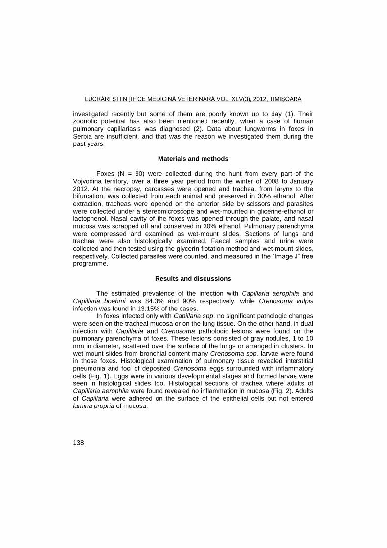
LUCRĂRI ŞTIINŢIFICE MEDICINĂ VETERINARĂ VOL. XLV(3), 2012, TIMIŞOARA
138
investigated recently but some of them are poorly known up to day (1). Their zoonotic potential has also been mentioned recently, when a case of human pulmonary capillariasis was diagnosed (2). Data about lungworms in foxes in Serbia are insufficient, and that was the reason we investigated them during the past years.
Materials and methods
Foxes (N = 90) were collected during the hunt from every part of the
Vojvodina territory, over a three year period from the winter of 2008 to January 2012. At the necropsy, carcasses were opened and trachea, from larynx to the bifurcation, was collected from each animal and preserved in 30% ethanol. After extraction, tracheas were opened on the anterior side by scissors and parasites were collected under a stereomicroscope and wet-mounted in glicerine-ethanol or lactophenol. Nasal cavity of the foxes was opened through the palate, and nasal mucosa was scrapped off and conserved in 30% ethanol. Pulmonary parenchyma were compressed and examined as wet-mount slides. Sections of lungs and trachea were also histologically examined. Faecal samples and urine were collected and then tested using the glycerin flotation method and wet-mount slides, respectively. Collected parasites were counted, and measured in the ―Image J‖ free programme.
Results and discussions
The estimated prevalence of the infection with Capillaria aerophila and
Capillaria boehmi was 84.3% and 90% respectively, while Crenosoma vulpis
infection was found in 13.15% of the cases. In foxes infected only with Capillaria spp. no significant pathologic changes
were seen on the tracheal mucosa or on the lung tissue. On the other hand, in dual infection with Capillaria and Crenosoma pathologic lesions were found on the pulmonary parenchyma of foxes. These lesions consisted of gray nodules, 1 to 10 mm in diameter, scattered over the surface of the lungs or arranged in clusters. In wet-mount slides from bronchial content many Crenosoma spp. larvae were found in those foxes. Histological examination of pulmonary tissue revealed interstitial pneumonia and foci of deposited Crenosoma eggs surrounded with inflammatory cells (Fig. 1). Eggs were in various developmental stages and formed larvae were seen in histological slides too. Histological sections of trachea where adults of Capillaria aerophila were found revealed no inflammation in mucosa (Fig. 2). Adults of Capillaria were adhered on the surface of the epithelial cells but not entered lamina propria of mucosa.
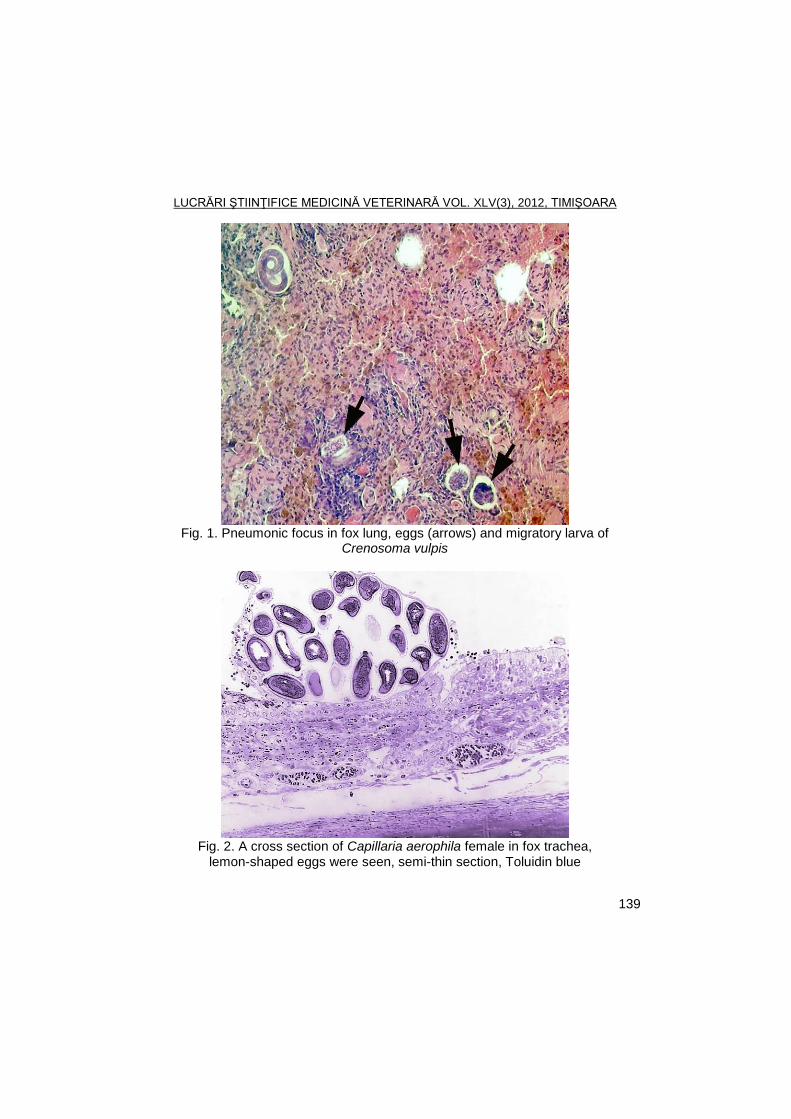
LUCRĂRI ŞTIINŢIFICE MEDICINĂ VETERINARĂ VOL. XLV(3), 2012, TIMIŞOARA
139
Fig. 1. Pneumonic focus in fox lung, eggs (arrows) and migratory larva of
Crenosoma vulpis
Fig. 2. A cross section of Capillaria aerophila female in fox trachea,
lemon-shaped eggs were seen, semi-thin section, Toluidin blue
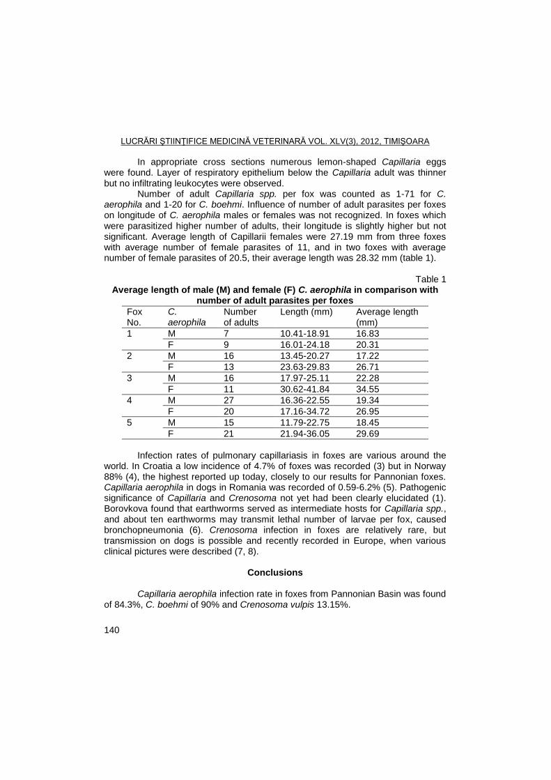
LUCRĂRI ŞTIINŢIFICE MEDICINĂ VETERINARĂ VOL. XLV(3), 2012, TIMIŞOARA
140
In appropriate cross sections numerous lemon-shaped Capillaria eggs were found. Layer of respiratory epithelium below the Capillaria adult was thinner but no infiltrating leukocytes were observed.
Number of adult Capillaria spp. per fox was counted as 1-71 for C. aerophila and 1-20 for C. boehmi. Influence of number of adult parasites per foxes on longitude of C. aerophila males or females was not recognized. In foxes which were parasitized higher number of adults, their longitude is slightly higher but not significant. Average length of Capillarii females were 27.19 mm from three foxes with average number of female parasites of 11, and in two foxes with average number of female parasites of 20.5, their average length was 28.32 mm (table 1).
Table 1
Average length of male (M) and female (F) C. aerophila in comparison with number of adult parasites per foxes
Fox No.
C. aerophila
Number of adults
Length (mm) Average length (mm)
1 M 7 10.41-18.91 16.83
F 9 16.01-24.18 20.31
2 M 16 13.45-20.27 17.22
F 13 23.63-29.83 26.71
3 M 16 17.97-25.11 22.28
F 11 30.62-41.84 34.55
4 M 27 16.36-22.55 19.34
F 20 17.16-34.72 26.95
5 M 15 11.79-22.75 18.45
F 21 21.94-36.05 29.69
Infection rates of pulmonary capillariasis in foxes are various around the
world. In Croatia a low incidence of 4.7% of foxes was recorded (3) but in Norway 88% (4), the highest reported up today, closely to our results for Pannonian foxes. Capillaria aerophila in dogs in Romania was recorded of 0.59-6.2% (5). Pathogenic significance of Capillaria and Crenosoma not yet had been clearly elucidated (1). Borovkova found that earthworms served as intermediate hosts for Capillaria spp., and about ten earthworms may transmit lethal number of larvae per fox, caused bronchopneumonia (6). Crenosoma infection in foxes are relatively rare, but transmission on dogs is possible and recently recorded in Europe, when various clinical pictures were described (7, 8).
Conclusions
Capillaria aerophila infection rate in foxes from Pannonian Basin was found
of 84.3%, C. boehmi of 90% and Crenosoma vulpis 13.15%.
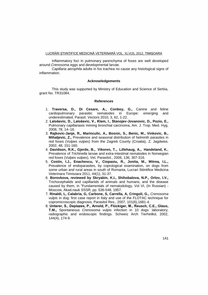
LUCRĂRI ŞTIINŢIFICE MEDICINĂ VETERINARĂ VOL. XLV(3), 2012, TIMIŞOARA
141
Inflammatory foci in pulmonary parenchyma of foxes are well developed around Crenosoma eggs and developmental larvae.
Capillaria aerophila adults in fox trachea no cause any histological signs of inflammation.
Acknowledgements
This study was supported by Ministry of Education and Science of Serbia,
grant No. TR31084.
References
1. Traversa, D., Di Cesare, A., Conboy, G., Canine and feline cardiopulmonary parasitic nematodes in Europe: emerging and underestimated, Parasit. Vectors 2010, 3, 62, 1-22.
2. Laloševic, D., Laloševic, V., Klem, I., Stanojev-Jovanovic, D., Pozio, E., Pulmonary capillariasis miming bronchial carcinoma, Am. J. Trop. Med. Hyg, 2008, 78, 14–16.
3. Rajkovic-Janje, R., Marinculic, A., Bosnic, S., Benic, M., Vinkovic, B., Mihaljevic, Z., Prevalence and seasonal distribution of helminth parasites in red foxes (Vulpes vulpes) from the Zagreb County (Croatia), Z. Jagdwiss. 2002, 48, 151-160.
4. Davidson, R.K., Gjerde, B.,, Vikoren, T.,, Lillehaug, A.,, Handeland, K., Prevalence of Trichinella larvae and extra-intestinal nematodes in Norwegian red foxes (Vulpes vulpes), Vet. Parasitol., 2006, 136, 307-316.
5. Costin, I.J., Enachescu, V., Ciopasiu, R., Jonita, M., Mitrea, I.L., Prevalence of endoparasites, by coprological examination, on dogs from some urban and rural areas in south of Romania, Lucrari Stiintifice Medicina Veterinara Timisoara 2011, 44(1), 31-37.
6. Borovkova, reviewed by Skryabin, K.I., Shihobalova, N.P., Orlov, I.V., Trichocephalids and capillariids of animals and humans, and the disease caused by them, in "Fundamentals of nematodology, Vol VI, (In Russian) - Moscow, Akad.nauk SSSR, pp. 536-548, 1957.
7. Rinaldi, L, Calabria, G, Carbone, S, Carrella, A, Cringoli, G., Crenosoma vulpis in dog: first case report in Italy and use of the FLOTAC technique for copromicroscopic diagnosis, Parasitol Res., 2007, 101(6),1681-4.
8. Unterer, S., Deplazes, P., Arnold, P., Flückiger, M., Reusch, C.E., Glaus, T.M., Spontaneous Crenosoma vulpis infection in 10 dogs: laboratory, radiographic and endoscopic findings. Schweiz Arch Tierheilkd, 2002, 144(4), 174-9.
LUCRĂRI ŞTIINŢIFICE MEDICINĂ VETERINARĂ VOL. XLV(3), 2012, TIMIŞOARA
142
THE DYNAMICS OF BLOWFLIES OF CALLIPHORA, LUCILIA AND PROTOPHORMIA GENERA DURING 2004 IN
NORTHERN TIMIȘOARA
S. MORARIU1, GH. DĂRĂBUȘ
1, I. OPRESCU
1, NARCISA MEDERLE
1,
M.S. ILIE1, FLORICA MORARIU
2
1Banat's University of Agricultural Sciences and Veterinary Medicine Timisoara,
Faculty of Veterinary Medicine, 300645, Aradului Street No. 119, Timisoara, Romania
2Banat's University of Agricultural Sciences and Veterinary Medicine Timisoara,
Faculty of Animal Sciences and Biotechnologies E-mail: [email protected]
Summary
This paper describes the dynamics of the three important blowfly genera from the western Romania. During 2004 the blowflies of Lucilia, Calliphora and Protophormia genera were monitored between April 20
th and October 30
th. For Calliphora genus six population
peaks were recorded. First one was achieved on May 03rd
, at an air average temperature of 17.6ºC, a soil average temperature of 24.3ºC and 58% relative humidity, and the last one on October 01
st, at an air average temperature 14.6ºC, soil average temperature 16.4ºC and
75% relative humidity, respectively. For Lucilia genus five population peaks were recorded, with May 04
th the first peak date, at an air average temperature of 18.1ºC, a soil average
temperature of 19.8ºC and 60% relative humidity, and September 24th
the last capture date. For Protophormia genus were also five population peaks recorded, as for Lucilia one. First peak was noticed on May 03
rd at an air average temperature of 17.6ºC, a soil average
temperature of 24.3ºC and 58% relative humidity, and the last one like that for Lucilia genus. The shortest life cycle interval was recorded in Lucilia blowflies.
Keywords: Blowflies, Calliphora, Lucilia, Protophormia, dynamics
Lucilia sericata, the greenbottle blowfly, and Wohlfahrtia magnifica, the spotted flesh fly, are detrimental to the livestock industries in temperate areas, causing the potentially fatal disease condition known as sheep strike (5, 6, 10, 12).
Blowflies are also important in forensic science, mainly to estimate the time of death. Some of these insects are Calliphoridae (Diptera), including species of Calliphora, Protophormia and Lucilia genera. In many European countries Calliphora species are considered the most important, with C. erythrocephala and C. vomitoria being widely distributed (1).
Blowflies are poikilothermic, so their rate of development is governed by ambient temperature. Knowing the environmental temperature and their developmental progress, an estimation of time since the eggs of blowflies were laid can be made.
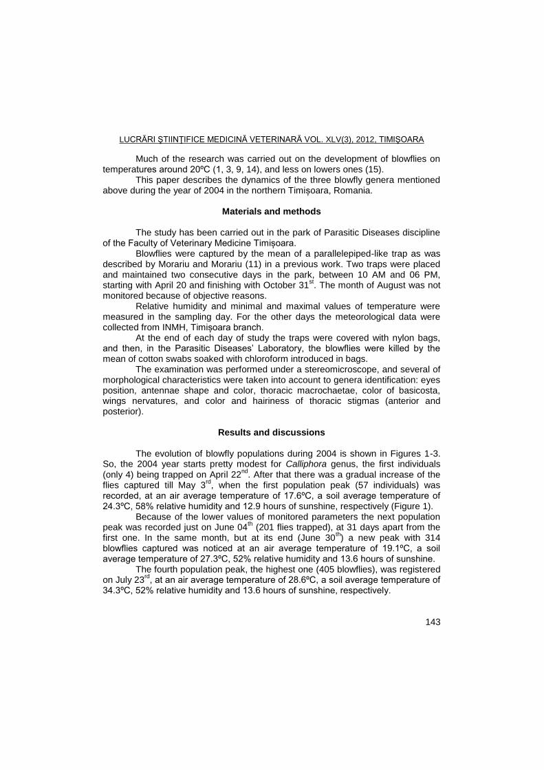
LUCRĂRI ŞTIINŢIFICE MEDICINĂ VETERINARĂ VOL. XLV(3), 2012, TIMIŞOARA
143
Much of the research was carried out on the development of blowflies on temperatures around 20ºC (1, 3, 9, 14), and less on lowers ones (15).
This paper describes the dynamics of the three blowfly genera mentioned above during the year of 2004 in the northern Timișoara, Romania.
Materials and methods
The study has been carried out in the park of Parasitic Diseases discipline
of the Faculty of Veterinary Medicine Timișoara. Blowflies were captured by the mean of a parallelepiped-like trap as was
described by Morariu and Morariu (11) in a previous work. Two traps were placed and maintained two consecutive days in the park, between 10 AM and 06 PM, starting with April 20 and finishing with October 31
st. The month of August was not
monitored because of objective reasons. Relative humidity and minimal and maximal values of temperature were
measured in the sampling day. For the other days the meteorological data were collected from INMH, Timișoara branch.
At the end of each day of study the traps were covered with nylon bags, and then, in the Parasitic Diseases’ Laboratory, the blowflies were killed by the mean of cotton swabs soaked with chloroform introduced in bags.
The examination was performed under a stereomicroscope, and several of morphological characteristics were taken into account to genera identification: eyes position, antennae shape and color, thoracic macrochaetae, color of basicosta, wings nervatures, and color and hairiness of thoracic stigmas (anterior and posterior).
Results and discussions
The evolution of blowfly populations during 2004 is shown in Figures 1-3.
So, the 2004 year starts pretty modest for Calliphora genus, the first individuals (only 4) being trapped on April 22
nd. After that there was a gradual increase of the
flies captured till May 3rd
, when the first population peak (57 individuals) was recorded, at an air average temperature of 17.6ºC, a soil average temperature of 24.3ºC, 58% relative humidity and 12.9 hours of sunshine, respectively (Figure 1).
Because of the lower values of monitored parameters the next population peak was recorded just on June 04
th (201 flies trapped), at 31 days apart from the
first one. In the same month, but at its end (June 30th) a new peak with 314
blowflies captured was noticed at an air average temperature of 19.1ºC, a soil average temperature of 27.3ºC, 52% relative humidity and 13.6 hours of sunshine.
The fourth population peak, the highest one (405 blowflies), was registered on July 23
rd, at an air average temperature of 28.6ºC, a soil average temperature of
34.3ºC, 52% relative humidity and 13.6 hours of sunshine, respectively.
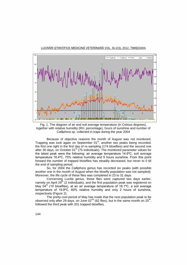
LUCRĂRI ŞTIINŢIFICE MEDICINĂ VETERINARĂ VOL. XLV(3), 2012, TIMIŞOARA
144
-20
0
20
40
60
80
100
120
1 2 3 4 I 5 6 7 8 9 10 11 12
0
50
100
150
200
250
300
350
400
450
T medie aer T medie sol UR % Soare Nr diptere
Fig. 1. The diagram of air and soil average temperature (in Celsius degrees),
together with relative humidity (RH, percentage), hours of sunshine and number of Calliphora sp. collected in traps during the year 2004
Because of objective reasons the month of August was not monitored.
Trapping was took again on September 01st, another two peaks being recorded:
the first one right in the first day of re-sampling (174 blowflies) and the second one after 30 days, on October 01
st (75 individuals). The monitored parameter values for
the latest peak were the following: air average temperature 14.6ºC, soil average temperature 16.4ºC, 75% relative humidity and 5 hours sunshine. From this point forward the number of trapped blowflies has steadily decreased, but never to 0 till the end of sampling period.
So, for 2004 the Calliphora genus has recorded six peaks (with possible another one in the month of August when the blowfly population was not sampled). Moreover, the life cycle of these flies was completed in 23 to 31 days.
Concerning Lucilia genus, these flies were captured two days earlier, namely on April 29
th (2 individuals), and the first population peak was registered on
May 04th (10 blowflies), at an air average temperature of 18.1ºC, a soil average
temperature of 19.8ºC, 60% relative humidity and only 2 hours of sunshine, respectively (Figure 2).
The pretty cool period of May has made that the next population peak to be observed only after 29 days, on June 02
nd (62 flies), but in the same month on 25
th,
followed the third peak with 201 trapped blowflies.
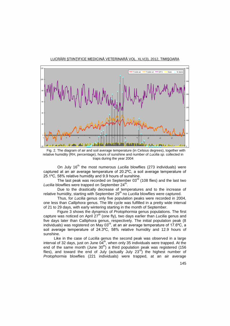
LUCRĂRI ŞTIINŢIFICE MEDICINĂ VETERINARĂ VOL. XLV(3), 2012, TIMIŞOARA
145
-20
0
20
40
60
80
100
120
1 2 3 4 I 5 6 7 8 9 10 11 12
0
50
100
150
200
250
300
T medie aer T medie sol UR % Soare Nr diptere
Fig. 2. The diagram of air and soil average temperature (in Celsius degrees), together with relative humidity (RH, percentage), hours of sunshine and number of Lucilia sp. collected in
traps during the year 2004
On July 16
th the most numerous Lucilia blowflies (273 individuals) were
captured at an air average temperature of 20.2ºC, a soil average temperature of 25.1ºC, 58% relative humidity and 9.9 hours of sunshine.
The last peak was recorded on September 03rd
(108 flies) and the last two Lucilia blowflies were trapped on September 24
th.
Due to the drastically decrease of temperatures and to the increase of relative humidity, starting with September 29
th no Lucilia blowflies were captured.
Thus, for Lucilia genus only five population peaks were recorded in 2004, one less than Calliphora genus. The life cycle was fulfilled in a pretty wide interval of 21 to 29 days, with early wintering starting in the month of September.
Figure 3 shows the dynamics of Protophormia genus populations. The first capture was noticed on April 27
th (one fly), two days earlier than Lucilia genus and
five days later than Calliphora genus, respectively. The initial population peak (8 individuals) was registered on May 03
rd, at an air average temperature of 17.6ºC, a
soil average temperature of 24.3ºC, 58% relative humidity and 12.9 hours of sunshine.
Like in the case of Lucilia genus the second peak was observed in a large interval of 32 days, just on June 04
th, when only 35 individuals were trapped. At the
end of the same month (June 30th) a third population peak was registered (156
flies), and toward the end of July (actually July 23rd
) the highest number of Protophormia blowflies (221 individuals) were trapped, at an air average
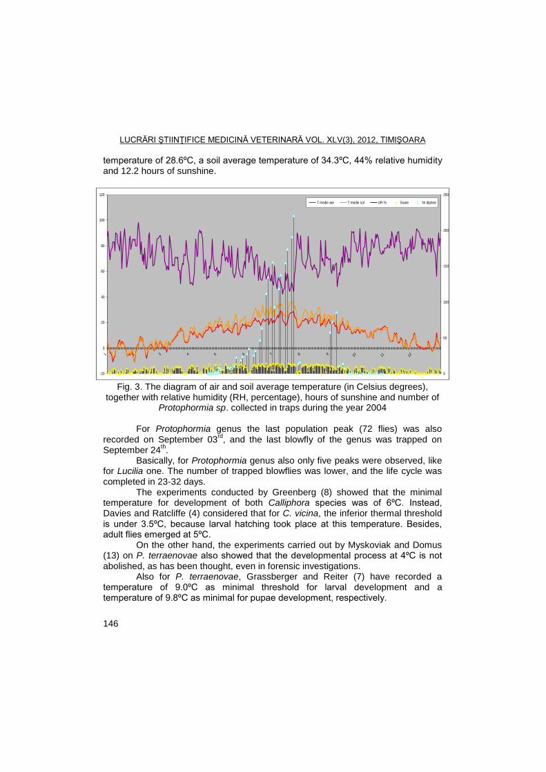
LUCRĂRI ŞTIINŢIFICE MEDICINĂ VETERINARĂ VOL. XLV(3), 2012, TIMIŞOARA
146
temperature of 28.6ºC, a soil average temperature of 34.3ºC, 44% relative humidity and 12.2 hours of sunshine.
-20
0
20
40
60
80
100
120
1 2 3 4 5 6 7 8 9 10 11 12
0
50
100
150
200
250
T medie aer T medie sol UR % Soare Nr diptere
Fig. 3. The diagram of air and soil average temperature (in Celsius degrees),
together with relative humidity (RH, percentage), hours of sunshine and number of Protophormia sp. collected in traps during the year 2004
For Protophormia genus the last population peak (72 flies) was also
recorded on September 03rd
, and the last blowfly of the genus was trapped on September 24
th.
Basically, for Protophormia genus also only five peaks were observed, like for Lucilia one. The number of trapped blowflies was lower, and the life cycle was completed in 23-32 days.
The experiments conducted by Greenberg (8) showed that the minimal temperature for development of both Calliphora species was of 6ºC. Instead, Davies and Ratcliffe (4) considered that for C. vicina, the inferior thermal threshold is under 3.5ºC, because larval hatching took place at this temperature. Besides, adult flies emerged at 5ºC.
On the other hand, the experiments carried out by Myskoviak and Domus (13) on P. terraenovae also showed that the developmental process at 4ºC is not abolished, as has been thought, even in forensic investigations.
Also for P. terraenovae, Grassberger and Reiter (7) have recorded a temperature of 9.0ºC as minimal threshold for larval development and a temperature of 9.8ºC as minimal for pupae development, respectively.
LUCRĂRI ŞTIINŢIFICE MEDICINĂ VETERINARĂ VOL. XLV(3), 2012, TIMIŞOARA
147
Conclusions For Lucilia and Protophormia genera, five population peaks were
registered, while Calliphora one recorded six peaks. The highest population peak was seen on July for all monitored genera. The shortest life cycle interval was noticed for Lucilia blowflies (21 to 29
days).
References
1. Ames, C., Turner, B., Low temperature episodes in development of blowflies: implications for postmortem interval estimation. Med Vet Entomol, 2003, 17, 178-186.
2. Anderson, G.S., Minimum and maximum development rate of some forensically important Calliphoridae (Diptera), J Forensic Sci, 2000, 45, 824-832.
3. Dallwitz, R., The influence of constant and fluctuating temperatures on development rate and survival of pupae of the Australian sheep blowfly Lucilia cuprina. Entomol Exp Appl, 1984, 36, 89-95.
4. Davies, L., Ratcliffe, G.G., Development rates of some pre-adult stages in blowflies with reference to low temperatures, Med Vet Entomol, 1994, 8, 245–254.
5. Farkas, R., Hall, M.J.R., Kelemen, F., Wound myiasis of sheep in Hungary, Vet Parasitol, 1997, 69, 133–144.
6. Giangaspero, A., Traversa, D., Trentini, R., Scala, A., Otranto, D., Traumatic myiasis by Wohlfahrtia magnifica in Italy. Vet Parasitol, 2011, 175, 109-112.
7. Grassberger, M., Reiter, C., Effect of temperature on Lucilia sericata (Diptera: Calliphoridae) development with special reference to the isomegalen- and isomorphen-diagram, Forensic Sci Int, 2001, 120, 32–36.
8. Greenberg, B., Flies as forensic indicators, J Med Entomol, 1991, 28, 565-577.
9. Greenberg, B., Tantawi, T., Different developmental strategies in two boreal blow flies (Diptera: Calliphoridae), J Med Entomol, 1993, 30, 481-484.
10. Hall, M.J.R., Wall, R., Myiasis of humans and domestic animals. Adv. Parasitol., 1995, 35, 257–334.
11. Morariu, S., Morariu, Florica, The role of olfaction in orientation of blowflies to hosts or odorous baited traps, Lucr Șt Zoot Biotehn Timișoara, 2010, 43, 2, 112-115.
12. Morariu, S., Morariu, Florica, The dynamics of blowflies of Calliphora, Lucilia and Protophormia genera during 2003 in northern Timisoara, Lucr Șt Zoot Biotehn Timișoara, 2011, 44, 1, 497-500.
LUCRĂRI ŞTIINŢIFICE MEDICINĂ VETERINARĂ VOL. XLV(3), 2012, TIMIŞOARA
148
13. Myskowiak, J.B., Doums, C., Effects of refrigeration on the biometryand development of Protophormia terraenovae (Robineau Desvoidy) (Diptera: Calliphoridae) and its consequences in estimating post-mortem interval in forensic investigations, Forensic Sci Int, 2002, 125, 254–261.
14. Tarone, A.M., Picard, C.J, Spiegelman, C., Foran, D.R., Population and temperature effects on Lucilia sericata (Diptera: Calliphoridae) body size and minimum development time, J Med Entomol, 2011, 48, 1062-1068.
15. Vinogradova, E.B., Marchenko, M.J., The use of temperature parameters of fly growth in the medico-legal practice. Sud-Med Ekspert, 1984, 27, 16–19.
LUCRĂRI ŞTIINŢIFICE MEDICINĂ VETERINARĂ VOL. XLV(3), 2012, TIMIŞOARA
149
EFFICACY TESTING OF FLUKIVER COMBI (JANSSEN PHARMACEUTICA NV) IN NATURAL MIXED INFECTION WITH
FASCIOLA HEPATICA AND GASTROINTESTINAL NEMATODES, IN SHEEPS FROM BILBOR, HARGHITA
I. OPRESCU, S. MORARIU, NARCISA MEDERLE, M.S. ILIE, D. INDRE, MIRELA
IMRE, DENISA SORESCU, M. ŢIFREA, , GH. DĂRĂBUŞ
Banat's University of Agricultural Sciences and Veterinary Medicine Timisoara, Faculty of Veterinary Medicine, 300645, Aradului Street No. 119, Timisoara, Romania
E-mail: [email protected]
Summary
The study efficacy for Flukiver Combi was conducted between March-April 2010 on sheeps natural infected with Fasciola hepatica and gastrointestinal nemathodes owned by private breeders from Bilbor village, Harghita, County.
The animals were divided in two groups (OV1 and OV2), each one composed of 10 individuals (n=10); the group OV1 was treated with Flukiver Combi, at the dose of 1 ml/5 kg body weight (10 mg/kg closantel + 15 mg mebendazole) and the group OV2 was the witness group, untreated. Fecal samples were harvested from all animals in day 0 and day 12 post-treatment establishing the EPG using McMaster and Brumpt method. The efficacy was established using Presidente and Boorgsteede relations.
Following experiment, we establish a mean efficacy of 100% for Flukiver Combi and we consider the product safe and effective.
Keywords: efficacy, Flukiver Combi, Fasciola hepatica, gastrointestinal nemathods
Prophylaxis of liver fluke infestation is related to the awareness of
epizootological status of the flock/farm. In assessing the epizootiological status, investigations should be carried out by a veterinarian and morphoclinical and laboratory aspects should be considered.
The literature states that, in endemic areas, two treatments per year are recommended: in spring, two weeks before entering the pasture, treatment for adulticid effect, and in fall 2-3 weeks after entering into stalls, when the use of anthelmintics that target both adult and young Fasciola is recommended (6).
The aim of this study was to test the efficacy of commercial product Flukiver Combi in natural mixed infection with Fasciola hepatica and gastrointestinal nematodes, in sheeps.
Materials and methods
The study was conducted between March-April 2010. The aim of the research
was to investigate the effectiveness of the product Flukiver Combi (Jenssen Pharmaceutica NV) on populations of Fasciola hepatica and gastrointestinal
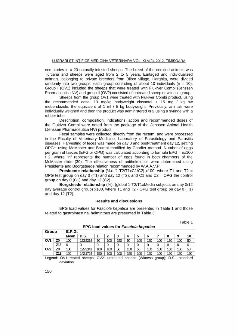
LUCRĂRI ŞTIINŢIFICE MEDICINĂ VETERINARĂ VOL. XLV(3), 2012, TIMIŞOARA
150
nematodes in a 20 naturally infested sheeps. The breed of the enrolled animals was Țurcana and sheeps were aged from 2 to 5 years. Eartaged and individualized animals, belonging to private breeders from Bilbor village, Harghita, were divided randomly into two groups, each group consisting of about 10 individuals (n = 10). Group I (OV1) included the sheeps that were treated with Flukiver Combi (Jenssen Pharmaceutica NV) and group II (OV2) consisted of untreated sheep or witness group.
Sheeps from the group OV1 were treated with Flukiver Combi product, using the recommended dose: 10 mg/kg bodyweight closantel + 15 mg / kg bw mebendazole, the equivalent of 1 ml / 5 kg bodyweight. Previously, animals were individually weighed and then the product was administered oral using a syringe with a rubber tube.
Description, composition, indications, action and recommended doses of the Flukiver Combi were noted from the package of the Jenssen Animal Health (Jenssen Pharmaceutica NV) product.
Fecal samples were collected directly from the rectum, and were processed in the Faculty of Veterinary Medicine, Laboratory of Parasitology and Parasitic diseases. Harvesting of feces was made on day 0 and post-treatment day 12, setting OPG's using McMaster and Brumpt modified by Charlier method. Number of eggs per gram of faeces (EPG or OPG) was calculated according to formula EPG = nx100 / 2, where "n" represents the number of eggs found in both chambers of the McMaster slide (30). The effectiveness of antihelmintics were determined using Presidente and Boorgsteede relation recommended by W.A.A.V.P.
Presidente relationship (%): [1-T2/T1xC1/C2] x100, where T1 and T2 = OPG test group on day 0 (T1) and day 12 (T2), and C1 and C2 = OPG the control group on day 0 (C1) and day 12 (C2).
Borgsteede relationship (%): (global 1-T2/T1xMedia subjects on day 0/12 day average control group) x100, where T1 and T2 - OPG test group on day 0 (T1) and day 12 (T2).
Results and discussions
EPG load values for Fasciola hepatica are presented in Table 1 and those related to gastrointestinal helminthes are presented in Table 3.
Table 1
EPG load values for Fasciola hepatica
Group E.P.G. Mean D.S. 1 2 3 4 5 6 7 8 9 10
OV1 Z0 100 123.3214 50 100 150 50 100 150 100 150 100 50
Z12 0 0 0 0 0 0 0 0 0 0 0 0
OV2 Z0 100 125.3341 100 100 50 150 50 100 100 150 150 50
Z12 120 142.1724 150 100 100 150 100 150 100 150 150 150
Legend: OV1-treated sheeps; OV2- untreated sheeps (Witness group); D.S.- standard deviation
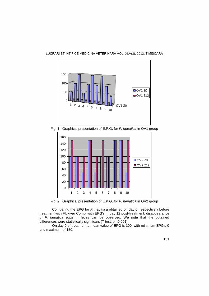
LUCRĂRI ŞTIINŢIFICE MEDICINĂ VETERINARĂ VOL. XLV(3), 2012, TIMIŞOARA
151
1 2 3 4 5 6 7 8 9 10
OV1 Z0
0
50
100
150
OV1 Z0
OV1 Z12
Fig. 1. Graphical presentation of E.P.G. for F. hepatica in OV1 group
0
20
40
60
80
100
120
140
160
1 2 3 4 5 6 7 8 9 10
OV2 Z0
OV2 Z12
Fig. 2. Graphical presentation of E.P.G. for F. hepatica in OV2 group
Comparing the EPG for F. hepatica obtained on day 0, respectively before
treatment with Flukiver Combi with EPG's in day 12 post-treatment, disappearance of F. hepatica eggs in feces can be observed. We note that the obtained differences were statistically significant (T test, p <0.001).
On day 0 of treatment a mean value of EPG is 100, with minimum EPG's 0 and maximum of 150.
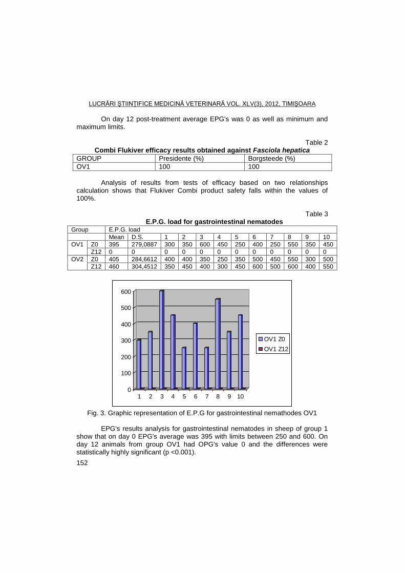
LUCRĂRI ŞTIINŢIFICE MEDICINĂ VETERINARĂ VOL. XLV(3), 2012, TIMIŞOARA
152
On day 12 post-treatment average EPG's was 0 as well as minimum and maximum limits.
Table 2 Combi Flukiver efficacy results obtained against Fasciola hepatica
GROUP Presidente (%) Borgsteede (%)
OV1 100 100
Analysis of results from tests of efficacy based on two relationships
calculation shows that Flukiver Combi product safety falls within the values of 100%.
Table 3
E.P.G. load for gastrointestinal nematodes Group E.P.G. load
Mean D.S. 1 2 3 4 5 6 7 8 9 10
OV1 Z0 395 279,0887 300 350 600 450 250 400 250 550 350 450
Z12 0 0 0 0 0 0 0 0 0 0 0 0
OV2 Z0 405 284,6612 400 400 350 250 350 500 450 550 300 500
Z12 460 304,4512 350 450 400 300 450 600 500 600 400 550
0
100
200
300
400
500
600
1 2 3 4 5 6 7 8 9 10
OV1 Z0
OV1 Z12
Fig. 3. Graphic representation of E.P.G for gastrointestinal nemathodes OV1
EPG's results analysis for gastrointestinal nematodes in sheep of group 1
show that on day 0 EPG's average was 395 with limits between 250 and 600. On day 12 animals from group OV1 had OPG's value 0 and the differences were statistically highly significant (p <0.001).
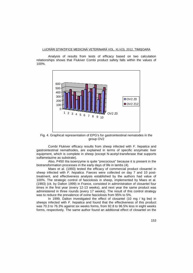
LUCRĂRI ŞTIINŢIFICE MEDICINĂ VETERINARĂ VOL. XLV(3), 2012, TIMIŞOARA
153
Analysis of results from tests of efficacy based on two calculation relationships shows that Flukiver Combi product safety falls within the values of 100%.
1 2 3 4 5 6 7 8 9 10
OV2 Z0
0
100
200
300
400
500
600
OV2 Z0
OV2 Z12
Fig. 4. Graphical representation of EPG's for gastrointestinal nematodes in the
group OV2
Combi Flukiver efficacy results from sheep infected with F. hepatica and gastrointestinal nemathodes, are explained in terms of specific enzymatic liver equipment, which is complete in sheep (except N-acetyl-transferase that supports sulfametazine as substrate).
Also, P450 IIIa isoenzyme is quite "precocious" because it is present in the biotransformation processes in the early days of life in lambs (4).
Maes et al. (1993) tested the efficacy of commercial product closantel in sheep infected with F. hepatica. Faeces were collected on day 7 and 10 post-treatment, and effectiveness analysis established by the authors had value of 100%. The strategic control of fasciolosis in sheep, implemented by Maes et al. (1993) (cit. by Dalton 1999) in France, consisted in administration of closantel four times in the first year (every 12-13 weeks), and next year the same product was administered in three rounds (every 17 weeks). The result of this control strategy was to reduce the prevalence of ovine fasciolosis from 95% to 5%.
In 1999, Dalton investigated the effect of closantel (10 mg / kg bw) in sheeps infected with F. hepatica and found that the effectiveness of this product was 70.3 to 76.3% against six weeks forms, from 92.8 to 96.5% less in eight weeks forms, respectively. The same author found an additional effect of closantel on the
LUCRĂRI ŞTIINŢIFICE MEDICINĂ VETERINARĂ VOL. XLV(3), 2012, TIMIŞOARA
154
development of F. hepatica eggs that had been a reduction in egg production for 13 weeks after treatment.
Almeida et al. (2010) end Kempler et al. (2010) studied the effect of anthelmintic products (moxidectin, triclorfon, closantel, levamisole, albendazole and ivermectin) in sheeps naturally infected with H. contortus and T. colubriformis. The author calculated average EPG's for each group of animals treated compared with average EPG's from the control group. Only triclorfon and moxidectin significantly reduced the number of eggs in feces. In terms of effectiveness of ivermectin, the author states that it has significantly reduced the number of eggs in feces.
Conclusions
EPG's progress in the days 0 and 12 post-treatment in OV1 group shows that Flukiver Combi is safe in the infestation with F. hepatica and gastrointestinal nematodes in sheep.
The mean Combi Flukiver calculated efficacy in sheep was 100%. None of helminthes species investigated were found to be resistant to
Combi Flukiver.
References
1. Almeida, F.A., Garcia, K.C.O.D., Torgerson, P.R., Amarante, A.F.T., Multiple resistance to antihelmintics by Haemonchus contortus and Trichostrongylus colubriformis in sheep in Brazil, Parasitol., 2010, 10, 10-16.
2. Cosoroabă, I., Dărăbuş, Gh., Oprescu, I., Compendiu de parazitologie veterinară, vol. I şi II. Ed. Mirton, Timisoara, 1995.
3. Cosoroabă, I., Dărăbuş, Gh., Oprescu, I., Morariu, S., Diagnostic paraclinic şi tehnici experimentale în parazitologie. Ed. Mirton, Timişoara, 2002.
4. Cristina, R.T., Introducere în farmacologia şi terapia veterinară. Ed. Solness, Timişoara, 2006.
5. Dalton, J.P., Fasciolosis, CABI Publishing, Cambridge, UK, 1999. 6. Dărăbuş, Gh., Oprescu, I., Morariu, S., Mederle, Narcisa, Parazitologie şi
boli parazitare. Ed. Mirton, Timişoara, 2006. 7. Indre, D., Cercetări privind strategiile de control în trichostrongilidoze la
ovine în vestul României. Teză de doctorat. U.S.A.M.V.B. Timisoara, 2011. 8. Kempler, K.E., Palmer, D.G., Liu, S.M., Greeff, J.C., Bishop, S.C.,
Karlsson, L.J.E., Reduction of faecal worm egg count, worm numbers and worm fecundity in sheep selected for worm resistance following artificial infection with Teladorsagia circumcincta and Trichostrongylus colubriformis. Veterinary Parasitology, 2010, 171, 238-246.
LUCRĂRI ŞTIINŢIFICE MEDICINĂ VETERINARĂ VOL. XLV(3), 2012, TIMIŞOARA
155
SEASON DISTRIBUTION OF GASTROINTESTINAL HELMINTHS OF SMALL RUMINANTS IN SPREAD BELGRADE AREA
I. PALOVIŠ
1, SNEŢANA IVANOVIŠ
1, GORDANA ŢUGIŠ
2, DUBRAVKA JOVŢIŠ
3,
J. BOJKOVSKI4, M. PAJIŠ
5
1Scientific Veterinary Institute, Belgrade, Serbia
2Medicines and Medical Devices Agency of Serbia, Belgrade, Serbia
3Faculty of Apply Ecology ―Futura‖, University Singidunum, Belgrade, Serbia
4Faculty of Veterinary Medicine, Belgrade, Serbia
5Faculty of Agronomy, Belgrade, Serbia
E-mail: [email protected]
Summary
The study about season distribution of gastrointestinal helminthes of small ruminant
at spread Belgrade area was performed in period March 2009 to January 2010. Coprological and post-mortem examination revealed the following gastrointestinal helminthes prevalence: Ostertagia circumcincta (95.23%), O.trifurcata (91.53%), O. ostertagi (23.33%), Trichostrongylus axei (100%), T.colubriformis (89.57%), T.capricola (62.85%), Nematodirus spathiger (100%), N. filicolis (43,31%), Haemonchus contortus (88.95%), Marshallagia marshalli (23.77%), Skrjabinema caprae (13.28%), Skrjabinema ovis (5.26%), Bunostomum trigonocephalum (13.28%), Chabertia ovina (64.14%), Oesophagostomum venulosum (28.39%), и Cooperia curticei (60.52%). The dynamics of the first occurrence of established species of gastro-intestinal strongilida was as follows: in March: Ostertagia circumcincta, Ostertagia ostertagi, Trichostrongylus colubriformis, T.vitrinus, Nematodirus filicoliis and N.spathiger, in May: Ostertagia occidentalis, Trichostrongylus axei, Bunostomum trigonocephalum and Chabertia ovina; in June: Skrjabinema ovis and S. capra; in July: Haemonchus contortus, Cooperia curticei and Oesophagostomum venulosum and in November: Marshallagia marshalli Species in the genus Ostertagia, Trichostrongylus and Nematodirus were present after the first appearance of those present during the entire study period. Haemonchus contortus is ordered in animals during the warmer and Marshallagia marshali during the colder period of the year. Species in the genus Cooperia, and Oesophagostomum. Bunostomum were often present in lambs sacrificed during all the
monitoring period. Key words:sheeps, goats, helminths
Way of breeding usually at sheep and goats breeding had prerequisite to a
lot of infections including parasitoses. They are usually kept under extensive conditions and graze or brows on any land that is not being cultivated. After harvesting, the animals are turned onto wheat and barley stubble from which they obtained nourishment. Pasture breeding make possible contact within sheep and eggs, larvae stages and intermediate host of parasites. Those induce that there are no one sheep without parasites.
LUCRĂRI ŞTIINŢIFICE MEDICINĂ VETERINARĂ VOL. XLV(3), 2012, TIMIŞOARA
156
Breeding of sheep and goats were increased during last decade on Belgrade area. Today, small flocks of sheep and goats play an important role in providing animal protein for diet, especially for those people who live in village at mountains part of Belgrade area. Both, sheep and goats are milked and they produce the bulk milk supply, together with a large proportion of the meat that is consumed. Along with the increasing number of flocks began on examination of their health status. During those examination especially was paid to the parasitic infections (16, 19, 20, 21).
Materials and methods
The study about season distribution of gastrointestinal helminthes of small
ruminant at spread Belgrade area was started in March 2009 and finished in January 2010. During study we examined a total of 91 flocks of goats and sheep from 6 Belgrade districs Mladenovac, Lazarevac, Obrenovac, Grocka, and Vozdovac (from the village Mladenovac, Vlaska, Mala Krsna, Velika Krsna, Medjuluzje, Senjak, Velika Ivanča, Orašac, Mala Vrbica, Rajkovac, Dubona, Šepšin, Resnik, Velike Granice, Granice, Koracica, Jagnjilo, Markovac, Lazarevac, Arapovac, Junkovac, Leskovac, Sokolovo Rabrovac, Vrbovno, Zvecka, Krtinska, and Stepojevac).
Geographical and climate data about examined area was next: Belgrade is situated in South-Eastern Europe, on the Balkan Peninsula. It lies at the point where the river Sava merges into the Danube, on the slope between two alluvial planes. The river waters surround it from three sides, and that is why since ancient times it has been the guardian of river passages.
Belgrade lies 116.75 metres above sea level and is located at confluence of the Danube and Sava rivers. The city has an urban area of 360 square kilometres, while together with its metropolitan area it covers 3,223 km
2. On the
right bank of the Sava (examined area), central Belgrade has a hilly terrain, while the highest point of Belgrade proper is Torlak hill at 303 m. The mountains of Avala (511 m) and Kosmaj (628 m) lies in south of the city (4). Across the Sava and Danube, the land is mostly flat, consisting of alluvial plains and loessial plateaus.
Belgrade's climate exhibits influences of oceanic, humid continental and humid subtropical zones, (10) with four seasons and uniformly spread precipitation. Monthly averages range from 0.4 °C in January to 21.8 °C in July, with an annual mean of 12.2 °C. There are, on average, 31 days a year when the temperature is above 30 °C, and 95 days when the temperature is above 25 °C. Belgrade receives about 680 millimeters of precipitation a year, with late spring being wettest (22).
During study we collected fecal samples at monthly intervals. A total of 910 fecal samples were analyzed using standard coprological techniques (2,18). A total of 67 goats and 89 sheep’s we were analyzed by post-mortem examination. Total differential worm counts were performed on all the alimentary tract and lungs using the technique described by Pavlović and AnĎelić-Buzadţić (18).
LUCRĂRI ŞTIINŢIFICE MEDICINĂ VETERINARĂ VOL. XLV(3), 2012, TIMIŞOARA
157
Determination of adult helminthes and eggs of parasites were done by keys given by Euzeby (2).
Results and discussions
The faecal samples were obtained from a different source all together as
they were collected from flocks in the field, and the results support the other findings. These counts were also of value in providing some information on the egg rise. The number of guts and lungs examined in this survey thought small in number, but in combination with results of coprological examination, samples appeared to represent the population adequately.
During the period 2009-2010 the next helminth species were found: The prevalence of individual species was the next: Ostertagia circumcincta (95.23%), O.trifurcata (91.53%), O.ostertagi (23.33%), Trichostrongylus axei (100%), T.colubriformis (89.57%), T.capricola (62.85%), Nematodirus spathiger (100%), N. filicolis (43.31%), Haemonchus contortus (88,95%), Marshallagia marshalli (23.77%), Skrjabinema caprae (13.28%), Skrjabinema ovis (5.26%), Bunostomum trigonocephalum (13.28%), Chabertia ovina (64.14%), Oesophagostomum venulosum (28.39%), and Cooperia curticei (60.52%).
The dynamics of the first occurrence of established species of gastro-intestinal strongilida was as follows:
- In March: Ostertagia circumcincta, Ostertagia ostertagi, Trichostrongylus colubriformis, T. vitrinus, Nematodirus filicoliis and N. spathiger
- In May: Ostertagia occidentalis, Trichostrongylus axei, Bunostomum trigonocephalum and Chabertia ovina;
- In June: Skrjabinema ovis and S. capra; - In July: Haemonchus contortus, Cooperia curticei and Oesophagostomum venulosum; - In November: Marshallagia marshalli
Species in the genus Ostertagia, Trichostrongylus and Nematodirus were present after the first appearance of those present during the entire study period. Haemonchus contortus is ordered in animals during the warmer and Marshallagia marshali during the colder period of the year. Species in the genus Cooperia, Oesophagostomum, and Bunostomum were often present in lambs sacrificed during all the monitoring period. Species in the genus Cooperia, and Oesophagostomum, Bunostomum were often present in lambs sacrificed during the monitoring period,
At the beginning of our research, conducted in March, the real extent of gastrointestinal infections strongilidae was 83.33%, after which he soon reached a level of 100% in the same way and moved to the end of follow-up period.
Extensity of infection established genera gastrointestinal strongilidae was different. The distribution of parasites of the genera Ostertagia, Trichostrongylus
LUCRĂRI ŞTIINŢIFICE MEDICINĂ VETERINARĂ VOL. XLV(3), 2012, TIMIŞOARA
158
and Nematodirus was reached during the monitoring period almost the maximum level.
The distribution of species within the established genera also varied. Within the genus Ostertagia most abundant were dominated by Ostertagia circumcincta and O. trifurcata. Prevalence of infection with Ostertagia ostertagi and Ostertagia occidentalis was higher during the colder periods of the year.
Among the species of the genus Trichostrongylus was the most prevalent Trichostrongylus colubriformis. Extensity of infection with Trichostrongylus axei and T.vitrinus varied, without any regularity. Extensity of infection with Nematodirus filicollis and N.spathiiger demonstrated a tendency to increase and leveled off at the highest level of the whole study period.
When we compared our results to the similar survey at mountain area of Serbia that have been done at Šara Mountain (11,14), Homolje (9), and at East Serbia (7,12,13), we were concluded that season distribution were different with our results. During those examination at East Serbia (including Šara Mountain) strongillidae of genera Haemonchus, Ostertagia, and Nematodirus was most abundant at spring and summer and strongillidae of genera Trichostrongylus and Marshallagia during outmn and winter period. Similar results were obtained during the studies in neighboring countries. In Macedonia Iliev (6) and later Georgievski at all. (5) occured Trichostrongylуs specis at spring months, and Marshallagia marshalli, Chabertia ovina and Oesophagostomуm species only at winter period. Simillary results were established in Montenegro by Karanfilovski (8), in Bulgaria by Denev and Kostov (2) and in Romania by Ardelaeanu et al. (1).
The obtain results confirm that the seasonal distribution of gastrointestinal helminths of small ruminants depends on the microclimate of environmental conditions.
References
1. Ardeleanu, D., Pivodă, C., Neacşu, M., Ida, A., Bio-ecolgical phenomenon
of poly-parasitism–actual major problem in breeding of sheep and goats, Lucr. St. Zootehnie si Biotehnologii, 2007, 40(2), 309-317.
2. Denev, I., Kostov R., Seasonal variations in the principal helminth infections on a sheep-grazing complex. Veterinarno-meditsinski Nauki, 1984, 21, 81-88.
3. Euzeby, J., Diagnostic experimental de helminthoses animals", ITVC, Paris, France, 1981.
4. "Geographical Position". Official website.www.beograd.rs 5. Georgievski B., Rasprostranjenost parazitskih infekcija na području opštine
Prilep, BSc thesis, Faculty of Veterinary Medicine in Belgrade, 1991. 6. Ilijev, A., Fauna endoparazita ovaca na područja pelagonje SR Makedonja .
phd Diseration Faculty of Veterinary Medicine in Belgrade, 1974.
LUCRĂRI ŞTIINŢIFICE MEDICINĂ VETERINARĂ VOL. XLV(3), 2012, TIMIŞOARA
159
7. Jovanoviš, D., Iliš, G., Nešiš, D., Pavloviš, I., Valter, D., Parasitoses of sheep in Timok district during 1986-1989, 1
th International Summer
Conference for Advancement of Sheep and Goat Production 1991, Ohrid, Macedonia, 383-385.
8. Karanfilovski, G., Stete od parazita u ovcarstvu, Pljevlja, 1991. 9. Lepojev, O., Helminti ovaca sa područja Homolja i mere za njiihovo
suzbijanje. Vet.glasnik, 1970, 12, 1001-1015. 10. "Natural Features". Official site. Retrieved 2007-07-16. www.beograd.rs 11. Pavlovic, D., Uloga različcitih starosnih kategorija ovaca u kontaminaciji
pasnjaka i odrzavanju i sirenju infekcija zeludacno-crevnim stronrgilidama tokom godine na planinskim pasnjacima Sare. Vet.glasnik, 1975, 2, 127-135.
12. Pavloviš,I., Nešiš,D., Iliš,G., Jovanoviš,D., Vlater,D., Parasitoses of sheep in Timok district during 1990. Proceeding of 1
th International Summer
Conference for Advancement of Sheep and Goat Production, Ohrid, Macedonia,1991, 387-389;
13. Pavloviš,I., Kulišiš,Z., Nešiš,D., Romaniš,S., Endoparasites of sheep and goats in Prizren district. Proceedeing of 3
rd International Conference of
Sheep and Goats Production, Ohrid, Macedonia, 1995, 106-110. 14. Pavloviš, I., Jakiš-Dimiš, D., Ivanoviš, S., The effect of parasitic
infection on sheep body weight. Biotech. Animal Husband, 2003, 19,145-148.
15. Pavloviš, I., Saviš, B., Ivetiš, V., Radanoviš, O., Ţutiš, M., Jakiš-Dimiš, D., Bojkovski, J.,The effect of parasitic infections to production results of sheep Proceeding of IV Balkan Conference of Animal Science BALNIMALCON 2009, Challanges of the Balkan Animal industry and the Role of science and Cooperation,. Stara Zagora, Bulgaria, 2009, 389-391
16. Pavloviš, I., Ivetiš, V., Saviš, B., Radanoviš, O., Ţutiš, M., Ivanoviš, S., Ţeludačno-crevna strongilidoza koza i ovaca na području Beograda Zbornik naučnih radova Instituta PKB Agroekonomik, 2009, 15 (3-4),123-127.
17. Pavloviš, I., Ivanoviš, S., Ţujoviš M., Tomiš Z., Plućna strongilidoza koza Zbornik naučnih radova Instituta PKB Agroekonomik, 2010, 16 (3-4), 171-177.
18. Pavloviš, I., AnŤeliš-Buzadţiš, G., Osnovi dijagnostike parazitskih bolesti ţivotinja za studente visoke poljoprivredne škole strukovnih studija u Šapcu studijski program: strukovna veterina Naučni institut za veterinarstvo Srbije, Šabac; Srbija, 2011.
19. Pavloviš I., Ivanoviš S., Ţujoviš M., Tomiš Z., Helminthoses of goats breeding at Belgrade area, Biotech. Animal Husband, 2011, 27, 1499-1504.
20. Pavloviš, I., Kneţeviš, N., Nematode parasites of sheep in spread
Belgrade area at measure to its contol Congress Proceeding of 19th
LUCRĂRI ŞTIINŢIFICE MEDICINĂ VETERINARĂ VOL. XLV(3), 2012, TIMIŞOARA
160
International Congress of Mediterranean federation of Health and Production of Ruminants, Beograd, 2011, 328-330.
21. Pavloviš, I., Ivanoviš, S, Ţujoviš,M., Tomiš Z., Memiši, N., Endoparasites of goats in spread Belgrade area in period 2009-2010. V
TH
international conference: BALNIMALCON 2011 & Xth International Symposium of Animal Biology and Nutrition & 40th International Session of Scientific Communications of the Faculty of Animal Science, Bucharest, Romania, Lucrari Stiintifice serial D, 2011,Vol. LIV.
22. Peel, M.C., Finlayson, B.L., McMahon, T.A., Updated world map of the Köppen-Geiger climate classification, Hydrol. Earth Syst. Sci., 2007, 11, 1641.
LUCRĂRI ŞTIINŢIFICE MEDICINĂ VETERINARĂ VOL. XLV(3), 2012, TIMIŞOARA
161
SEROPREVALENCE OF NEOSPORA CANINUM IN DAIRY COWS WITH REPRODUCTIVE DISORDERS IN VOJVODINA PROVINCE,
SERBIA
M. SAVOVIŠ1, VESNA LALOŠEVIŠ
2, S. SIMIN
2, LJILJANA PAVIŢIŠ
2,
S. BOBOŠ2
1PVS „MSV Medicus D.O.O.―, Milice Stojadinović Srpkinje No. 1, 21209, Bukovac,
Serbia 2 Faculty of Agriculture, Department for Veterinary Medicine, Novi Sad, Serbia
E-mail: [email protected]
Summary
Neospora caninum, a causative agent of neosporosis is obligate intra-cellular protozoa recognised all over the world as a major cause of abortions and other reproductive disorders in cattle. With regard to the lack of sufficient epizootiological data on this parasite in Serbia, the aim of our study was to asses the seroprevalence to N.caninum in dairy cows
with reported reproductive disorders in Vojvodina, northern province of the country. Total of 52 dairy cows with various reproductive disorders were sampled during
2011 and 2012. Collected sera were diluted in phosphate-buffered saline (PBS; pH=7.4) to 1:200 (cut off value) and examined using commercial slides for indirect immunofluorescence antibody test (IFAT) (VMRD Inc, USA). Positive samples were further titrated using two-fold serial dilutions to determine the highest titer. Cows were classified in two groups: cows with abortion (AB group) and without abortion (WAB group). Statistical analysis was performed in Quantitative Parasitology 3.0 free software.
The overall seroprevalence of N. caninum in dairy cows with reproductive disorders was 17.3% (9/52, CI 95: 9.1% - 29.7%). Antibodies were detected in three out of twelve aborting cows (AB group: 18.8%, CI 95: 5.3% - 43.6%) and in six out of forty animals without abortions (WAB group: 16.7%, CI 95: 7.5% - 32.0%). Statistical analysis of the results obtained in our study showed no significant difference between seroprevalences of AB group and WAB group (p=1.000). Antibody levels in seropositive animals ranged from 1:200 to 1:1600, with the highest level recorded in one cow from AB group.
Although further investigations, which would exclude other abortifacient agents and involve histopathological examination of aborted foetuses, are needed, results of our study suggest reasonable doubt that N. caninum could be one of the major causes of abortions and other reproductive disorders in dairy cattle from Vojvodina.
Key words: Neospora caninum, seroprevalence, reproductive disorders, dairy
cows, Serbia
Neospora caninum, a causative agent of neosporosis is an obligate intra-
cellular protozoa which is proved to invade a wide range of domestic and wild animals (8, 5, 1, 20).
Canids act as definitive hosts of N. caninum, and herbivores are recognized as its intermediate host. The most affected domestic animals are dogs
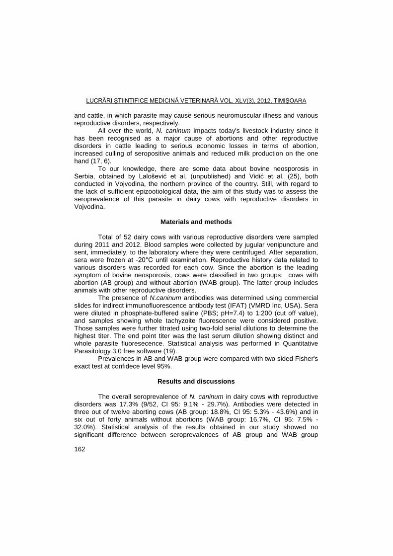
LUCRĂRI ŞTIINŢIFICE MEDICINĂ VETERINARĂ VOL. XLV(3), 2012, TIMIŞOARA
162
and cattle, in which parasite may cause serious neuromuscular illness and various reproductive disorders, respectively.
All over the world, N. caninum impacts today's livestock industry since it has been recognised as a major cause of abortions and other reproductive disorders in cattle leading to serious economic losses in terms of abortion, increased culling of seropositive animals and reduced milk production on the one hand (17, 6).
To our knowledge, there are some data about bovine neosporosis in Serbia, obtained by Lalošević et al. (unpublished) and Vidić et al. (25), both conducted in Vojvodina, the northern province of the country. Still, with regard to the lack of sufficient epizootiological data, the aim of this study was to assess the seroprevalence of this parasite in dairy cows with reproductive disorders in Vojvodina.
Materials and methods
Total of 52 dairy cows with various reproductive disorders were sampled
during 2011 and 2012. Blood samples were collected by jugular venipuncture and sent, immediately, to the laboratory where they were centrifuged. After separation, sera were frozen at -20°C until examination. Reproductive history data related to various disorders was recorded for each cow. Since the abortion is the leading symptom of bovine neosporosis, cows were classified in two groups: cows with abortion (AB group) and without abortion (WAB group). The latter group includes animals with other reproductive disorders. The presence of N.caninum antibodies was determined using commercial slides for indirect immunofluorescence antibody test (IFAT) (VMRD Inc, USA). Sera were diluted in phosphate-buffered saline (PBS; pH=7.4) to 1:200 (cut off value), and samples showing whole tachyzoite fluorescence were considered positive. Those samples were further titrated using two-fold serial dilutions to determine the highest titer. The end point titer was the last serum dilution showing distinct and whole parasite fluoresecence. Statistical analysis was performed in Quantitative Parasitology 3.0 free software (19).
Prevalences in AB and WAB group were compared with two sided Fisher's exact test at confidece level 95%.
Results and discussions
The overall seroprevalence of N. caninum in dairy cows with reproductive
disorders was 17.3% (9/52, CI 95: 9.1% - 29.7%). Antibodies were detected in three out of twelve aborting cows (AB group: 18.8%, CI 95: 5.3% - 43.6%) and in six out of forty animals without abortions (WAB group: 16.7%, CI 95: 7.5% - 32.0%). Statistical analysis of the results obtained in our study showed no significant difference between seroprevalences of AB group and WAB group
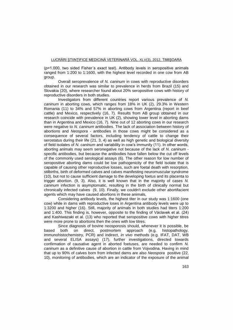
LUCRĂRI ŞTIINŢIFICE MEDICINĂ VETERINARĂ VOL. XLV(3), 2012, TIMIŞOARA
163
(p=1.000, two sided Fisher´s exact test). Antibody levels in seropositive animals ranged from 1:200 to 1:1600, with the highest level recorded in one cow from AB group. Overall seroprevalence of N. caninum in cows with reproductive disorders obtained in our research was similar to prevalence in herds from Brazil (15) and Slovakia (20), where researcher found about 20% seropositive cows with history of reproductive disorders in both studies.
Investigators from different countries report various prevalence of N. caninum in aborting cows, which ranges from 18% in UK (2), 29.3% in Western Romania (11) to 34% and 57% in aborting cows from Argentina (report in beef cattle) and Mexico, respectively (16, 7). Results from AB group obtained in our research coincide with prevalence in UK (2), showing lower level in aborting dams than in Argentina and Mexico (16, 7). Nine out of 12 aborting cows in our research were negative to N. caninum antibodies. The lack of association between history of abortions and Neospora - antibodies in those cows might be considered as a consequence of several factors, including tendency of cattle to change their serostatus during their life (21, 3, 4) as well as high genetic and biological diversity of field isolates of N. caninum and variability in cow’s immunity (11). In other words, aborting animals may seem seronegative not because of the lack of N. caninum - specific antibodies, but because the antibodies have fallen below the cut off levels of the commonly used serological assays (6). The other reason for low number of seropositive aborting dams could be low pathogenicity of the field isolate that is capable of causing other reproductive losses, such are foetal death with resorption, stilbirths, birth of deformed calves and calves manifesting neuromuscular syndrome (10), but not to cause sufficient damage to the developing foetus and its placenta to trigger abortion. (9, 3). Also, it is well known that in the majority of cases N. caninum infection is asymptomatic, resulting in the birth of clinically normal but chronically infected calves (9, 10). Finally, we couldn't exclude other abortifacient agents which may have caused abortions in these animals,
Considering antibody levels, the highest titer in our study was 1:1600 (one cow) while in dams with reproductive loses in Argentina antibody levels were up to 1:3200 and higher (16). Still, majority of animals in both studies had titers 1:200 and 1:400. This finding is, however, opposite to the finding of Václavek et al. (24) and Kashiwazaki et al. (13) who reported that seropositive cows with higher titres were more prone to abortions then the ones with low titres.
Since diagnosis of bovine neosporosis should, whenever it is possible, be based both on direct, postmortem approach (e.g. histopathology, immunohistochemistry, PCR) and indirect, in vivo methods (e.g. IFAT, DAT, WB and several ELISA assays) (17), further investigations, directed towards confirmation of causative agent in aborted foetuses, are needed to confirm N. caninum as a definitive cause of abortion in cattle from Vojvodina. Having in mind that up to 90% of calves born from infected dams are also Neospora positive (22, 10), monitoring of antibodies, which are an indicator of the exposure of the animal
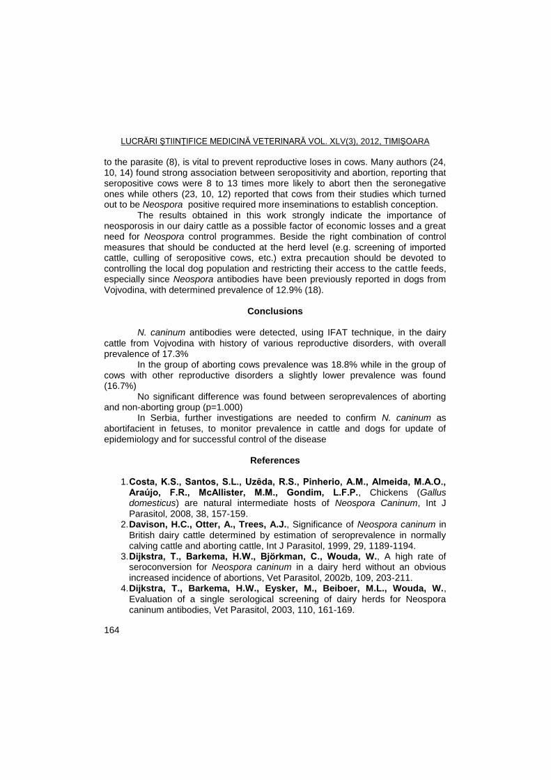
LUCRĂRI ŞTIINŢIFICE MEDICINĂ VETERINARĂ VOL. XLV(3), 2012, TIMIŞOARA
164
to the parasite (8), is vital to prevent reproductive loses in cows. Many authors (24, 10, 14) found strong association between seropositivity and abortion, reporting that seropositive cows were 8 to 13 times more likely to abort then the seronegative ones while others (23, 10, 12) reported that cows from their studies which turned out to be Neospora positive required more inseminations to establish conception.
The results obtained in this work strongly indicate the importance of neosporosis in our dairy cattle as a possible factor of economic losses and a great need for Neospora control programmes. Beside the right combination of control measures that should be conducted at the herd level (e.g. screening of imported cattle, culling of seropositive cows, etc.) extra precaution should be devoted to controlling the local dog population and restricting their access to the cattle feeds, especially since Neospora antibodies have been previously reported in dogs from Vojvodina, with determined prevalence of 12.9% (18).
Conclusions
N. caninum antibodies were detected, using IFAT technique, in the dairy
cattle from Vojvodina with history of various reproductive disorders, with overall prevalence of 17.3%
In the group of aborting cows prevalence was 18.8% while in the group of cows with other reproductive disorders a slightly lower prevalence was found (16.7%)
No significant difference was found between seroprevalences of aborting and non-aborting group (p=1.000)
In Serbia, further investigations are needed to confirm N. caninum as abortifacient in fetuses, to monitor prevalence in cattle and dogs for update of epidemiology and for successful control of the disease
References
1. Costa, K.S., Santos, S.L., Uzȇda, R.S., Pinherio, A.M., Almeida, M.A.O.,
Araújo, F.R., McAllister, M.M., Gondim, L.F.P., Chickens (Gallus domesticus) are natural intermediate hosts of Neospora Caninum, Int J Parasitol, 2008, 38, 157-159.
2. Davison, H.C., Otter, A., Trees, A.J., Significance of Neospora caninum in British dairy cattle determined by estimation of seroprevalence in normally calving cattle and aborting cattle, Int J Parasitol, 1999, 29, 1189-1194.
3. Dijkstra, T., Barkema, H.W., Björkman, C., Wouda, W., A high rate of seroconversion for Neospora caninum in a dairy herd without an obvious increased incidence of abortions, Vet Parasitol, 2002b, 109, 203-211.
4. Dijkstra, T., Barkema, H.W., Eysker, M., Beiboer, M.L., Wouda, W., Evaluation of a single serological screening of dairy herds for Neospora caninum antibodies, Vet Parasitol, 2003, 110, 161-169.
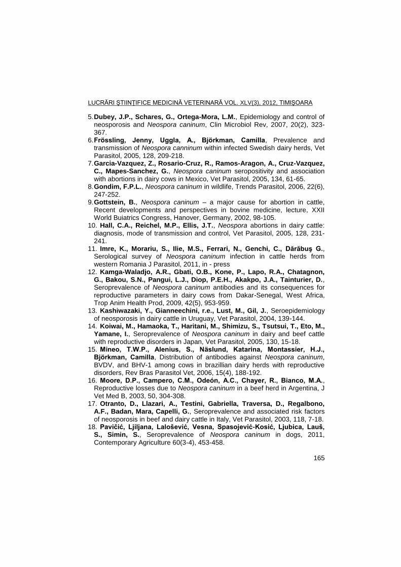
LUCRĂRI ŞTIINŢIFICE MEDICINĂ VETERINARĂ VOL. XLV(3), 2012, TIMIŞOARA
165
5. Dubey, J.P., Schares, G., Ortega-Mora, L.M., Epidemiology and control of neosporosis and Neospora caninum, Clin Microbiol Rev, 2007, 20(2), 323-367.
6. Frössling, Jenny, Uggla, A., Björkman, Camilla, Prevalence and transmission of Neospora canninum within infected Swedish dairy herds, Vet Parasitol, 2005, 128, 209-218.
7. Garcia-Vazquez, Z., Rosario-Cruz, R., Ramos-Aragon, A., Cruz-Vazquez, C., Mapes-Sanchez, G., Neospora caninum seropositivity and association with abortions in dairy cows in Mexico, Vet Parasitol, 2005, 134, 61-65.
8. Gondim, F.P.L., Neospora caninum in wildlife, Trends Parasitol, 2006, 22(6), 247-252.
9. Gottstein, B., Neospora caninum – a major cause for abortion in cattle, Recent developments and perspectives in bovine medicine, lecture, XXII World Buiatrics Congress, Hanover, Germany, 2002, 98-105.
10. Hall, C.A., Reichel, M.P., Ellis, J.T., Neospora abortions in dairy cattle: diagnosis, mode of transmission and control, Vet Parasitol, 2005, 128, 231-241.
11. Imre, K., Morariu, S., Ilie, M.S., Ferrari, N., Genchi, C., Dărăbuş G., Serological survey of Neospora caninum infection in cattle herds from western Romania J Parasitol, 2011, in - press
12. Kamga-Waladjo, A.R., Gbati, O.B., Kone, P., Lapo, R.A., Chatagnon, G., Bakou, S.N., Pangui, L.J., Diop, P.E.H., Akakpo, J.A., Tainturier, D., Seroprevalence of Neospora caninum antibodies and its consequences for reproductive parameters in dairy cows from Dakar-Senegal, West Africa, Trop Anim Health Prod, 2009, 42(5), 953-959.
13. Kashiwazaki, Y., Gianneechini, r.e., Lust, M., Gil, J., Seroepidemiology of neosporosis in dairy cattle in Uruguay, Vet Parasitol, 2004, 139-144.
14. Koiwai, M., Hamaoka, T., Haritani, M., Shimizu, S., Tsutsui, T., Eto, M., Yamane, I., Seroprevalence of Neospora caninum in dairy and beef cattle with reproductive disorders in Japan, Vet Parasitol, 2005, 130, 15-18.
15. Mineo, T.W.P., Alenius, S., Näslund, Katarina, Montassier, H.J., Björkman, Camilla, Distribution of antibodies against Neospora caninum, BVDV, and BHV-1 among cows in brazillian dairy herds with reproductive disorders, Rev Bras Parasitol Vet, 2006, 15(4), 188-192.
16. Moore, D.P., Campero, C.M., Odeón, A.C., Chayer, R., Bianco, M.A., Reproductive losses due to Neospora caninum in a beef herd in Argentina, J Vet Med B, 2003, 50, 304-308.
17. Otranto, D., Llazari, A., Testini, Gabriella, Traversa, D., Regalbono, A.F., Badan, Mara, Capelli, G., Seroprevalence and associated risk factors of neosporosis in beef and dairy cattle in Italy, Vet Parasitol, 2003, 118, 7-18.
18. Paviţiš, Ljiljana, Laloševiš, Vesna, Spasojeviš-Kosiš, Ljubica, Lauš, S., Simin, S., Seroprevalence of Neospora caninum in dogs, 2011, Contemporary Agriculture 60(3-4), 453-458.
LUCRĂRI ŞTIINŢIFICE MEDICINĂ VETERINARĂ VOL. XLV(3), 2012, TIMIŞOARA
166
19. Reiczigel, J., Rosza, L., Quantitative Parasitology 3.0, Budapest, 2005. 20. Reiterová, K., Špilovská, S., Antolová, D., Dubinský, P., Neospora
caninum, potential cause of abortions in dairy cows: The current serological follow-up in Slovakia, Vet Parasitol, 2009, 159, 1-6.
21. Sager, H., Fischer, I., Furrer, K., Strasser, M., Waldvogel, A., Boerlin, P., Audigé, L., Gottstein, B., A Swiss case-control study to assess Neospora caninum – associated bovine abortions by PCR, histopathology and serology, Vet Parasitol, 2001, 102(1-2), 1-15.
22. Schares, G., Bärwald, A., Staubach, C., Ziller, M., Klöss, D., Schröder, R., Labohm, R., Dräger, K., Fasen, W., Hess, R.G., Conraths, F.J., Potential risk factors for bovine Neospora caninum infection in Germany are not under the control of the farmers, Parasitology, 2004, 129, 301-309.
23. Stenlund, Sussane, Kindahi, H., Uggla, A., Björkman, Camilla, A long-term study of Neospora caninum infection in a Swedish dairy herd, Acta Vet Scand, 2003, 44(2), 63-71.
24. Václavek, P., Koudela, B., Modrý, D., Sedlák, K., Seroprevalence of Neospora caninum in aborting dairy cattle in Czech Republic, Vet Parasitol, 2003, 115, 239-245.
25. Vidiš, Branka, Grgiš, Ţ., Saviš, Sara, Bugarski, D., Neospora caninum causes abortions in cows, Scientific Symposium ―Reproduction of domestic animals‖, Naučna KMD, Belgrade, Divčibare, Serbia, 2011.
LUCRĂRI ŞTIINŢIFICE MEDICINĂ VETERINARĂ VOL. XLV(3), 2012, TIMIŞOARA
167
THE PARASITISM WITH GIARDIA SPP. IN CALVES FROM CARAS-SEVERIN COUNTY
IONELA DENISA SORESCU, I. OPRESCU, S. MORARIU, NARCISA MEDERLE,
M.S. ILIE, IONELA HOTEA, GH. DĂRĂBUŞ
Banat's University of Agricultural Sciences and Veterinary Medicine Timisoara, Faculty of Veterinary Medicine, 300645, Aradului Street No. 119, Timisoara, Romania
E-mail: [email protected]
Summary
The aim of the present study was to determine the prevalence of the infections in
calves in farms from Caras-Severin County to analyze the potential risk factors that support this infection (gender, age, race). For this, 204 calves aged between 4 days and 5 month from farms of Caras-Severin County were investigated. Giardia spp. (trophozoites and/or cysts) were revealed by Willis and Lügol staining methods and then analyzed by Giardiasis Ag ELISA kit (Cypress Diagnostics). The prevalence of Giardia spp. infection was 33.82%. Frequently, Giardia spp. infection was associated to the presence of Eimeria spp., and Cryptosporidium spp. respectively.
Key words: Giardia spp., calves, Lügol staining, ELISA. Giardiosis or lambliosis is a parasitical disease of the small intestine, found
in a wide range of mammal and bird species, caused by a single-celled organism of the genus Giardia. It can develop into a zoonotic disease. Giardia spp. is a cosmopolitan protozoan which leads to a syndrome of malabsorption found in animals and humans (2, 3). About rent animals, cattle are most often infected and most studies have focused on this species. Giardia is common both in meat and dairy cattle in the world and numerous studies have shown the prevalence rate of 100% (9, 10, 12, 18). In ruminants, giardiosis has emerged as an important gastrointestinal disease due to high prevalence and pathogenicity in young animals. High prevalence of G. duodenalis species in ruminants is a public health problem because of the possibility of zoonotic transmission from animals to humans through contaminated food or water with viable cysts. Assemblage E (called as ungulates animals’ assemblage) is the predominant isotype of G. duodenalis in calves (16, 17) and sheep (5, 13). Human assemblage, genotype A and B were also identified in two species of ruminants (5, 13, 14, 15).
The epidemiological surveillance regarding the prevalence of giardiosis and data regarding the main risk factors that contribute to the presence of the disease in calves in Caras-Severin County are unknown or incomplete.
Giardia spp. infection can evolve as unique pathogen agent or associated with other enteropathogen agents, including Cryptosporidium spp. (6). Giardia has
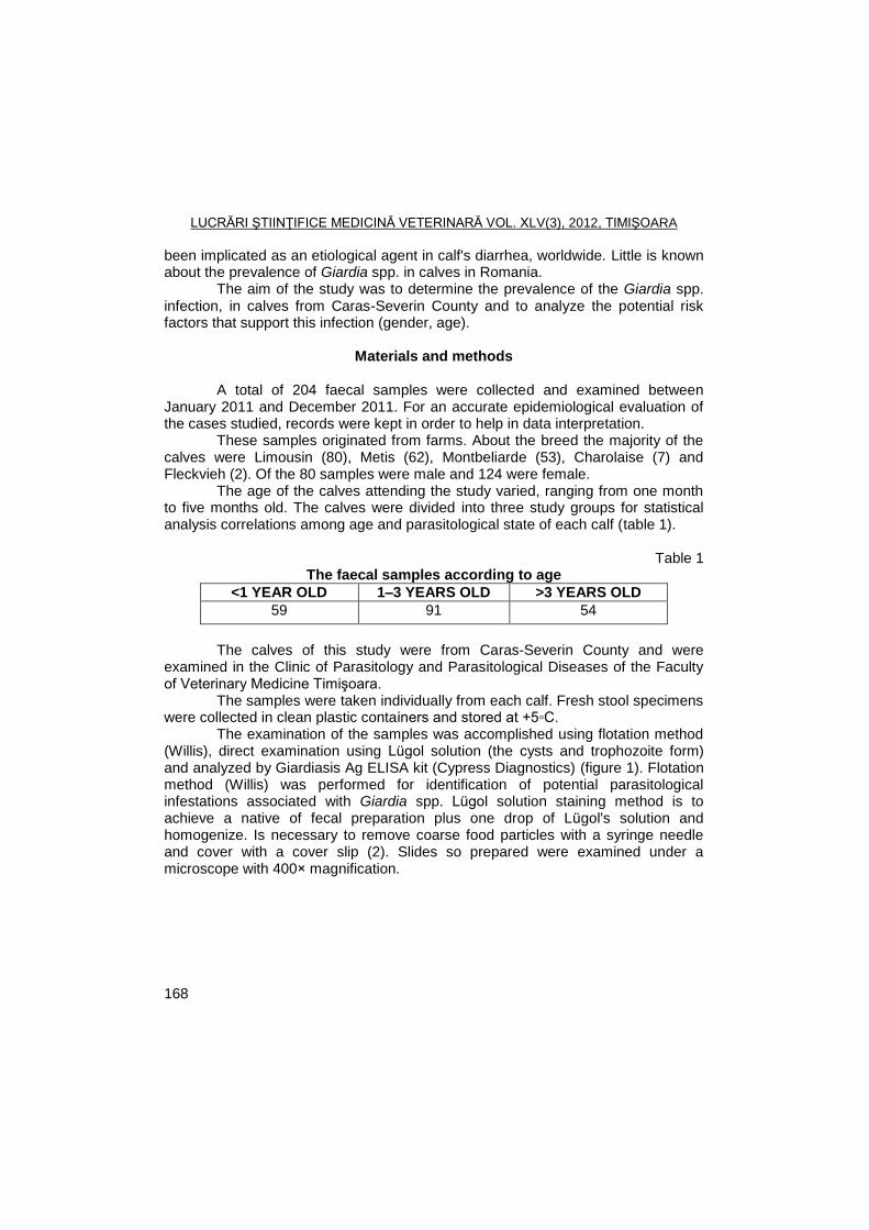
LUCRĂRI ŞTIINŢIFICE MEDICINĂ VETERINARĂ VOL. XLV(3), 2012, TIMIŞOARA
168
been implicated as an etiological agent in calf's diarrhea, worldwide. Little is known about the prevalence of Giardia spp. in calves in Romania.
The aim of the study was to determine the prevalence of the Giardia spp. infection, in calves from Caras-Severin County and to analyze the potential risk factors that support this infection (gender, age).
Materials and methods
A total of 204 faecal samples were collected and examined between January 2011 and December 2011. For an accurate epidemiological evaluation of the cases studied, records were kept in order to help in data interpretation. These samples originated from farms. About the breed the majority of the calves were Limousin (80), Metis (62), Montbeliarde (53), Charolaise (7) and Fleckvieh (2). Of the 80 samples were male and 124 were female. The age of the calves attending the study varied, ranging from one month to five months old. The calves were divided into three study groups for statistical analysis correlations among age and parasitological state of each calf (table 1).
Table 1 The faecal samples according to age
<1 YEAR OLD 1–3 YEARS OLD >3 YEARS OLD
59 91 54
The calves of this study were from Caras-Severin County and were
examined in the Clinic of Parasitology and Parasitological Diseases of the Faculty of Veterinary Medicine Timişoara.
The samples were taken individually from each calf. Fresh stool specimens were collected in clean plastic containers and stored at +5◦C.
The examination of the samples was accomplished using flotation method (Willis), direct examination using Lügol solution (the cysts and trophozoite form) and analyzed by Giardiasis Ag ELISA kit (Cypress Diagnostics) (figure 1). Flotation method (Willis) was performed for identification of potential parasitological infestations associated with Giardia spp. Lügol solution staining method is to achieve a native of fecal preparation plus one drop of Lügol's solution and homogenize. Is necessary to remove coarse food particles with a syringe needle and cover with a cover slip (2). Slides so prepared were examined under a microscope with 400× magnification.
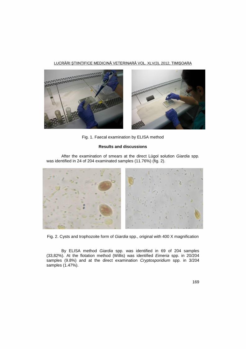
LUCRĂRI ŞTIINŢIFICE MEDICINĂ VETERINARĂ VOL. XLV(3), 2012, TIMIŞOARA
169
Fig. 1. Faecal examination by ELISA method
Results and discussions
After the examination of smears at the direct Lügol solution Giardia spp.
was identified in 24 of 204 examinated samples (11.76%) (fig. 2).
Fig. 2. Cysts and trophozoite form of Giardia spp., original with 400 X magnification
By ELISA method Giardia spp. was identified in 69 of 204 samples (33,82%). At the flotation method (Willis) was identified Eimeria spp. in 20/204 samples (9.8%) and at the direct examination Cryptosporidium spp. in 3/204 samples (1.47%).
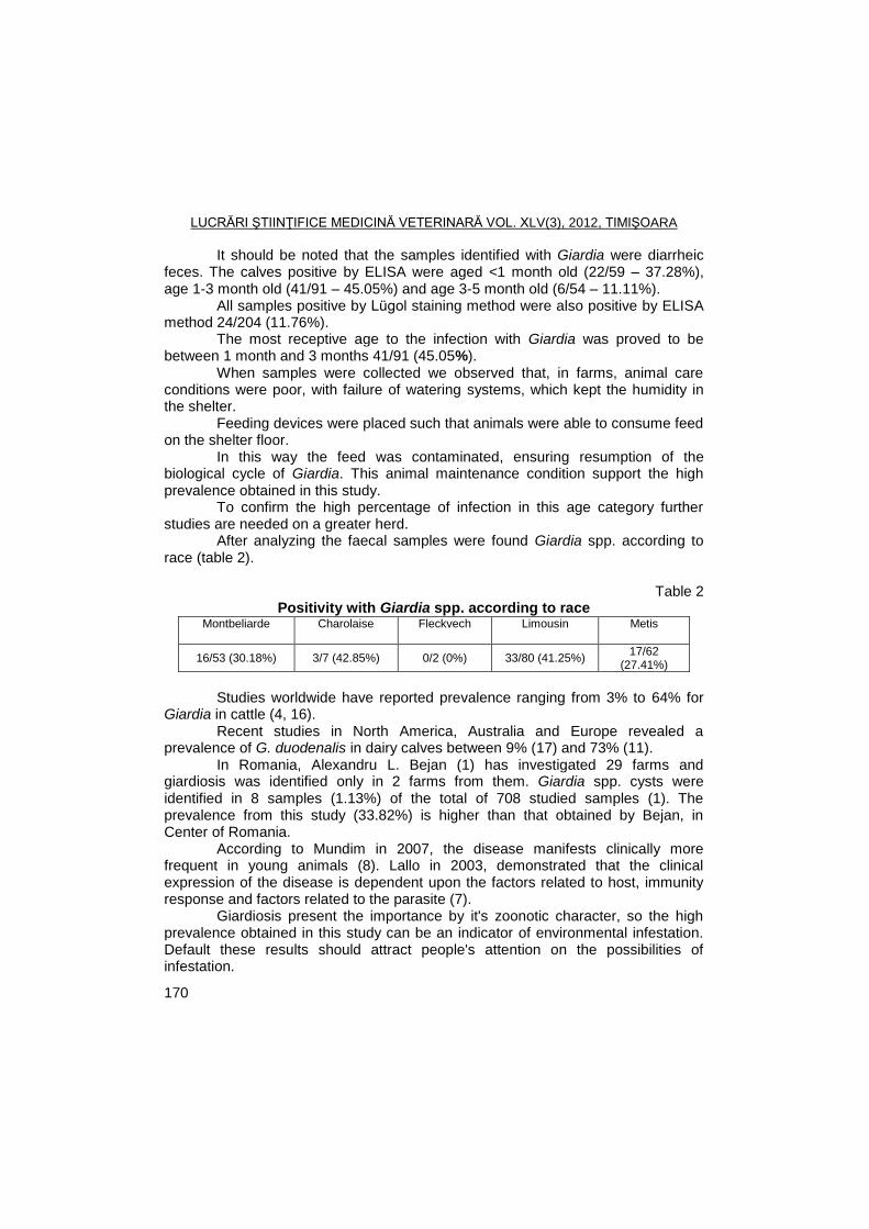
LUCRĂRI ŞTIINŢIFICE MEDICINĂ VETERINARĂ VOL. XLV(3), 2012, TIMIŞOARA
170
It should be noted that the samples identified with Giardia were diarrheic feces. The calves positive by ELISA were aged <1 month old (22/59 – 37.28%), age 1-3 month old (41/91 – 45.05%) and age 3-5 month old (6/54 – 11.11%).
All samples positive by Lügol staining method were also positive by ELISA method 24/204 (11.76%).
The most receptive age to the infection with Giardia was proved to be between 1 month and 3 months 41/91 (45.05%).
When samples were collected we observed that, in farms, animal care conditions were poor, with failure of watering systems, which kept the humidity in the shelter.
Feeding devices were placed such that animals were able to consume feed on the shelter floor.
In this way the feed was contaminated, ensuring resumption of the biological cycle of Giardia. This animal maintenance condition support the high prevalence obtained in this study.
To confirm the high percentage of infection in this age category further studies are needed on a greater herd.
After analyzing the faecal samples were found Giardia spp. according to race (table 2).
Table 2
Positivity with Giardia spp. according to race Montbeliarde
Charolaise
Fleckvech
Limousin
Metis
16/53 (30.18%) 3/7 (42.85%) 0/2 (0%) 33/80 (41.25%) 17/62
(27.41%)
Studies worldwide have reported prevalence ranging from 3% to 64% for Giardia in cattle (4, 16). Recent studies in North America, Australia and Europe revealed a prevalence of G. duodenalis in dairy calves between 9% (17) and 73% (11).
In Romania, Alexandru L. Bejan (1) has investigated 29 farms and giardiosis was identified only in 2 farms from them. Giardia spp. cysts were identified in 8 samples (1.13%) of the total of 708 studied samples (1). The prevalence from this study (33.82%) is higher than that obtained by Bejan, in Center of Romania.
According to Mundim in 2007, the disease manifests clinically more frequent in young animals (8). Lallo in 2003, demonstrated that the clinical expression of the disease is dependent upon the factors related to host, immunity response and factors related to the parasite (7).
Giardiosis present the importance by it's zoonotic character, so the high prevalence obtained in this study can be an indicator of environmental infestation. Default these results should attract people's attention on the possibilities of infestation.
LUCRĂRI ŞTIINŢIFICE MEDICINĂ VETERINARĂ VOL. XLV(3), 2012, TIMIŞOARA
171
Conclusions
Giardia spp. cysts were identified in faecal samples from calves in Caras-
Severin County for the first time. The prevalence of Giardia spp. infection established by Lügol solution
staining method was 24 of 204 samples (11.76%) and at ELISA kit was 69 of 204 samples (33.82%).
Parasitic fauna, less Giardia spp., of calves from Caras-Severin County was represented by Cryptosporidium spp. 3/204 samples (1.47%) and Eimeria spp. 20/204 samples (9.80%).
It is a proven fact that the age up between 1 month and 3 months is an important risk factor (41/91-45.05%).
References
1. Bejan, L.A., Criptosporidioza viteilor si iezilor: cercetari privind diagnosticul,
epidemiologia si etiopatogeneza, Teza de doctorat, Facultatea de Medicina Veterinara Cluj-Napoca, 2009.
2. Cosoroabă, I., Zoonoze parazitare. Ed. First/Artpress., Timişoara, 2005. 3. Dărăbuş, Gh., Oprescu, I., Morariu, S., Mederle, Narcisa, Parazitologie şi
boli parazitare, Ed. Mirton, Timişoara, 2006. 4. Geurden, T., Geldhof, P., Levecke, B., Martens, C., Berkvens, D.,
Casaert, S., Vercruysse, J., Claerebout E., Mixed Giardia duodenalis assemblage A and E infections in calves. Int. J. Parasitol., 2008, 38, 259–264.
5. Giangaspero, A., Paoletti, B., Iorio, R., Traversa, D., Prevalence and molecular characterization of Giardia duodenalis from sheep in central Italy. Parasitol. Res., 2005, 96, 32–37.
6. Hamnes, I.S., Bjørn, K., Robertson, Gjerde, A longitudinal study on the occurrence of Cryptosporidium and Giardia in dogs during their first year of life. Acta Veterinaria Scandinavica, 2007, 49, 22.
7. Lallo, M.A., Rodrigues, L.C.S., Bondan, E.F., Giardíase em cães e gatos- revisão. Clín. Vet., 2003, 43, 40–44.
8. Mundim, M.J.S., Rosa, L.A.G., Hortencio, S.M., Faria, E.S.M., Rodrigues, R.M., Cury, M.C., Prevalence of Giardia duodenalis and Cryptosporidium spp. in dogs from different living conditions in Uberlaˆndia, Brazil. Vet. Parasitol., 2007, 144, 356–359.
9. O’handley, R.M., Giardia in farm animals. In: Olson, B.E., Olson, M.E., Wallis, P.M. (Eds.), Giardia: The cosmopolitan parasite. CAB International, Wallingford, UK, 2002, 97-105.
10. Oliveira-Sequeira, T.C.G., Amarante, A.F.T., Ferrari, T.B., Nunes, L.C., Prevalence of intestinal parasites in dogs from Sao Paulo. Vet. Parasitol., 2002, 103, 19–27.
LUCRĂRI ŞTIINŢIFICE MEDICINĂ VETERINARĂ VOL. XLV(3), 2012, TIMIŞOARA
172
11. Olson, M.E., Guselle, N.J., O’Handley, R.M., Swift, M.L., McAllister, T.A., Jelinski M.D., Morck D.W., Giardia and Cryptosporidium in dairy calves in British Columbia. Can. Vet. J., 1997, 38, 703–706.
12. Ralston, B.J., Cockwill, C., Guselle, N., Van Herk, F.H., Mcallister, T.A., Olson, M.E., Prevalence of Giardia and Cryptosporidium andersoni and their effect on performance in feedlot beef calves. Can. J. Anim. Sci., 2003, 83, 153-159.
13. Santin, M., Trout, J.M., Fayer, R., Prevalence and molecular characterization of Cryptosporidium and Giardia species and genotypes in sheep in Maryland. Vet. Parasitol., 2007, 146, 17–24.
14. Traub, R., Wade, S., Read, C., Thompson, A., Mohammed, H., Molecular characterization of potentially zoonotic isolates of Giardia duodenalis in horses. Vet. Parasitol., 2005, 130, 317–321.
15. Trout J.M., Santin M., Fayer R., Prevalence of Giardia duodenalis genotypes in adult dairy cows. Vet. Parasitol., 2007, 147, 205–209.
16. Trout, J.M., Santin, M., Greiner, E., Fayer, R., Prevalence and genotypes of Giardia duodenalis in post-weaned dairy calves. Vet. Parasitol., 2005, 130, 177–183.
17. Wade, S.E., Mohammed, H.O., Schaaf, S.L., Prevalence of Giardia sp., Cryptosporidium parvum and Cryptosporidium muris (C. andersoni) in 109 dairy herds in five counties of southeastern New York. Vet. Parasitol., 2000, 93, 1–11.
18. Xiao, L., Fayer, R., Molecular characterisation of species and genotypes of Cryptosporidium and Giardia and assessment of zoonotic transmission. International Journal for Parasitology, 2008, 38, 1239-1255.
LUCRĂRI ŞTIINŢIFICE MEDICINĂ VETERINARĂ VOL. XLV(3), 2012, TIMIŞOARA
173
EFFICACY TESTING OF DOVENIX (NITROXINIL, MERIAL) IN NATURAL INFESTATION WITH FASCIOLA HEPATICA
AT SHEEP
M. ŢIFREA, I. OPRESCU, D. INDRE, GH. DĂRĂBUŞ
Banat's University of Agricultural Sciences and Veterinary Medicine Timisoara, Faculty of Veterinary Medicine, 300645, Aradului Street No. 119, Timisoara, Romania
E-mail: [email protected]
Summary
The efficacy testing of Dovenix was achieved on 20 sheep, Turcana and Tigaie
breeds, naturally infested with F. hepatica. The animals were randomly divided in two groups of 10 animals and treated with
Dovenix in day 0 (1 ml/25 kg b.w., s.c.). In day 0 and 12 post-treatment sheep were coproscopical examined using Brumpt method establishing EPG. The product efficacy was established using Presidente and Borgsteede methods.
Results obtained confirmed that Dovenix product had a maximum efficacy (100%) in animals naturally infected with F. hepatica.
Key words: efficacy, Dovenix, Fasciola hepatica
Prophylaxis in fasciolosis must be based on concrete knowledge of the
epizootiological situation of each farm and flock. This can be achieved only by conjunction of epizootiological surveys data and coproscopic as well as morfoclinic examinations. A good control is based on the knowledge of the parasitological level in animals, the contamination of pastures, breeding system, the possibilities of rotation, location of pasture, pasture and grazing management (1, 4, 8).
In endemic areas, chemoprevention is made at least twice a year: • 2-3 weeks after the entry into stalls, with anthelmintics that affects both
young and adult fasciola; • 2 weeks before entering the pasture, when medication acting on the adult
forms is used. If strong parasitic infestation is present it is necessary to perform two
treatments in spring and two, in fall-winter at 2-6 weeks intervals, depending on the structure and biology of parasites. Parasitological control success depends on treating all susceptible animals. Sometimes, on the same pasture, treated flocks meat untreated flocks, the last category compromising the therapeutic activity (2, 3, 8).
If sheep and cattle are grazing together the treatment applies to both species. As a general rule, in sheep, treatment will be more frequent than in cattle because are more susceptible to parasitism and the grazing has been made a longer period of time (2, 5, 7).
In the antihelmintic treatment it should be assumed that whatever antiparasitary is used, it has a degree of toxicity for the host. Given this,
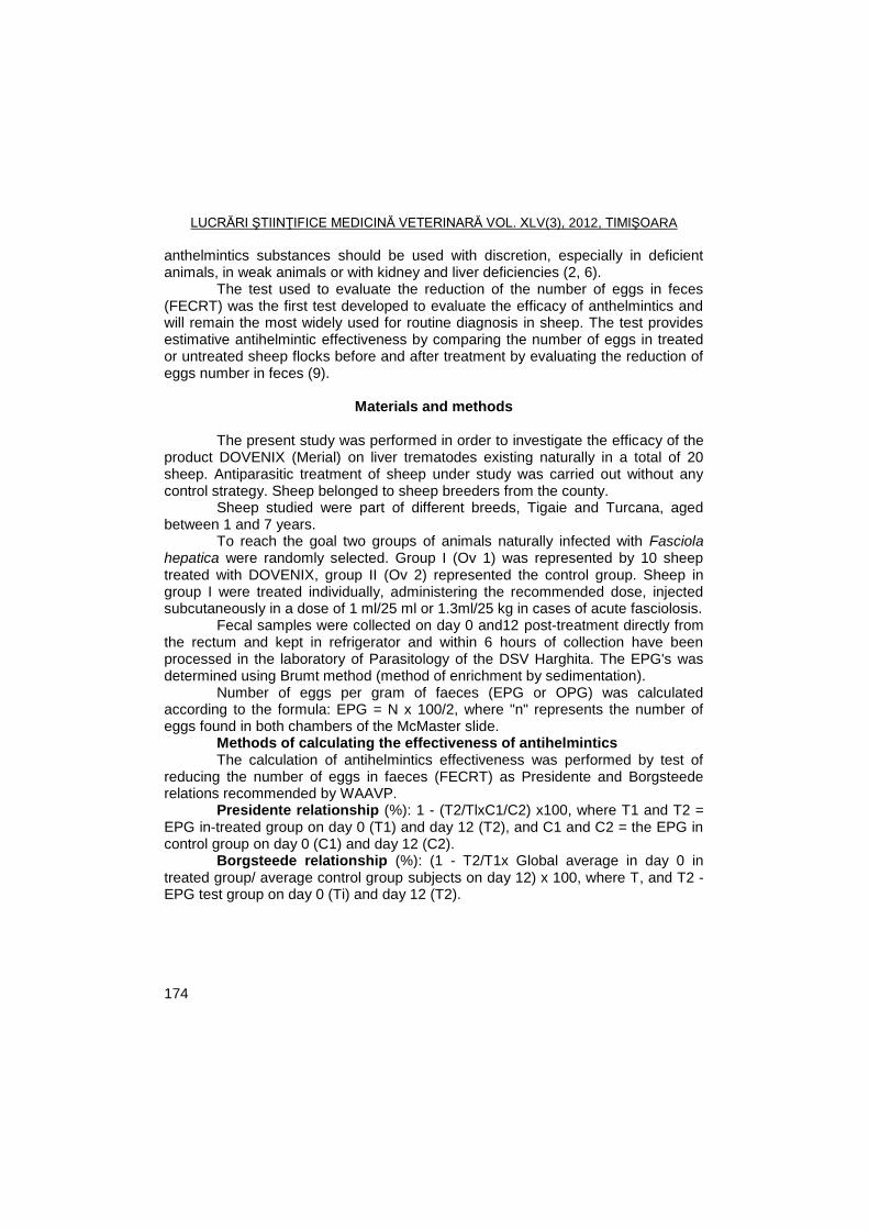
LUCRĂRI ŞTIINŢIFICE MEDICINĂ VETERINARĂ VOL. XLV(3), 2012, TIMIŞOARA
174
anthelmintics substances should be used with discretion, especially in deficient animals, in weak animals or with kidney and liver deficiencies (2, 6).
The test used to evaluate the reduction of the number of eggs in feces (FECRT) was the first test developed to evaluate the efficacy of anthelmintics and will remain the most widely used for routine diagnosis in sheep. The test provides estimative antihelmintic effectiveness by comparing the number of eggs in treated or untreated sheep flocks before and after treatment by evaluating the reduction of eggs number in feces (9).
Materials and methods
The present study was performed in order to investigate the efficacy of the
product DOVENIX (Merial) on liver trematodes existing naturally in a total of 20 sheep. Antiparasitic treatment of sheep under study was carried out without any control strategy. Sheep belonged to sheep breeders from the county.
Sheep studied were part of different breeds, Tigaie and Turcana, aged between 1 and 7 years.
To reach the goal two groups of animals naturally infected with Fasciola hepatica were randomly selected. Group I (Ov 1) was represented by 10 sheep treated with DOVENIX, group II (Ov 2) represented the control group. Sheep in group I were treated individually, administering the recommended dose, injected subcutaneously in a dose of 1 ml/25 ml or 1.3ml/25 kg in cases of acute fasciolosis.
Fecal samples were collected on day 0 and12 post-treatment directly from the rectum and kept in refrigerator and within 6 hours of collection have been processed in the laboratory of Parasitology of the DSV Harghita. The EPG's was determined using Brumt method (method of enrichment by sedimentation).
Number of eggs per gram of faeces (EPG or OPG) was calculated according to the formula: EPG = N x 100/2, where "n" represents the number of eggs found in both chambers of the McMaster slide.
Methods of calculating the effectiveness of antihelmintics The calculation of antihelmintics effectiveness was performed by test of
reducing the number of eggs in faeces (FECRT) as Presidente and Borgsteede relations recommended by WAAVP.
Presidente relationship (%): 1 - (T2/TlxC1/C2) x100, where T1 and T2 = EPG in-treated group on day 0 (T1) and day 12 (T2), and C1 and C2 = the EPG in control group on day 0 (C1) and day 12 (C2).
Borgsteede relationship (%): (1 - T2/T1x Global average in day 0 in treated group/ average control group subjects on day 12) x 100, where T, and T2 - EPG test group on day 0 (Ti) and day 12 (T2).
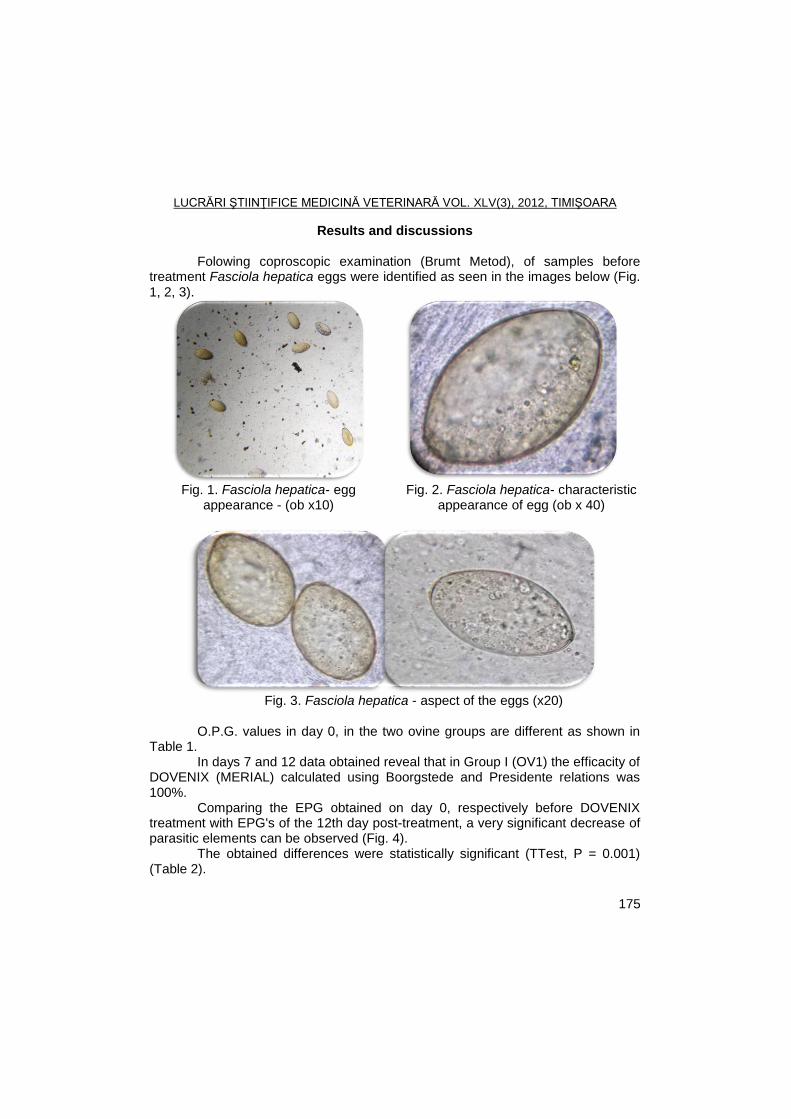
LUCRĂRI ŞTIINŢIFICE MEDICINĂ VETERINARĂ VOL. XLV(3), 2012, TIMIŞOARA
175
Results and discussions
Folowing coproscopic examination (Brumt Metod), of samples before treatment Fasciola hepatica eggs were identified as seen in the images below (Fig. 1, 2, 3).
Fig. 1. Fasciola hepatica- egg appearance - (ob x10)
Fig. 2. Fasciola hepatica- characteristic appearance of egg (ob x 40)
Fig. 3. Fasciola hepatica - aspect of the eggs (x20)
O.P.G. values in day 0, in the two ovine groups are different as shown in
Table 1. In days 7 and 12 data obtained reveal that in Group I (OV1) the efficacity of
DOVENIX (MERIAL) calculated using Boorgstede and Presidente relations was 100%.
Comparing the EPG obtained on day 0, respectively before DOVENIX treatment with EPG's of the 12th day post-treatment, a very significant decrease of parasitic elements can be observed (Fig. 4).
The obtained differences were statistically significant (TTest, P = 0.001) (Table 2).
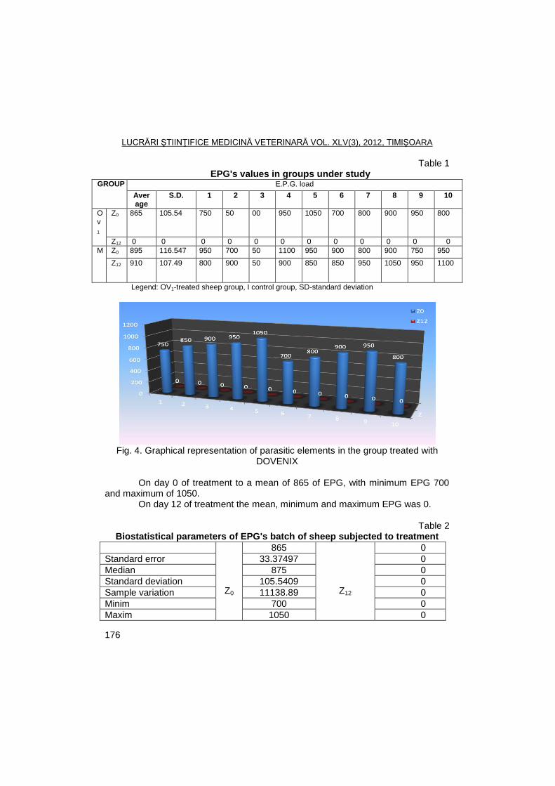
LUCRĂRI ŞTIINŢIFICE MEDICINĂ VETERINARĂ VOL. XLV(3), 2012, TIMIŞOARA
176
Table 1 EPG's values in groups under study
GROUP E.P.G. load
Average
S.D. 1 2 3 4 5 6 7 8 9 10
Ov
1
Z0 865 105.54 750 50 00 950 1050 700 800 900 950 800
Z12 0 0 0 0 0 0 0 0 0 0 0 0
M Z0 895 116.547 950 700 50 1100 950 900 800 900 750 950
Z12 910 107.49 800 900 50 900 850 850 950 1050 950 1100
Legend: OV1-treated sheep group, I control group, SD-standard deviation
Fig. 4. Graphical representation of parasitic elements in the group treated with
DOVENIX
On day 0 of treatment to a mean of 865 of EPG, with minimum EPG 700 and maximum of 1050.
On day 12 of treatment the mean, minimum and maximum EPG was 0.
Table 2 Biostatistical parameters of EPG's batch of sheep subjected to treatment
Average
Z0
865
Z12
0
Standard error 33.37497 0
Median 875 0
Standard deviation 105.5409 0
Sample variation 11138.89 0
Minim 700 0
Maxim 1050 0
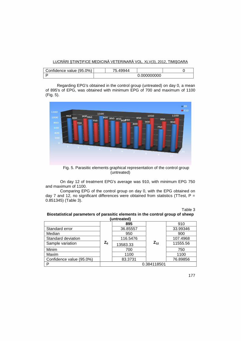
LUCRĂRI ŞTIINŢIFICE MEDICINĂ VETERINARĂ VOL. XLV(3), 2012, TIMIŞOARA
177
Confidence value (95.0%) 75.49944 0
P 0.000000000
Regarding EPG's obtained in the control group (untreated) on day 0, a mean
of 895's of EPG, was obtained with minimum EPG of 700 and maximum of 1100 (Fig. 5).
Fig. 5. Parasitic elements graphical representation of the control group
(untreated)
On day 12 of treatment EPG's average was 910, with minimum EPG 750 and maximum of 1100.
Comparing EPG of the control group on day 0, with the EPG obtained on day 7 and 12, no significant differences were obtained from statistics (TTest, P = 0.851345) (Table 3).
Table 3
Biostatistical parameters of parasitic elements in the control group of sheep (untreated)
Average
Z0
895
Z12
910
Standard error 36.85557 33.99346
Median 950 900
Standard deviation 116.5476 107.4968
Sample variation 13583.33
11555.56
Minim 700 750
Maxim 1100 1100
Confidence value (95.0%) 83.3731 76.89856
P 0.384118501
LUCRĂRI ŞTIINŢIFICE MEDICINĂ VETERINARĂ VOL. XLV(3), 2012, TIMIŞOARA
178
Careful analysis of results from efficacy tests based on two relationships shows that in sheep, DOVENIX product falls within the safety value of 100%.
Dărăbuş (2003) states that antihelmintics efficacy is either on adult or larval forms of the parasite. Thus, clorsulon and albendazole has efficacy only on adults older than 2 months, while triclabendazole is effective also in the young fasciola. Knowing this is important to choose knowingly, drugs to be administered for treatment of winter and spring, when young fasciolae respectively adult fasciola predominate.
Also Dărăbuş (2003) recommends the use antihelmintics taking into account the therapeutic index (ratio of toxic dose / therapeutic dose), some medical preparations having a very close toxic dose to the therapy dose. Therefore, the dosage will be strictly indicated, with accurate assessment of body weight of herbivores. Low therapeutic index have drugs included in the halogenated phenols (nitroxinil, closantel, niclosamide, etc..) group.
Despite having presented the chemoresistance phenomenon as found, Fairweather and Boray, (1998) claim that chimioresistence is not yet a major problem in fasciolosis. Of salicilanilide, closantel and rafoxanidul were wide used in the treatment sheep fasciolosis. Therefore, the selection of resistant strains to this treatment was reported. However, these fasciolicide chimiorezistence not cross with other salicylate such as oxiclozanide.
Mehlhorn (2001) reported the chemoresistence phenomenon for triclabendazole, luxabendazol and clorsulon. However, in laboratory test selection of resistant strains of F. hepatica was not successful.
Good efficacy of this product proves it can still be used since it was installed a resistance to therapy of chronic infestation.
Conclusions
In sheep naturally infected with Fasciola hepatica average efficacy of DOVENIX (MERIAL) was 100%.
Careful analysis of results from efficacy tests based on two relationships of 100%.
The evolution of EPG in day 12 post-treatment shows that in sheep, DOVENIX product falls within the safety for natural Fasciola hepatica infestation in sheep.
References
1. Cristina, R.T., Introducere în farmacologia şi terapia veterinară. Editura Solness, Timişoara, 2006
2. Indre, D., Cercetări privind strategiile de control în trichostrongilidoze la ovine în vestul României, Teză de doctorat, USAMVB Timișoara, 2011
LUCRĂRI ŞTIINŢIFICE MEDICINĂ VETERINARĂ VOL. XLV(3), 2012, TIMIŞOARA
179
3. Maes, L., Veys, P., Geerts, H., Chiarisoli, O., Essai de terrain du Closantel dans une stratégie de contrôle intégre de la fasciolose ovine, Revue Méd. Vét., 2003, 144, 781-786.
4. Martinez-Valladares, M., Cordero-Perez, C., Castanon-Ordonez, L., Famularo, M.R., Fernandez-Pato, F.A., Efficacy of a moxidectin/ triclabendazole formulation against mixed infections of Fasciola hepatica and gastrointestinal nematodes in sheep, Vet. Parasitol., 2010, 174, 166-169.
5. Meaney, M., Allister, J., Mc Kinstry, B., Mc Laughlin, M.K., Brennan, G.P., Forbes, A.B., Fairweather, I., Fasciola hepatica: ultrastructural effects of a combination of triclabendazole and clorsulon against mature fluke, Parasitol. Res., 2006, 100, 1091-1104.
6. Montenegro, Vera., Velarde-Ibarra, F., Hernandez, E.L., Romero, H.Q., Bocanegra, R.C., Campos, A.H., Galvan, P.O. Efficacy of an experimental fasciolicide against immature and mature Fasciola hepatica in artificially infected calves, Parasitol. Res., 2004, 92, 211-214.
7. Scarcella, S., Fiel,C., Guzman, M., Alzola, R., Felipe, A., Hanna, R.E.B., Faiweather, I., Mc Connell, S., Solana, H., Reproductive disruption in Fasciola hepatica associated with incomplete efficacy of a new experimental formulation of triclabendazol, Vet. Parasitol., 2010, 55, 234-242.
8. Toner, E., Brennan, G.P., Hanna, R.E.B., Edgar, H.W., Faiweather, I., Tegumental surface changes in adult Fasciola hepatica in response to treatment in vivo with triclabndazole in the sheep host, Vet. Parasitol., 2011, 172, 3-4, 238-248.
9. Wood, I.B., Amaral, N.K., Bairden, K., Duncan, J.L., Kassai, T., Malone, J.B., Pankavich, J.A., Reinecke, R., Slocombe, O., Taylor, S.M., Vercruysse, J., World Association for the Advancement of Veterinary Parasitology (W.A.A.V.P.) second edition of guidelines for evaluating the efficacy of anthelmintics in ruminant (bovine, ovine, caprine), Vet. Parasitol., 1995, 53, 181-213.
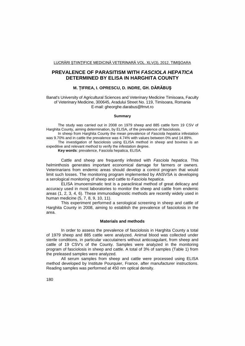
LUCRĂRI ŞTIINŢIFICE MEDICINĂ VETERINARĂ VOL. XLV(3), 2012, TIMIŞOARA
180
PREVALENCE OF PARASITISM WITH FASCIOLA HEPATICA DETERMINED BY ELISA IN HARGHITA COUNTY
M. ȚIFREA, I. OPRESCU, D. INDRE, GH. DĂRĂBUŞ
Banat's University of Agricultural Sciences and Veterinary Medicine Timisoara, Faculty
of Veterinary Medicine, 300645, Aradului Street No. 119, Timisoara, Romania E-mail: [email protected]
Summary
The study was carried out in 2008 on 1979 sheep and 885 cattle form 19 CSV of
Harghita County, aiming determination, by ELISA, of the prevalence of fasciolosis. In sheep from Harghita County the mean prevalence of Fasciola hepatica infestation
was 9.70% and in cattle the prevalence was 4.74% with values between 0% and 14.89%. The investigation of fasciolosis using ELISA method in sheep and bovines is an
expeditive and relevant method to verify the infestation degree. Key words: prevalence, Fasciola hepatica, ELISA.
Cattle and sheep are frequently infested with Fasciola hepatica. This
helminthosis generates important economical damage for farmers or owners. Veterinarians from endemic areas should develop a control program that would limit such losses. The monitoring program implemented by ANSVSA is developing a serological monitoring of sheep and cattle to Fasciola hepatica.
ELISA imunoensimatic test is a paraclinical method of great delicacy and accuracy used in most laboratories to monitor the sheep and cattle from endemic areas (1, 2, 3, 4, 6). These immunodiagnostic methods are recently widely used in human medicine (5, 7, 8, 9, 10, 11).
This experiment performed a serological screening in sheep and cattle of Harghita County in 2008, aiming to establish the prevalence of fasciolosis in the area.
Materials and methods
In order to assess the prevalence of fasciolosis in Harghita County a total of 1979 sheep and 885 cattle were analyzed. Animal blood was collected under sterile conditions, in particular vaccutainers without anticoagulant, from sheep and cattle of 19 CSV's of the County. Samples were analyzed in the monitoring program of fasciolosis in sheep and cattle. A total of 3% of samples (Table 1) from the preleased samples were analyzed.
All serum samples from sheep and cattle were processed using ELISA method developed by Institute Pourquier, France, after manufacturer instructions. Reading samples was performed at 450 nm optical density.
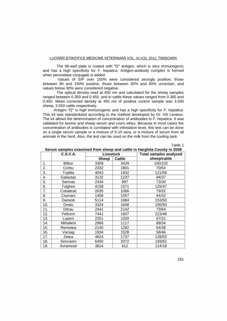
LUCRĂRI ŞTIINŢIFICE MEDICINĂ VETERINARĂ VOL. XLV(3), 2012, TIMIŞOARA
181
The 96-well plate is coated with "f2" antigen, which is very immunogenic and has a high specificity for F. hepatica. Antigen-antibody complex is formed when peroxidase conjugate is added.
Values of S/P over 150% were considered strongly positive; those between 80 and 150% positive, those between 30% and 80% uncertain, and values below 30% were considered negative.
The optical density read at 450 nm and calculated for the sheep samples ranged between 0.350 and 0.450, and in cattle these values ranged from 0.365 and 0.450. Mean corrected density at 450 nm of positive control sample was 3.500 sheep, 3.550 cattle respectively.
Antigen "f2" is high immunogenic and has a high specificity for F. hepatica. This kit was standardized according to the method developed by Dr. HA Levieux. The kit allows the determination of concentration of antibodies to F. hepatica. It was validated for bovine and sheep serum and cow's whey. Because in most cases the concentration of antibodies is correlated with infestation level, this test can be done on a single serum sample or a mixture of 5-10 sera, or a mixture of serum from all animals in the herd. Also, the test can be used on the milk from the cooling tank.
Table 1
Serum samples examined from sheep and cattle in Harghita County in 2008
. o. C.S.V.A. Livestock Total samples analyzed sheep/cattle Sheep Cattle
1. Bilbor 3309 3429 100/102
2. Corbu 2332 1801 70/54
3. Toplita 4043 1932 121/58
4. Galautas 3132 1237 94/37
5. Sarmas 2434 997 73/30
6. Tulghes 4158 1571 125/47
7. Cobatesti 2635 1066 79/32
8. Ciumani 1458 1057 44/32
9. Danesti 5114 1684 153/50
10. Dealu 3324 1648 100/50
11. Ditrau 2441 2142 73/64
12. Feliceni 7441 1607 223/48
13. Lupeni 2251 1020 67/31
14. Mihaileni 2966 1117 89/34
15. Remetea 2140 1282 64/38
16. Varsag 1934 1528 58/46
17. Zetea 4624 1737 139/52
18. Sincraeni 6450 2072 193/62
19. Avramesti 3814 612 114/18
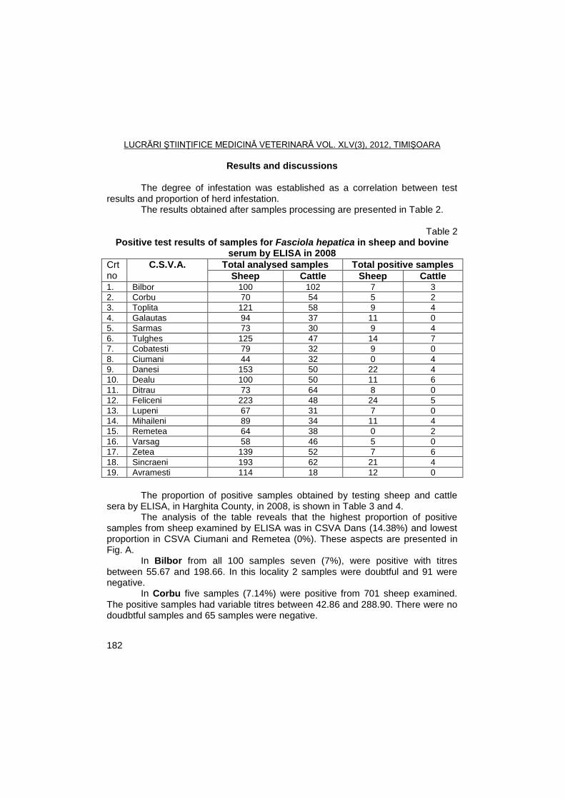
LUCRĂRI ŞTIINŢIFICE MEDICINĂ VETERINARĂ VOL. XLV(3), 2012, TIMIŞOARA
182
Results and discussions
The degree of infestation was established as a correlation between test results and proportion of herd infestation.
The results obtained after samples processing are presented in Table 2.
Table 2 Positive test results of samples for Fasciola hepatica in sheep and bovine
serum by ELISA in 2008
Crtno.
C.S.V.A. Total analysed samples Total positive samples
Sheep Cattle Sheep Cattle 1. Bilbor 100 102 7 3
2. Corbu 70 54 5 2
3. Toplita 121 58 9 4
4. Galautas 94 37 11 0
5. Sarmas 73 30 9 4
6. Tulghes 125 47 14 7
7. Cobatesti 79 32 9 0
8. Ciumani 44 32 0 4
9. Danesi 153 50 22 4
10. Dealu 100 50 11 6
11. Ditrau 73 64 8 0
12. Feliceni 223 48 24 5
13. Lupeni 67 31 7 0
14. Mihaileni 89 34 11 4
15. Remetea 64 38 0 2
16. Varsag 58 46 5 0
17. Zetea 139 52 7 6
18. Sincraeni 193 62 21 4
19. Avramesti 114 18 12 0
The proportion of positive samples obtained by testing sheep and cattle
sera by ELISA, in Harghita County, in 2008, is shown in Table 3 and 4. The analysis of the table reveals that the highest proportion of positive
samples from sheep examined by ELISA was in CSVA Dans (14.38%) and lowest proportion in CSVA Ciumani and Remetea (0%). These aspects are presented in Fig. A.
In Bilbor from all 100 samples seven (7%), were positive with titres between 55.67 and 198.66. In this locality 2 samples were doubtful and 91 were negative.
In Corbu five samples (7.14%) were positive from 701 sheep examined. The positive samples had variable titres between 42.86 and 288.90. There were no doudbtful samples and 65 samples were negative.
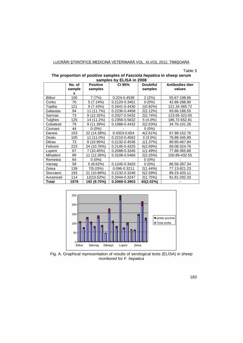
LUCRĂRI ŞTIINŢIFICE MEDICINĂ VETERINARĂ VOL. XLV(3), 2012, TIMIŞOARA
183
Table 3 The proportion of positive samples of Fasciola hepatica in sheep serum
samples by ELISA in 2008
Lo No. of sample
s
Positive samples
CI 95% Doubtful samples
Antibodies titer values
Bilbor 100 7 (7%) 0.224-0.4538 2 (2%) 55.67-198.66
Corbu 70 5 (7.14%) 0.2120-0.3451 0 (0%) 42.86-288.90
Toplita 121 9 (7.43%) 0.2641-0.4430 1(0.82%) 121.34-365.72
Galautas 94 11 (11.7%) 0.2236-0.4458 2(2.12%) 93.66-186.55
Sarmas 73 9 (12.32%) 0.3327-0.5432 2(2.74%) 123.65-322.65
Tulghes 125 14 (11.2%) 0.2356-0.5632 5 (4.0%) 186.72-652.81
Cobatesti 79 9 (11.39%) 0.1988-0.4432 2(2.53%) 34.76-101.28
Ciumani 44 0 (0%) - 0 (0%) -
Danesi 153 22 (14.38%) 0.4323-0.654 4(2.61%) 67.98-152.76
Dealu 100 11 (11.0%) 0.2210-0.4562 3 (3.0%) 78,88-345.89
Ditrau 73 8 (10.95%) 0.2132-0.4536 1(1.37%) 89.95-467.84
Feliceni 223 24 (10.76%) 0.2145-0.4325 6(2.69%) 69.08-324.76
Lupeni 67 7 (10.45%) 0.2088-0.3245 1(1.49%) 77.88-365.88
Mihaileni 89 11 (12.36%) 0.3108-0.5466 2(2.25%) 100.89-432.55
Remetea 64 0 (0%) - 0 (0%) -
Varsag 58 5 (8.62%) 0.1245-0.3420 0 (0%) 86.56-287.34
Zetea 139 7(5.03%) 0.096-0.3211 2(1.44%) 77.13-821.23
Sincraeni 193 21 (10.88%) 0.2132-0.3248 5(2.59%) 89.23-420.11
Avramesti 114 12(10.52%) 0.2044-0.3247 2(1.75%) 91.81-292.33
Total 1979 192 (9.70%) 0.2068-0.3903 40(2.02%) -
0
50
100
150
200
250
Bilbor Sărmaş Dăneşti Lupeni Zetea
probe pozitive
Total probe
Fig. A. Graphical representation of results of serological tests (ELISA) in sheep
monitored for F. hepatica
LUCRĂRI ŞTIINŢIFICE MEDICINĂ VETERINARĂ VOL. XLV(3), 2012, TIMIŞOARA
184
In Toplita nine samples (7.43%) were positive, with titers between 121.34 and 365.72. One sample was uncertain (0.82%) and 111 samples were negative.
In Galautas, 11 samples of 94 were positive with titers that ranged between 93.66 and 186.55. In this village two samples (2.12%) were uncertain, while the remaining 81 samples were negative.
In Sarmas, nine of 73 samples were positive, with titers ranging from 123.65 and 322.65. In this village two samples (2.74%) were uncertain, and other samples (62) were negative.
In Tulghes 14 samples were positive (11.20%), with titers ranging from 186,72 and 652,81. In this locality five samples (4%) were uncertain and 106 were negative.
In Cobatesti from 79 samples examined nine (11.39%) were positive with titers between 34.76 and 101.28. There two uncertain samples (2.53%) and 68 negative.
In Ciumani and Remetea there were no positive or uncertain samples all samples being negative.
In Danesti from the 153 samples 22 were positive (14.38%), titers of positive samples were within the limits of 67.98 and 152.76, 127 samples were negative and four uncertain (2.61%).
In Dealu of the 100 samples collected and examined serologically 11 (11%) were positive; their titers ranged between 89.95 and 467.84, three samples were uncertain and 86 negative.
In Ditrau of the 73 samples collected and examined serologically, eight were positive, which represent 10.95%, and titers of these samples ranged between 89.95 and 467.84. One sample was uncertain, and the differences (64) were negative samples.
In Feliceni of the 223 samples collected and examined serologically, 24 were positive (10.76%) and their titers were between 69.08 and 324.76. Negative samples were 193 and six (2.69%) were uncertain.
In Lupeni of the 67 samples collected and examined serologically, seven were positive (10.45%), and titers ranged between 77.88 and 365.88. One sample was uncertain, and the difference was negative samples.
In Mihaileni 89 samples were collected, and of them 11 were positive (12.36%) with titers between 100.89 and 432.55 and two samples were uncertain, while the remaining 76 samples were negative.
In Varsag, of the 58 samples collected, five were positive (8.62%) and had antibody limits between 86.56 and 287.34. In this village there were no uncertain samples.
In Zetea of the 139 samples collected, seven were positive (5.03%), with antibodies limits ranged between 77.13 and 821.23. In this village two samples were uncertain and 130 negative.
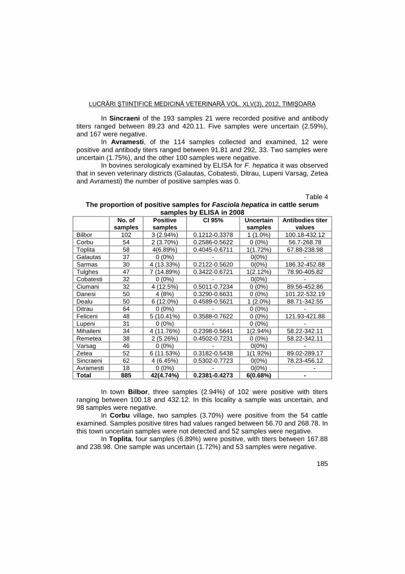
LUCRĂRI ŞTIINŢIFICE MEDICINĂ VETERINARĂ VOL. XLV(3), 2012, TIMIŞOARA
185
In Sincraeni of the 193 samples 21 were recorded positive and antibody titers ranged between 89.23 and 420.11. Five samples were uncertain (2.59%), and 167 were negative.
In Avramesti, of the 114 samples collected and examined, 12 were positive and antibody titers ranged between 91.81 and 292, 33. Two samples were uncertain (1.75%), and the other 100 samples were negative.
In bovines serologicaly examined by ELISA for F. hepatica it was observed that in seven veterinary districts (Galautas, Cobatesti, Ditrau, Lupeni Varsag, Zetea and Avramesti) the number of positive samples was 0.
Table 4 The proportion of positive samples for Fasciola hepatica in cattle serum
samples by ELISA in 2008
Locality No. of samples
Positive samples
CI 95% Uncertain samples
Antibodies titer values
Bilbor 102 3 (2.94%) 0.1212-0.3378 1 (1.0%) 100.18-432.12
Corbu 54 2 (3.70%) 0.2586-0.5622 0 (0%) 56.7-268.78
Toplita 58 4(6.89%) 0.4045-0.6711 1(1.72%) 67.88-238.98
Galautas 37 0 (0%) - 0(0%) -
Sarmas 30 4 (13.33%) 0.2122-0.5620 0(0%) 186.32-452.88
Tulghes 47 7 (14.89%) 0.3422-0.6721 1(2.12%) 78.90-405.82
Cobatesti 32 0 (0%) - 0(0%) -
Ciumani 32 4 (12.5%) 0.5011-0.7234 0 (0%) 89.56-452.86
Danesi 50 4 (8%) 0.3290-0.6631 0 (0%) 101.22-532.19
Dealu 50 6 (12.0%) 0.4589-0.5621 1 (2.0%) 88.71-342.55
Ditrau 64 0 (0%) - 0 (0%) -
Feliceni 48 5 (10.41%) 0.3588-0.7622 0 (0%) 121.93-421.88
Lupeni 31 0 (0%) - 0 (0%) -
Mihaileni 34 4 (11.76%) 0.2398-0.5641 1(2.94%) 58.22-342.11
Remetea 38 2 (5.26%) 0.4502-0.7231 0 (0%) 58.22-342.11
Varsag 46 0 (0%) - 0(0%) -
Zetea 52 6 (11.53%) 0.3182-0.5438 1(1.92%) 89.02-289.17
Sincraeni 62 4 (6.45%) 0.5302-0.7723 0(0%) 78.23-456.12
Avramesti 18 0 (0%) - 0(0%) -
Total 885 42(4.74%) 0.2381-0.4273 6(0.68%) -
In town Bilbor, three samples (2.94%) of 102 were positive with titers
ranging between 100.18 and 432.12. In this locality a sample was uncertain, and 98 samples were negative.
In Corbu village, two samples (3.70%) were positive from the 54 cattle examined. Samples positive titres had values ranged between 56.70 and 268.78. In this town uncertain samples were not detected and 52 samples were negative.
In Toplita, four samples (6.89%) were positive, with titers between 167.88 and 238.98. One sample was uncertain (1.72%) and 53 samples were negative.
LUCRĂRI ŞTIINŢIFICE MEDICINĂ VETERINARĂ VOL. XLV(3), 2012, TIMIŞOARA
186
In Sarmas, four of 30 samples were positive, with titers ranging from 186.32 and 452.88. In this town there were no uncertain samples and the remaining samples (26) were negative.
In Tulghes, seven samples were positive (14.89%), titers of these samples ranged between 78.90 and 405.82. In this locality, a sample (2.12%) was uncertain and 39 were negative.
In Ciumani there were four positive samples (12.50%), with variable limits between 89.56 and 452.86. There were no uncertain samples.
In Danesti, from the 50 samples prelevated, four were positive (8.00%), titers of positive samples were within the limits of 101.22 and 532.19; negative samples were 46 and none uncertain.
In Dealu of the 50 samples collected and examined serologically, six (12%) were positive, their titers ranged between 88.71 and 342.55. A single sample was registered uncertain in this locality (2%).
In Feliceni, of the 48 samples collected and examined serologically, five were positive (10.41%), and their titers ranged between 121.93 and 421.88, 43 were negative samples and none uncertain.
In Mihaileni 34 samples were collected and four of them were positive (11.76%) with titers that had limits between 58.22 and 342.11. One sample was uncertain, while the remaining 29 samples were negative.
In town Remetea of the 38 samples collected, two were positive (5.26%) and had antibody limits between 58.22 and 342.11. In this village there were no uncertain samples.
In town Sincraeni, of the 62 positive samples, four were pozitive (6.45%), and antibody titers ranged between 78.23 and 456.12, none was uncertain and 58 were negative.
Conclusions
The average prevalence of infestation with Fasciola hepatica in sheep in
Harghita County, determined by ELISA, was 9.70%, with variations between 0% and 14.38%.
By localities the prevalence (%) of the infestation with Fasciola hepatica in sheep, was: zero Ciumani and Remetea; 5.03 in Zetea; Bilbor 7.0 in, 7.14 in Corbu village; 7.43 in Toplita; 8.62 in Varsag; 10.45 in Lupeni, in Avramesti 10.52, 10.88 in Sâncraieni; in Feliceni 10.76, 10.95 in Ditrau, 11.0 in Dealu, 11.2 in Tulghes, 11.39 in Cobatesti, 11.7 in Galautas; 12.32 in Sarmas, 12.36 in Mihaileni and 14.38 in Danesti.
In bovines the prevalence (%) of infestation with F. hepatica by localities was as follows: 0 in six localities (Avramesti, Ditrau, Lupeni, Galautas, Cobatesti and Varsag), 2.94 in Bilbor; 3.70 in Corbu, 5.26 in Remetea; 6.45 in Sâncraieni, 6.89 in Toplita, 8.70 in Danesti, 10.41 in Feliceni, 11.76 in Mihaileni, 12.0 in Dealu; 12.50 in Ciumani; 13.33 in Sarmas and 14.89 in Tulghes.
LUCRĂRI ŞTIINŢIFICE MEDICINĂ VETERINARĂ VOL. XLV(3), 2012, TIMIŞOARA
187
The average prevalence of infestation with Fasciola hepatica in cattle of Harghita County, determined by ELISA, was 4.74%, with variations between 0% and 14.89%.
Immunological diagnosis of fasciolosis by ELISA in sheep and cattle is a relevant and expedient method that can check the degree of infestation of animals.
References
1. Bohan, V.R., Hanks, R.D., Behrens, C.W., Phelps, D.A., Effects of liver flukes and abcesses on growth of feedlot cattle, Journ. An. Scie, 1979, 49, 183-184.
2. Bouix-Busson, D., Rondelaud, D., Combes, C.,- L’infestation de Lymnaea glabra( Müller) par Fasciola hepatica, Ann. Parasitol. Hum. Comp., 1985, 60 (1), 11-21.
3. Bourdoiseau, G., Les douves des ruminants. Point. Vét., 1997, 28: 15-19. 4. Bussieras, J., Chermette, R., Abrégé des Parasitologie vétérinaire.
Helminthologie. II.Ed. Rosset R. Bibliotéque Ecole Nationale Vétérinaire de Toulouse, 1992.
5. Charlier, J., Meulemeester, L., Claerebout, E., Williams, D., Vercruysse, J., Qualitative and quantitative evaluation of coprological and serological techniques for the diagnosis of fasciolosis in cattle. Vet. Parasitol., 2008, 153 (1/2), 44-51.
6. Cosoroaba, I., Cristina, R.T., Prevenirea si combaterea rezistentei la antihelmintice, Rev. Rom. M.V., 1995, 5 (2), 109-118.
7. Cringoli, G., Rinaldi, L., Veneziano, V., Capelli, G., Malone, J.B., A cross-sectional coprological survey of liver flukes in cattle and sheep from an area of the southern Italian Alpennines. Vet. Parasitol., 2002, 108: 137-143.
8. Darabus, GH., Controlul parazitologic in helmintozele ierbivorelor - partea I, Rev. Rom. Med. Vet., 2003, 13, 2, 70-81.
9. Gupta, A., Dixit, A.K., Dixit, P., Mahajan, C., Sharma R.L., Evalution of dispick-ELISA using 28 kDa Fasciola gigantica cathepsin L cystein proteinase (FgCL3) for serodiagnosis of fasciolosis in naturali infected goats. Veterinary Parasitology, 2011, 176, 2-3, 165-169.
10. Hammouda, N.A., Mansoury, S.T., Azzouni, M.Z., Hussein, E.D. Detection of circulating antigens in blood to evaluate treatment of fascioliasis. J. of the Egyptean Soc. Of Parasitol., 1997, 27, 365-371.
11. Osman, M.M., Shebab, A.Y., El Mashry, S.A., Helmy, M.H., Farag, H.F., Evaluation of Fasciola excretory-secretory (E/S) product in diagnosis of acute human fascioliasis by IgM ELISA, Trop. Med. And Parasit., 1995, 46, 115-118.
LUCRĂRI ŞTIINŢIFICE MEDICINĂ VETERINARĂ VOL. XLV(3), 2012, TIMIŞOARA
188
EFFECT OF "GELLIPRIM ORALE" ADMINISTRATION IN AN EXPERIMENTAL GROUP OF RABBITS WITH EIMERIOSIS
NARCISA MEDERLE
1, ELENA TILIBAŞA
1, GH. DĂRĂBUŞ
1, I. OPRESCU
1, S.
MORARIU1, M. S. ILIE
1, PAULA BOHATIR
2
1Banat's University of Agricultural Sciences and Veterinary Medicine Timisoara,
Faculty of Veterinary Medicine, 300645, Aradului Street No. 119, Timisoara, Romania
2 Banat's University of Agricultural Sciences and Veterinary Medicine Timisoara,
Faculty of Zootechny and Biotechnologies E-mail: [email protected]
Summary
In rabbit farms, especially young animals are affected by a large number of
infectious and parasitic diseases, including coccidiosis which is more common. Protection of rabbits during the critical period (after weaning) is achieved by chemoprophylaxis.
An experimental lot of rabbits diagnosed with Eimeriosis were treated with "Gelliprim orale", which contains sulfadiazine 20 g, trimethoprim 4 g.
The protocol consisted of administration of ―Gelliprim orale" in the week before the actual experiment. The same protocol was repeated next month.
Throughout the experiment, twice weekly, individual coproscopic examinations have been performed (Willis method).
At the end of the experiment (01.02.2012) 20 of the 22 individuals with eimeriosis were negative, when coproscopic examined, oocysts being present only in two rabbits.
Slaughtering the lot was not found liver damage in any of the individuals. We recommend "Gelliprim orale" as an effective therapeutic alternative in intestinal
coccidiosis of rabbits. Key words: Gelliprim orale, rabbits, Eimeria, treatment
In traditional rabbit farms, diseases such as bacterial and/or parasitical
have a high incidence and seriously affect the health of animals. This entails increased use of antibiotics and antiparasitic drugs, with repercussions on food safety. Use of prebiotics, probiotics, enzymes and fitoadditives as feed additives able to modulate the intestinal microflora and improve the health of rabbits, especially those kept in intensive farming system, is a current recommendation from specialists.
In this context the scope of this paper was to highlight the effect of "Gelliprim orale" on a group of rabbits, on which nutritional and bioproductive indices evolution following different categories of natural extracts administration were traced.
LUCRĂRI ŞTIINŢIFICE MEDICINĂ VETERINARĂ VOL. XLV(3), 2012, TIMIŞOARA
189
Materials and methods
During 23.11.2011 - 01.02.2012, a total of 24 rabbits was hospitalized, males and females, young weaned (28 days) from Chinchilla, English variegated, California Giant, Lion Head breeds in the Hospital of University Veterinary Clinics from Timişoara.
In the first week of experiment (23.11.2011 - 01.12.2011) young weaned was accustomed to feed administered and maintenance system. This week individual coproscopic examinations were performed to establish parasitic situation. Coproscopic examination consisted of flotation method (Willis and Mc Master).
Animals taken for this study were administered "Gelliprim orale" daily in drinking water 5 ml/60 kg, for a week, with anti-coccidian properties. This drug contains sulfadiazine 20 g, trimethoprim 4 g. Same protocol was repeated next month.
On 01.12.2011 young rabbits were divided into five groups and subjected to the experiment itself, which consisted of adding the drinking water with natural plant extracts, as follows:
Group 1 - horseradish root extract Group 2 - branches of rose hips extract Group 3 - red sea buckthorn extract Group 4 - grape seed Extract Group 5 – control Throughout the experiment, twice weekly, individual coproscopic
examinations have been performed (Willis and Mc Master methods). During the experiment, the animals studied were given a proper
environment, hygienic conditions, tracing temperature and humidity parameters three times a day.
On the last day of experiment (01.02.2012) young rabbits were euthanized. Nutrient and bioproductive index measurements were carried. We examined the liver and small intestine to find possible lesions caused by parasites.
Results and discussions
Individual coproscopic examinations carried out for the experimental group of rabbits in the first day of the experiment (23.11.2011) showed the presence of Eimeria oocysts in 22 individuals out of 24 (table 1).
There were no other genera or species of parasites reported on microscopic examination of faecal samples.
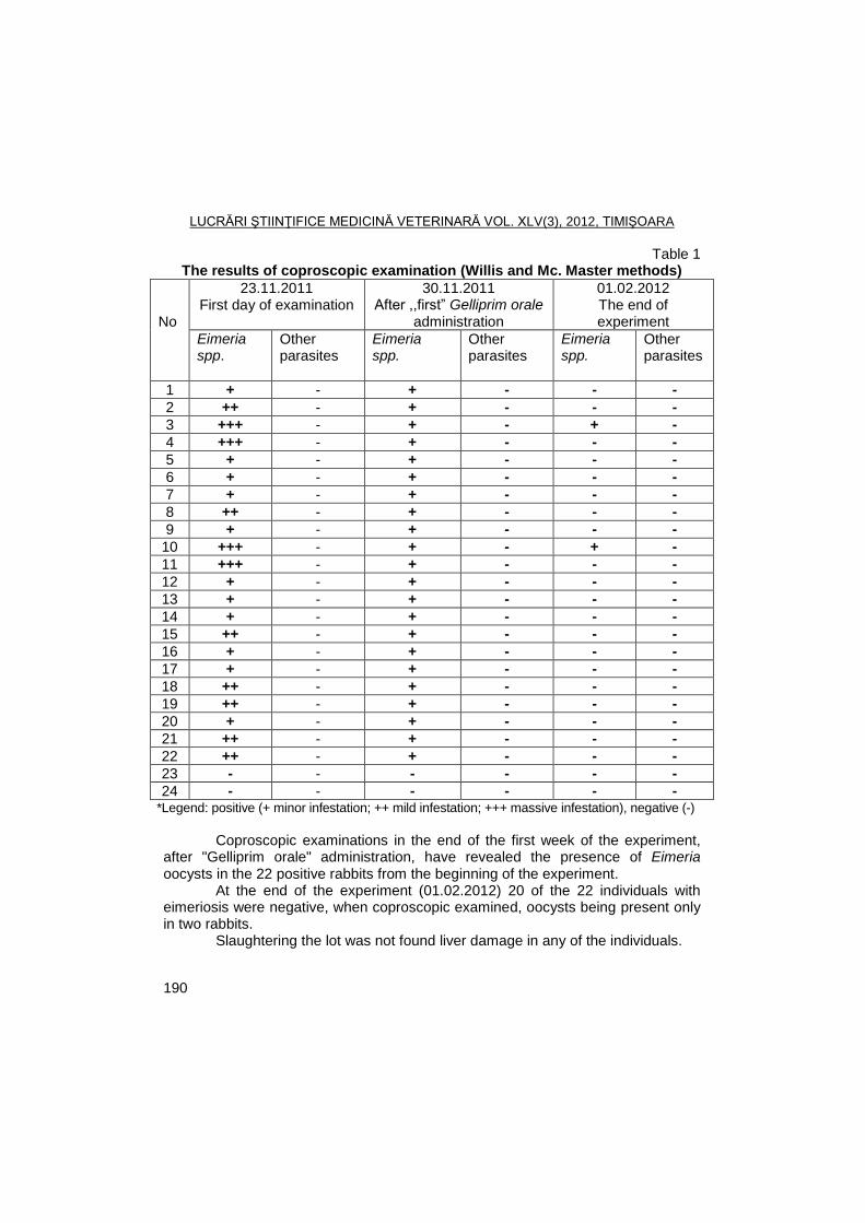
LUCRĂRI ŞTIINŢIFICE MEDICINĂ VETERINARĂ VOL. XLV(3), 2012, TIMIŞOARA
190
Table 1 The results of coproscopic examination (Willis and Mc. Master methods)
No
23.11.2011 First day of examination
30.11.2011 After ,,first‖ Gelliprim orale
administration
01.02.2012 The end of experiment
Eimeria spp.
Other parasites
Eimeria spp.
Other parasites
Eimeria spp.
Other parasites
1 + - + - - -
2 ++ - + - - -
3 +++ - + - + -
4 +++ - + - - -
5 + - + - - -
6 + - + - - -
7 + - + - - -
8 ++ - + - - -
9 + - + - - -
10 +++ - + - + -
11 +++ - + - - -
12 + - + - - -
13 + - + - - -
14 + - + - - -
15 ++ - + - - -
16 + - + - - -
17 + - + - - -
18 ++ - + - - -
19 ++ - + - - -
20 + - + - - -
21 ++ - + - - -
22 ++ - + - - -
23 - - - - - -
24 - - - - - - *Legend: positive (+ minor infestation; ++ mild infestation; +++ massive infestation), negative (-)
Coproscopic examinations in the end of the first week of the experiment, after "Gelliprim orale" administration, have revealed the presence of Eimeria oocysts in the 22 positive rabbits from the beginning of the experiment.
At the end of the experiment (01.02.2012) 20 of the 22 individuals with eimeriosis were negative, when coproscopic examined, oocysts being present only in two rabbits.
Slaughtering the lot was not found liver damage in any of the individuals.
LUCRĂRI ŞTIINŢIFICE MEDICINĂ VETERINARĂ VOL. XLV(3), 2012, TIMIŞOARA
191
Others authors and rabbit breeders also recommend the use of "Gelliprim orale" in prevention and treatment of eimeriosis.
Parasitic diseases of rabbits have been addressed in clinical and especially therapeutical perspective. Prophylaxis, early diagnosis, establishing the best therapeutic protocol is subjects of study and experiment for many authors (8, 14, 15, 16, 17, 18, 19, 20).
Sulfaveridin, Sulfacoccidin, Robenidine, Toltrazuril are just some of the medicinal preparations whose efficiency in eimeriosis of rabbits was studied in comparison with results obtained in this study (12, 13, 21).
Conclusions
The product Gelliprim orale have revealed a goof control of Eimeria in rabbit.
We recommend Gelliprim orale as an effective therapeutic alternative in intestinal coccidiosis of rabbits.
References
1. Bura, M., Proiect tehnologic pentru creşterea iepurilor de casă, Colecţia
revistei Ferma, Timişoara, 2001. 2. Bussieras, J., Chermette, R., Parasitologie veterinaire-Protozoologie,
Service de Parasitologie, E.N.V. d’Alfort, 1992. 3. Cosoroabă, I., Parazitologie veterinară, Ed. Mirton, Timişoara, 2000. 4. Coudert, P., Licois, D., Provot, F., Differential diagnosis of Eimeria species
from the rabbit, 4-th World Rabbit Congress, Budapest, Hungary, 480, 1988. 5. Coudert, P., Provot, F., Lasalocid – Tolerance for the rabbit and activity
against, 4-th World Rabbit Congress, Budapest, Hungary, 418-427, 1988. 6. Dărăbuş, Gh., Cătană, N., Bolile infecţioase şi parazitare ale iepurilor, Ed.
Brumar, Timişoara, 1996. 7. Dărăbuş, Gh., Oprescu I., Morariu S., Mederle Narcisa, Parazitologie şi
Boli Parazitare, Ed. Mirton, Timişoara, 2006. 8. Mehlhorn, H., Encyclopedic reference of parasitology, Second edition,
Spinger-Berlin, Heidelberg, 2001. 9. Mercier, P., Le traitement chez le lapin, Bull.G.T.V., 6, 83-94, 1990. 10. Mercier, P., Essay d’utilisation du decoquinate dans le contrôle de la
coccidiose chez le lapin à l’engraissement, Bull.G.T.V., 89-94, 1997. 11. Nesterov, V., Păstîrnac, N., Sîrbu, V., Bolile animalelor pentru blană, Ed.
Ceres, Bucureşti, 1981. 12. Orbulescu, Doina, Falcă, C., Bura, M., Eficienţa terapeutică a
Sulfaveridinului şi a Sulfacoccidinului în coccidioza iepurilor, Rev. de creşterea animalelor, 5, 48-49, 1982.
13. Peeters, J., E., Geermos, R., Halesn, PH., H., Epidemiology of coccidiosis in comercial rabbits (1982-1987) and resistance against
LUCRĂRI ŞTIINŢIFICE MEDICINĂ VETERINARĂ VOL. XLV(3), 2012, TIMIŞOARA
192
Robenidine, 4-th World Rabbit Congress, Budapest, Hungary, 399-408, 1988.
14. San Martin-Nuñez, B.V., Ordoñez-Escudero, D., Alunda, J.M., Preventive treatment of rabbit coccidiosis with α-difluoromethylornithine, Veterinary Parasitology, 1-10, 1988.
15. Stănescu, V., Bolile iepurelui de casă şi combaterea lor, Ed. Ceres, Bucureşti, 1984.
16. Şuteu, I., Cozma, V., Bolile parazitare la animalele domestice, Ed. Ceres, Bucureşti, 1998.
17. Şuteu, I., Cozma, V., Parazitologie clinică veterinară, vol. I, Ed. Risoprint, Cluj-Napoca, 2004.
18. Şuteu, I., Cozma, V. Parazitologie clinică veterinară, vol. II, Ed. Risoprint, Cluj-Napoca, 2004.
19. Şuteu, I., Vartic, N., Cozma, V., Diagnosticul şi tratamentul parazitozelor la animale, Ed. Ceres, Bucureşti, 1996.
20. Tacconi, G., Piergili-Fioretti, D., Moretti, A., Nobilini, N., Pasquali, P., Coccidia in Hare (Lepus europaeus) Reared in Umbria, Italy: Bioepidemiological Study, J. Protozool. Res., 5, 77-85, 1995.
21. Voros, G., Barna, J., Efficacy of Toltrazuril/Baycox in the prevention of hepatic coccidiosis in rabbits, 4-th World Rabbit Congress, Budapest, Hungary, 428-437, 1988.
LUCRĂRI ŞTIINŢIFICE MEDICINĂ VETERINARĂ VOL. XLV(3), 2012, TIMIŞOARA
193
AUTHORS INDEX
A Adanović O. – 89 Altamura G. – 10 Andrei Sidonia – 96, 121 Aniță Adriana – 5 B Barbuceanu Florica – 66 Belu C. – 66 Boboš S. – 161 Bocaneti Florentina – 10 Bohatir Paula – 188 Bojkovski J. – 18, 155 Borzacchiello G. – 10 Bozic M. – 137 Brudiu Ileana – 104 C Capo I. – 137 Cătană N. – 23. 27, 43, 48, 52 Cenușe Cătălina – 31, 37 Cocora Zoriţa Maria – 85 Colibar Olimpia – 104 Coman Alina – 23 Corteggio Annunziata – 10 Cristina R.T. – 43 Cumpănășoiu C. – 31, 37 D Daraban C. – 10 Dărăbuș Gh. – 96, 104, 121, 126, 131,
142, 149, 167, 173, 180, 188
Dégi J. – 27, 43, 57 Diaconu Cristina – 66 Dobai Gyöngyi – 85 F Fodor Ionica – 23, 27, 43, 48
G Georgescu, B. – 66 Gros R.V. – 31, 37 Groza Ioana – 48 H Herman V. – 43, 52, 81 Hotea Ionela – 104, 121, 131, 167 I Iacob Olimpia – 110 Ilie Alina – 131 Ilie M.S. – 96, 104, 121, 126, 131, 142,
149, 167, 188 Imre K. – 121, 126, 131 Imre Mirela – 104, 121, 126, 131, 149 Indre D. – 104, 149, 173, 180 Ivanovic N. – 137 Ivanović Sneţana – 155 Ivetić V. – 89 J Jeremic Svetlana – 77 Jovčić Dubravka – 155 L Laloşevic D. – 137 Laloşevic Vesna – 137, 161 Lazăr L. – 31 Lazău Al. – 57 M Mederle Narcisa – 142, 149, 167, 188 Mederle O. – 81 Mihali C. – 121, 131 Milicevic Vesna – 77 Morar Adriana – 126 Morariu Florica – 142 Morariu S. – 121, 131, 142, 149, 167, 188
LUCRĂRI ŞTIINŢIFICE MEDICINĂ VETERINARĂ VOL. XLV(3), 2012, TIMIŞOARA
194
N Nichita Ileana – 31, 37 O Obradovic N. – 137 Olariu-Jurca I. – 57 Onita P. – 121, 126 Oprescu I. – 121, 131, 142, 149, 167,
173, 180, 188 P Pajić M. – 155 Palca M. – 131 Palović I. – 155 Pascu Corina – 52 Pavičić Ljiljana – 161 Pavlović I. – 18 Petrujkić T. – 18 Popovici A. – 66 Praşovic S. – 137 Predoi G. – 66 Putic S. – 137 R Radosavljević V. – 77, 89 Raita Ştefania – 66 Relić R. – 18 Rogoţarski D. – 18 Roperto F. – 10
S Sala Claudia – 126 Savić B. – 18, 89 Savović M. – 161 Savuța G. – 5 Simin S. – 161 Simin Verica – 137 Sorescu Denisa – 104, 121, 149, 167 Stamate Daniela – 66 Surpat Anca Sofiana – 81
Ş Șereș Monica – 31, 37 T Tilibașa Elena – 188 Tîrziu E. – 31, 37 Turcitu M. – 66 Ț Țifrea M.- 149, 173, 180 Țibru I. – 85 U Uroş M. – 57 V Velescu Elena – 10 Z Ţugić Gordana – 155 Ţutić Jadranka – 77, 89 Ţutić M. – 89
LUCRĂRI ŞTIINŢIFICE MEDICINĂ VETERINARĂ VOL. XLV(3), 2012, TIMIŞOARA
195
CONTENT
ANIȚĂ ADRIANA, G. SAVUȚA
New possibilities of detection of swine hepatitis E infection
5
BOCANETI FLORENTINA, G. BORZACCHIELLO, G. ALTAMURA, ANNUNZIATA CORTEGGIO, F. ROPERTO, C. DARABAN, ELENA VELESCU
Detection of bovine Papillomavirus E5 by immunofluorescence in bovine cutaneous fibropapillomas from Eastern Romania
10
BOJKOVSKI J., B.SAVIŠ, R. RELIŠ, I. PAVLOVIŠ, D. ROGOŢARSKI, T. PETRUJKIŠ
Tuberculosis of the high producting dairy cows – case report –
18
CĂTANĂ N., ALINA COMAN,
IONICA FODOR
Research on the role of avian reovirus in transmissible viral etiology proventriculitis
23
CĂTANĂ N., FODOR IONICA, J. DÉGI
Using a simplified scheme for isolation and typing staphylococci isolated from animals
27
CENUȘE CĂTĂLINA, E. TÎRZIU, ILEANA NICHITA, C. CUMPĂNĂŞOIU, R.V. GROS, R. LAZĂR, MONICA ȘEREȘ
Changes in humoral compartment of the immune system associated with enzootic bovine leukosis
31
CENUȘE CĂTĂLINA, E. TÎRZIU, ILEANA NICHITA, C. CUMPĂNĂȘOIU, R.V. GROS, MONICA ȘEREȘ
Seroprevalence of bovine leukemia virus infection in cattle from Timis County between 2006 and 2010
37
DÉGI J., R.T.CRISTINA, IONICA FODOR, V. HERMAN, N. CĂTANĂ
Prevalence and antimicrobial susceptibility of staphylococci isolated from otitis externa of cats
43
LUCRĂRI ŞTIINŢIFICE MEDICINĂ VETERINARĂ VOL. XLV(3), 2012, TIMIŞOARA
196
FODOR IONICA, IOANA GROZA, NICOLAE CĂTANĂ
Research on the frequency of isolation strains of APEC in broilers
48
HERMAN V., N. CĂTANĂ, CORINA PASCU
Rabies cases in Romania during 2001 and 2010
52
LAZĂU AL., I. OLARIU-JURCA, M. UROŞ, J. DÉGI
Equine infectious anemia evolution in Western area of Romania during period 2007 and 2011
57
POPOVICI A., CRISTINA DIACONU, DANIELA STAMATE, ŞTEFANIA RAITA, M. TURCITU, B. GEORGESCU, C. BELU, G. PREDOI, FLORICA BARBUCEANU
Histopathological and ultrastructural aspects of the Gumboro disease virus Faragher 52/70 strain experimental infection in SPF chickens
66
RADOSAVLJEVIŠ V., SVETLANA JEREMIC, JADRANKA ZUTIC, VESNA MILICEVIC
Occurrence of Renibacterium salmoninarum in rainbow trout farm in Serbia
77
SURPAT ANCA SOFIANA, V. HERMAN, O. MEDERLE
Preliminary investigation about necrotic enteritis in weaned pigs
81
ŢIBRU I., GYÖNGYI DOBAI, ZORIŢA MARIA COCORA
Determining the Salmonella spp. carrier state in the case of fattening pigs
85
ŢUTIŠ JADRANKA, O. ADANOVIŠ, V. RADOSAVLJEVIŠ, B. SAVIŠ, V. IVETIŠ, M. ŢUTIŠ
Prevalence of F4 (K88) fimbrial adhesins in E. coli strains isolated from piglets in the suckling period
89
ANDREI SIDONIA, M.S. ILIE, GH. DĂRĂBUŞ
Intestinal parasite infections in dogs population from Timis and Arad Counties
96
LUCRĂRI ŞTIINŢIFICE MEDICINĂ VETERINARĂ VOL. XLV(3), 2012, TIMIŞOARA
197
HOTEA IONELA, M.S. ILIE, MIRELA IMRE, DENISA SORESCU, D. INDRE, ILEANA BRUDIU, OLIMPIA COLIBAR, GH. DĂRĂBUŞ
Toxoplasma gondii seroprevalence in cats and sheep from Caras-Severin County, Romania
104
IACOB OLIMPIA C.
The therapeutic efficacy of Gardal 10% (albendazole sulfoxide) against digestive and pulmonary parasitic fauna in naturally infested goats
110
M.S. ILIE, MIRELA IMRE, IONELA HOTEA, K.IMRE, IONELA DENISA SORESCU, SIDONIA ANDREI, P. ONITA, I. OPRESCU, S. MORARIU, C. MIHALI, GH. DĂRĂBUȘ
Prevalence of Hypoderma infestation in deer in Western Romania
121
IMRE K., CLAUDIA SALA, ADRIANA MORAR, M.S. ILIE, MIRELA IMRE, P. ONIȚĂ, GH. DĂRĂBUȘ
Occurence of Sarcocystis spp. in roe deer (Capreolus capreolus) from Western Romania - preliminary results
126
IMRE MIRELA, M.S. ILIE, C.V. MIHALI, I. OPRESCU, S. MORARIU, IONELA HOTEA, K. IMRE, ALINA ILIE, M. PALCA, GH. DĂRĂBUȘ
Prevalence of tick species in dogs using classical methods and SEM
131
LALOŞEVIC D., S. PRAŞOVIC, VESNA LALOŞEVIC, VERICA SIMIN, I. CAPO, N. OBRADOVIC, M. BOZIC, S. PUTIC, N. IVANOVIC
Verminous pneumonia and tracheobronchitis in foxes and their zoonotic potential
137
MORARIU S., GH. DĂRĂBUȘ, I. OPRESCU, NARCISA MEDERLE, M.S. ILIE, FLORICA MORARIU
The dynamics of blowflies of Calliphora, Lucilia and Protophormia genera during 2004 in Northern Timisoara
142
LUCRĂRI ŞTIINŢIFICE MEDICINĂ VETERINARĂ VOL. XLV(3), 2012, TIMIŞOARA
198
OPRESCU I., S. MORARIU, NARCISA MEDERLE, M.S. ILIE, D. INDRE, MIRELA IMRE, DENISA SORESCU, M. ŢIFREA, GH. DĂRĂBUŞ
Efficacy testing of Flukiver Combi (Janssen Pharmaceutica NV) in natural mixed infection with Fasciola hepatica and gastrointestinal nematodes, in sheeps from Bilbor, Harghita
149
PALOVIŠ I., SNEŢANA IVANOVIŠ, GORDANA ŢUGIŠ, DUBRAVKA JOVŢIŠ, J. BOJKOVSKI, M. PAJIŠ
Season distribution of gastrointestinal helminths of small ruminants in spread Belgrade area
155
SAVOVIŠ M., VESNA LALOŠEVIŠ, S. SIMIN, LJILJANA PAVIŢIŠ, S. BOBOŠ
Seroprevalence of Neospora caninum in dairy cows with reproductive disorders in Vojvodina Province, Serbia
161
SORESCU IONELA DENISA, I. OPRESCU, S. MORARIU, NARCISA MEDERLE, M.S. ILIE, IONELA HOTEA, GH. DĂRĂBUŞ
The parasitism with Giardia spp. in calves from Caras-Severin County
167
ŢIFREA M., I. OPRESCU, D. INDRE, GH. DĂRĂBUŞ
Efficacy testing of Dovenix (Nitroxinil, Merial) in natural infestation with Fasciola hepatica at sheep
173
ŢIFREA M., I. OPRESCU, D. INDRE, GH. DĂRĂBUŞ
Prevalence of parasitism with Fasciola hepatica determined by ELISA in Harghita County
180
NARCISA MEDERLE, ELENA TILIBAŞA, GH. DĂRĂBUŞ, I. OPRESCU, S. MORARIU, M.S. ILIE, PAULA BOHATIR
Effect of "Gelliprim orale" administration in an experimental group of rabbits with eimeriosis
188
AUTHORS INDEX 193