comparative studies between ferns gametophyte and sporophyte...
TRANSCRIPT
Muzeul Olteniei Craiova. Oltenia. Studii şi comunicări. Ştiinţele Naturii. Tom. 29, No. 1/2013 ISSN 1454-6914
120
COMPARATIVE STUDIES BETWEEN FERNS GAMETOPHYTE AND SPOROPHYTE BY BIDIMENSIONAL ELECTROPHORESIS
ALDEA Florentina, HELEPCIUC Florenţa Elena, BANCIU Cristian, BREZEANU Aurelia, SOARE Liliana-Cristina
Abstract. Athyrium filix-femina (L.) ROTH, Polypodium vulgare L. and Asplenium trichomanes L. are three valuable ferns species due to their potential as ornamental plants and secondary metabolite production and they originate fromValsan Valley protected area. Our experiments were focused on developing an optimal in vitro multiplication method and medium term conservation for these species. The reactivity of each species has been evaluated on the basis of biometric (morphometric and gravimetric) and biochemical determinations. The current study showed the differences between gametophytes and sporophytes of the mentioned species by comparing the proteomic profiles. We used gametophytes and sporophytes from A. filix-femina, P. vulgare and A. trichomanes obtained by in vitro culture and multiplicated on MS 1/2 medium. Proteins were isolated according to a phenol extraction procedure and the protein content was determined by Bradford assay. The protein samples were separated in two dimensional technique and stained with Coomassie. Although using PAGE technique we have not observed significant differences between gametophyte and sporophyte, the patterns obtained by two-dimensional gel electrophoresis showed the existence of distinct spots for both gametophyte and sporophyte extracts. Keywords: Athyrium filix-femina, Polypodium vulgare, Asplenium trichomanes, two-dimensional gel electrophoresis. Rezumat. Studii comparative privind gametofitul şi sporofitul ferigilor prin electroforeză bidimensională. A. filix-femina (L.) ROTH, P. vulgare L. şi A. trichomanes L. sunt trei specii de ferigi valoroase datorită potenţialului lor ca plante ornamentale şi sintezei de metaboliţi secundari, fiind originare din zona protejată Valea Vâlsanului. Experimentele noastre s-au axat pe dezvoltarea unei metode optime de multiplicare in vitro şi conservarea pe termen mediu pentru aceste specii. Reactivitatea fiecărei specii a fost evaluată pe baza determinărilor biometrice (morfometrice şi gravimetrice) şi biochimice. Studiul actual arată diferenţele dintre gametofiţii şi sporofitii speciilor menţionate prin compararea profilurilor proteomice. Am folosit gametofiţi şi sporofiţi de la A. filix-femina, P. vulgare şi A. trichomanes obţinuţi prin cultura in vitro şi multiplicaţi pe mediu MS 1/2. Proteinele au fost izolate în conformitate cu procedura de extracţie cu fenol iar concentraţia de proteine a fost determinată prin testul Bradford. Proteinele au fost separate prin electroforeză bidimensională şi colorate cu Coomassie. Deşi folosind electroforeza în gel de poliacrilamidă nu am observat diferenţe semnificative între gametofit şi sporofit, modelele obţinute prin electroforeză bidimensională au arătat existenţa unor spoturi distincte pentru ambele extracte din gametofit şi sporofit. Cuvinte cheie: A. filix-femina, P. mvulgare, A. trichomanes, electroforeză bidimensională.
INTRODUCTION
Pteridophytes represent a very important group of vascular plants. They have been known for more than 300
million years, and present an enormous diversity of forms, growing in many different habitats around the world. During the Carboniferous Period, they dominated the vegetation. While most of them became extinct, some evolved and their currently number is now around 12,000 species all over the world (FERNANDEZ & REVILLA, 2003).
In time, the ferns stimulated the interest of many research teams. After in vitro techniques developed, numerous interesting approaches from scientific as well as from biotechnological point of view appeared.
Using in vitro culture system, the fern life cycle was analysedin detail, representing a powerful tool for studying the mechanisms underlying plant development. In the same time many fern species present a great biotechnological interest because of their economic value for ornamental purposes (A. filix-femina) aswell as for the bioactive components produced. Among the metabolites synthetized by ferns there aretriterpenoids and flavonoids (ecdysone-20E, abutasterone, polypodine B, inokosterone, 24-hydroxyecdysone, catechin, saponin, osladin) isolated from P. vulgare and phenolic compounds (4-vinyl-phenol, 4-vinyl-phenol-1-O-[α-L-rhamnopyranosyl-(1->6)β-D-glucopyranose], arctigenin) extracted from A. trichomanes (HO et al., 2010).
Proteins execute and control essentially all functions in living organisms (FRAUENFELDER & MCMAHON, 1998). Proteomics allows to obtain a quantitative description of protein expression and its changes under the influences of biological perturbations, the occurrence of post-translational modifications and the distributions of specific proteins within the cell (LOPEZ, 2007). Two-dimensional gel electrophoresis is one of the most widely used techniques for resolving complex protein extracts (SHEORAN et al., 2009).
MATERIAL AND METHODS
The biological material was represented by gametophytes and sporophytes from A. filix-femina, P. vulgare and
A. trichomanes obtained by in vitro culture on MS ½ medium (MURASHIGE & SKOOG, 1962). Plants grew under the conditions of 20 ± 2 0C and a photoperiod of 16 hours of light and 8 hours of darkness.
ALDEA Florentina HELEPCIUC Florenţa Elena BANCIU Cristian BREZEANU Aurelia SOARE Liliana-Cristina
121
Proteins were isolated using a phenol-based extraction procedure (HURKMAN & TANAKA, 1986). The protein content was determined by BRADFORD (1976) assay. The protein samples were focused using 3-10 nonlinear IPG stripsfor the 1st dimension separated on 12.5% (v/v) acrylamide gel and stained with Colloidal Coomassie Brilliant Blue 250.
For molecular mass determination, there were used protein molecular weight markers in the size range of 14.4–116 k Dafrom Fermentas. Gels were scanned and calibrated with labscan 6 software (GE Healthcare). Image analysis was performed with Image Master 2D Platinum 6.0 (GE Healthcare).
For SDS-PAGE (sodium dodecyl sulphate-polyacrylamide gel electrophoresis) extraction of total proteins from cytosol was performed by grinding the tissue in 0.1 M phosphate buffer, pH 7 at 4oC. The supernatant obtained after centrifugation at 15,000 rpm for 10 minutes, was used for electrophoretic analysis. Analyses were based on the principle of electrophoretic migration of samples in the electrical field at 20 mA for 2 hours in a batch system. The samples were run in a 10% concentration polyacrylamide gel (SDS-PAGE), a 4% polyacrylamide stacking gel and a 0.05M Tris-Gly buffer, pH 8.3. Samples were loaded in the wells of stacking gel and subjected to electrophoretic migration process at a voltage of 10 mA through the stacking gel for 30 minutes and then at a voltage of 20 mA through the separating gel for 90 minutes, at 4oC. As a marker highlighting the front of migration, bromphenol blue was used. It was used a Biometra electrophoresis apparatus. The spectra of total proteins extracted from each sample were showed by gel staining with Coomassie Brilliant Blue solution. The gels were photographed and interpreted by comparison of the similar bands.
RESULTS AND DISCUSSIONS
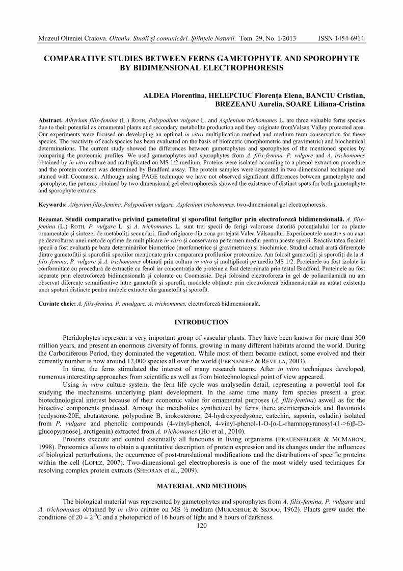
Spore-derived gametophytes maintained on MS medium were homogenized in aseptic conditions to obtain a high number of sporophytes (Fig. 1).
Figure 1. Sporophytes of ferms arising from homogenates of gametophytes: A, spores; B, gametophytes; C, cultures homogenates of gametophytes; D, formation of sporophytes; E, sporophytes; F, P. vulgare
(sporophytes); G, A. trichomanes (sporophyte); H, A. filix-femina (sporophyte).
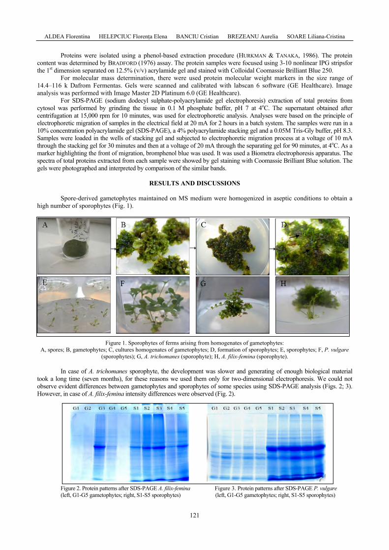
In case of A. trichomanes sporophyte, the development was slower and generating of enough biological material took a long time (seven months), for these reasons we used them only for two-dimensional electrophoresis. We could not observe evident differences between gametophytes and sporophytes of some species using SDS-PAGE analysis (Figs. 2; 3). However, in case of A. filix-femina intensity differences were observed (Fig. 2).
Figure 2. Protein patterns after SDS-PAGE A. filix-femina (left, G1-G5 gametophytes; right, S1-S5 sporophytes)
Figure 3. Protein patterns after SDS-PAGE P. vulgare (left, G1-G5 gametophytes; right, S1-S5 sporophytes)
Muzeul Olteniei Craiova. Oltenia. Studii şi comunicări. Ştiinţele Naturii. Tom. 29, No. 1/2013 ISSN 1454-6914
122
SDS-PAGE is a simple method used to estimate the molecular weight of proteins, but it cannot resolve more than 80-100 different protein components.
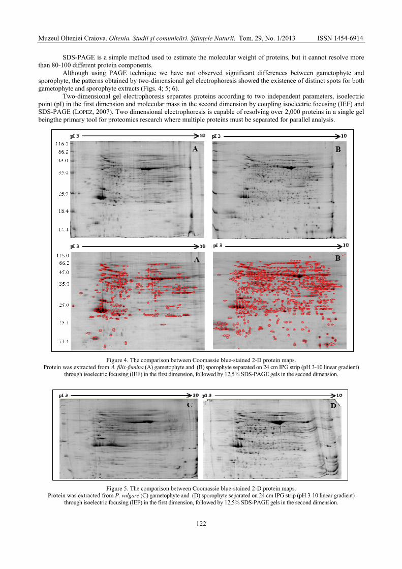
Although using PAGE technique we have not observed significant differences between gametophyte and sporophyte, the patterns obtained by two-dimensional gel electrophoresis showed the existence of distinct spots for both gametophyte and sporophyte extracts (Figs. 4; 5; 6).
Two-dimensional gel electrophoresis separates proteins according to two independent parameters, isoelectric point (pI) in the first dimension and molecular mass in the second dimension by coupling isoelectric focusing (IEF) and SDS-PAGE (LOPEZ, 2007). Two dimensional electrophoresis is capable of resolving over 2,000 proteins in a single gel beingthe primary tool for proteomics research where multiple proteins must be separated for parallel analysis.
Figure 4. The comparison between Coomassie blue-stained 2-D protein maps. Protein was extracted from A. filix-femina (A) gametophyte and (B) sporophyte separated on 24 cm IPG strip (pH 3-10 linear gradient)
through isoelectric focusing (IEF) in the first dimension, followed by 12,5% SDS-PAGE gels in the second dimension.
Figure 5. The comparison between Coomassie blue-stained 2-D protein maps. Protein was extracted from P. vulgare (C) gametophyte and (D) sporophyte separated on 24 cm IPG strip (pH 3-10 linear gradient)
through isoelectric focusing (IEF) in the first dimension, followed by 12,5% SDS-PAGE gels in the second dimension.
ALDEA Florentina HELEPCIUC Florenţa Elena BANCIU Cristian BREZEANU Aurelia SOARE Liliana-Cristina
123
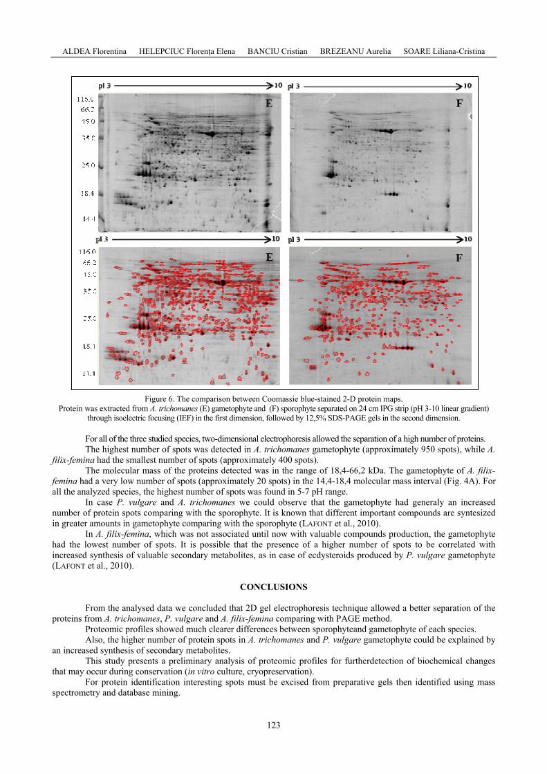
Figure 6. The comparison between Coomassie blue-stained 2-D protein maps. Protein was extracted from A. trichomanes (E) gametophyte and (F) sporophyte separated on 24 cm IPG strip (pH 3-10 linear gradient)
through isoelectric focusing (IEF) in the first dimension, followed by 12,5% SDS-PAGE gels in the second dimension. For all of the three studied species, two-dimensional electrophoresis allowed the separation of a high number of proteins. The highest number of spots was detected in A. trichomanes gametophyte (approximately 950 spots), while A.
filix-femina had the smallest number of spots (approximately 400 spots). The molecular mass of the proteins detected was in the range of 18,4-66,2 kDa. The gametophyte of A. filix-
femina had a very low number of spots (approximately 20 spots) in the 14,4-18,4 molecular mass interval (Fig. 4A). For all the analyzed species, the highest number of spots was found in 5-7 pH range.
In case P. vulgare and A. trichomanes we could observe that the gametophyte had generaly an increased number of protein spots comparing with the sporophyte. It is known that different important compounds are syntesized in greater amounts in gametophyte comparing with the sporophyte (LAFONT et al., 2010).
In A. filix-femina, which was not associated until now with valuable compounds production, the gametophyte had the lowest number of spots. It is possible that the presence of a higher number of spots to be correlated with increased synthesis of valuable secondary metabolites, as in case of ecdysteroids produced by P. vulgare gametophyte (LAFONT et al., 2010).
CONCLUSIONS
From the analysed data we concluded that 2D gel electrophoresis technique allowed a better separation of the
proteins from A. trichomanes, P. vulgare and A. filix-femina comparing with PAGE method. Proteomic profiles showed much clearer differences between sporophyteand gametophyte of each species. Also, the higher number of protein spots in A. trichomanes and P. vulgare gametophyte could be explained by
an increased synthesis of secondary metabolites. This study presents a preliminary analysis of proteomic profiles for furtherdetection of biochemical changes
that may occur during conservation (in vitro culture, cryopreservation). For protein identification interesting spots must be excised from preparative gels then identified using mass
spectrometry and database mining.
Muzeul Olteniei Craiova. Oltenia. Studii şi comunicări. Ştiinţele Naturii. Tom. 29, No. 1/2013 ISSN 1454-6914
124
ACKNOWLEDGEMENTS
We wish to thank Lucyna Domżalska and Damian Makowski for the help with two-dimensional gel electrophoresis technique.
REFERENCES
BRADFORD M. M. 1976. A rapid and sensitive method for the quantitation of microgram quantities of protein utilizing the principle of protein-dye binding. Analytical Biochemistry. http://hoffman.cm.utexas.edu/ courses/bradford_assay.pdf. 72: 248-254. (Accesed: August 07, 2012).
FRAUENFELDER H. & MCMAHON B. 1998. Dynamics and function of proteins: The search for general concepts. Proceedings of the National Academy of Sciences of the United States of America. http://www.pnas.org/content/95/9/4795.full.pdf+html. 95(9): 4795-4797. (Accesed: 20 March,2013).
FERNANDEZ HELENA & REVILLA MARIA ANGELES. 2003. In vitroculture of ornamental ferns. Plant Cell, Tissue and Organ Culture. Kluwer Academic Publishers. Netherlands. 73: 1-13.
HO R., TEAI T., BIANCHINI J., LAFONT R., PHILA RAHARIVELOMANANA. 2010. Ferns: From traditional Uses to Pharmaceutical development, Chemical Identification of Active Principles. In: Working with Ferns, Issues and Applications. Springer. New York: 321-347.
HURKMAN W. J. & TANAKA C. K. 1986. Solubilization of plant membrane proteins for analysis by two-dimensional gel electrophoresis. Plant Physiology. http://www.plantphysiol.org/content/81/3/802.full.pdf+html. 81(3): 802-806. (Accesed: December 03, 2012).
LAFONT R., HO R., PHILA RAHARIVELOMANANA, DINAN L. 2010. Ecdysteroids in Ferns: Distribution, Diversity, Biosynthesis, and Functions. In: Working with Ferns, Issues and Applications. Springer. New York: 305-321.
LÓPEZ J. L. 2007. Two-dimensional electrophoresis in proteome expression analysis. Journal of Chromatography B: “Analytical Tools for Proteomics”. http://www.sciencedirect.com/science/article/pii/ S1570023206010014. 849(12): 190-202. (Accesed: September 28, 2011).
SHEORAN I. S., ROSS A. R. S., OLSON D. J. H., SAWHNEY V. K. 2009. Compatibility of plant protein extraction methods with mass spectrometry for proteome analysis. Plant Science. http://www.sciencedirect.com/science /article/pii/S0168945208002768. 176: 99-104. (Accesed: March 21, 2013).
MURASHIGE T. & SKOOG F. 1965. A revised medium for rapid growth and bioassays with tobacco tissue cultures. Plant Physiology. 15: 437-497.
Aldea Florentina, Helepciuc Florenţa-Elena, Banciu Cristian, Brezeanu Aurelia Institute of Biology Bucharest, Romanian Academy,
296 Splaiul Independenţei, 060031 Bucharest, P.O.Box 56-53, Romania. E-mail: [email protected]
Soare Liliana-Cristina
University of Pitesti, Faculty of Sciences, 1 Târgul din Vale St., 110040, Piteşti, Argeş, Romania.
E-mail: [email protected] Received: March 26, 2013
Accepted: June 20, 2013